. 2022 Aug 10;18(31):5759-5769.
doi: 10.1039/d2sm00415a.

Self-assembly of globular proteins with intrinsically disordered protein polyelectrolytes and block copolymers

Affiliations

Self-assembly of globular proteins with intrinsically disordered protein polyelectrolytes and block copolymers

Justin M Horn et al. Soft Matter. .

Abstract

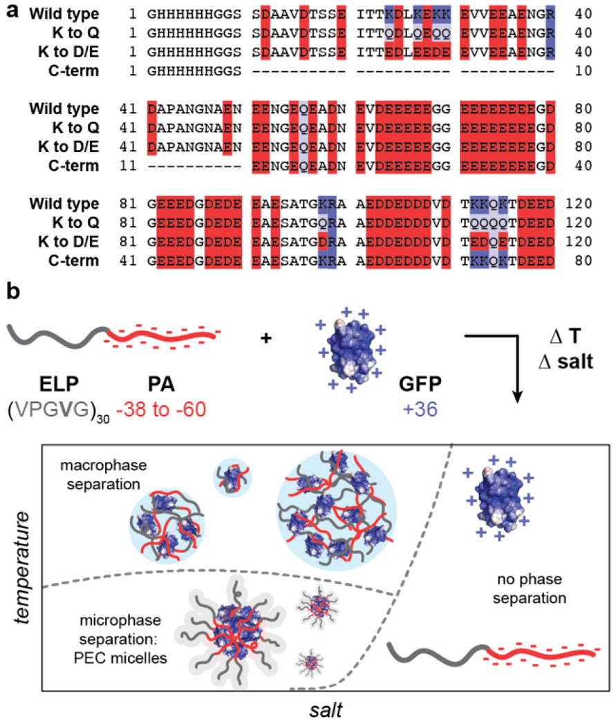
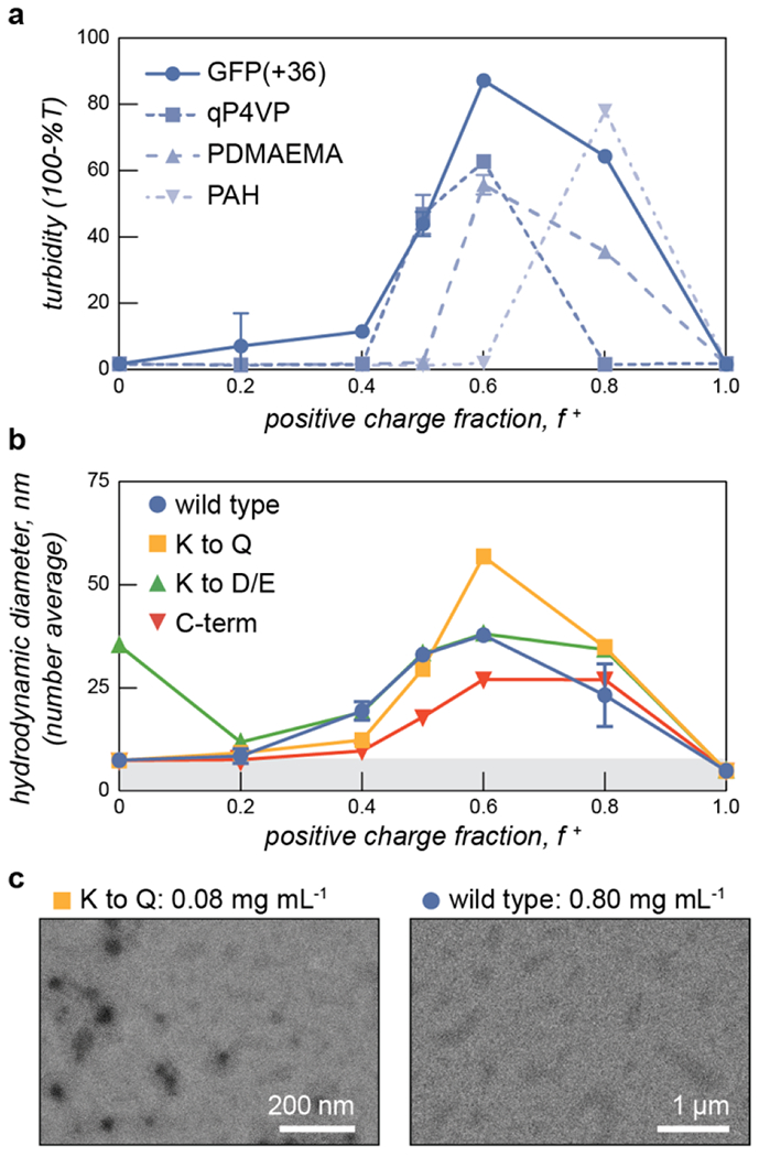
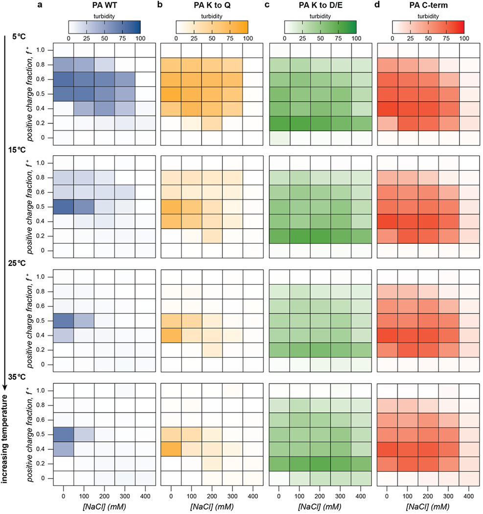
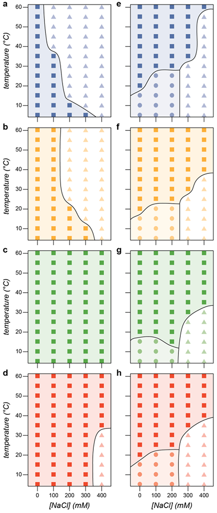
Intrinsically disordered polypeptides are a versatile class of materials, combining the biocompatibility of peptides with the disordered structure and diverse phase behaviors of synthetic polymers. Synthetic polyelectrolytes are capable of complex phase behavior when mixed with oppositely charged polyelectrolytes, facilitating nanoparticle formation and bulk phase separation. However, there has been limited exploration of intrinsically disordered protein polyelectrolytes as potential bio-based replacements for synthetic polyelectrolytes. Here, we produce negatively charged, intrinsically disordered polypeptides, capable of high-yield expression in E. coli and use this intrinsically disordered peptide to produce entirely protein-based polyelectrolyte complexes. The complexes display rich phase behavior, showing sensitivity to charge density, salt concentration, temperature, and charge fraction. We characterize this behavior through a combination of turbidity assays, dynamic light scattering, and transmission electron microscopy. The robust expression profile and stimuli-responsive phase behavior of the intrinsically disordered peptides demonstrates their potential as easily producible, biocompatible substitutes for synthetic polyelectrolytes.


Figures

Figure 1.
Figure 1.
Schematic of prothymosin alpha (PA) variants studied here, (a) Primary amino acid sequence of the four PA variants with acidic residues (D,E) highlighted in red, basic residues (K, R) highlighted in blue, and glutamine residues (Q) highlighted in purple, (b) Fusion of a charge neutral elastin-like polypeptide (ELP) to the N-terminus of the PA variants enables rich phase behavior when complexed with oppositely charged polycationic proteins. A schematic of the phase behavior as a function of salt and temperature is shown.
Figure 2.
Figure 2.
(a) Turbidity assays with PA WT with oppositely charged polyelectrolytes - GFP(+36), qP4VP, PDMAEMA, and PAH show that the protein polyanion phase separates with a range of polycations as evidenced by regions of increased turbidity. Samples were measured at 1.0 mg mL−1 total macromolecule concentration in 10 mM tris buffer, pH 7.4. (b) Dynamic light scattering of ELP-PA variants mixed with the cationic protein GFP(+36) at increasing positive charge fraction shows assemblies with a larger hydrodynamic radius than the consistutent proteins. Samples were measured at 0.8 mg mL−1 total macromolecule concentration in 10 mM tris buffer, pH 7.4. (c) Transmission electron micrographs of representative ELP-PA samples prepared at 5 °C and f + = 0.6 at the indicated sample concentration.
Figure 3.
Figure 3.
The phase behavior of PA variants was assessed by turbidity measurements (λ = 750 nm) as a function of charge fraction, salt concentration, and temperature. Select turbidity data at increasing temperatures has been plotted as heat maps with more saturated hues indicating higher turbidity. Mixtures of each PA variant and GFP(+36) were prepared at 1 mg mL−1 in 10 mM tris buffer, pH 7.4 and were allowed to equilibrate at each temperature for 15 min.
Figure 4.
Figure 4.
The macrophase separation behavior of ELP-PA variants was assessed by turbidity measurements (λ = 750 nm) as a function of charge fraction, salt concentration, and temperature. Select turbidity data at increasing temperatures has been plotted as heat maps with more saturated hues indicating higher turbidity. Mixtures of each ELP-PA variant and GFP(+36) were prepared at 0.8 mg mL−1 in 10 mM tris buffer, pH 7.4 and were allowed to equilibrate at each temperature for 15 min.
Figure 5.
Figure 5.
(a) The hydrodynamic diameter of assmeblies was monitored by DLS as a function of salt concentration for each ELP-PA variant. As salt concentration increased each variant transitioned from microphase separated particles lo turbid solutions. (b) A combination of DLS (solid data points) and turbidity (open data points) results were used to determine the transition temperature for microphase to macrophase separation for each ELP-PA variant as a function of ionic strength. Samples in (a) and (b) were mixed at f + = 0.6 and measured at 0.8 mg mL−1 total macromolecule concentration in 10 mM tris buffer, pH 7.4.
Figure 6.
Figure 6.
Phase diagram at f+ = 0.5 for (a) PA WT (blue), (b) PA K to Q (yellow), (c) PA K to D/E (green), and (d) PA C-term (red). Each of these proteins phase separates with GFP(+36) at low salt concentrations (squares), A single phase region (triangles) is observed in (a), (b), and (d). Phase diagram at f + = 0.6 for (e) ELP-PA WT, (f) ELP-PA K to Q, (g) ELP-PA K to D/E. and (h) ELP-PA C-term. Each of these protein block copolymers forms micelles with GFP(+36) at low temperatures and salt concentrations (circles) and undergoes an order-order transition from microphase separated micelles to macrophase separated particles (squares). Additionally, a single phase region was also observed at high ionic strengths and low temperatures (triangles).

References

    1. Gong J, Chen M, Zheng Y, Wang S and Wang Y, J. Controlled Release, 2012, 159, 312–323. - PubMed
    1. Ghezzi M, Pescina S, Padula C, Santi P, Del Favero E, Cantù L and Nicoli S, J. Controlled Release, 2021, 332, 312–336. - PubMed
    1. Feng Z, Lin L, Yan Z and Yu Y, Macromol. Rapid Commun, 2010, 31, 640–644. - PubMed
    1. Li G, Song S, Zhang T, Qi M and Liu J, Int. J. Biol. Macromol, 2013, 62, 203–210. - PubMed
    1. Kataoka K, Harada A and Nagasaki Y, Adv. Drug Deliv. Rev, 2012, 64, 37–48. - PubMed