. 2022 Jan-Dec:21:15347354221114337.
doi: 10.1177/15347354221114337.

Rosa hybrida Petal Extract Exhibits Antitumor Effects by Abrogating Tumor Progression and Angiogenesis in Bladder Cancer Both In Vivo and In Vitro

Affiliations

Rosa hybrida Petal Extract Exhibits Antitumor Effects by Abrogating Tumor Progression and Angiogenesis in Bladder Cancer Both In Vivo and In Vitro

Byungdoo Hwang et al. Integr Cancer Ther. 2022 Jan-Dec.

Abstract

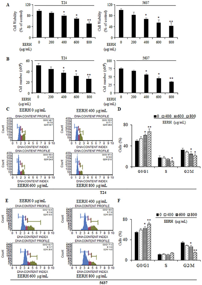
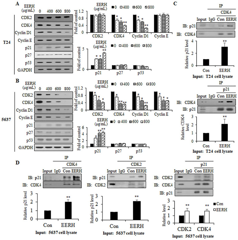
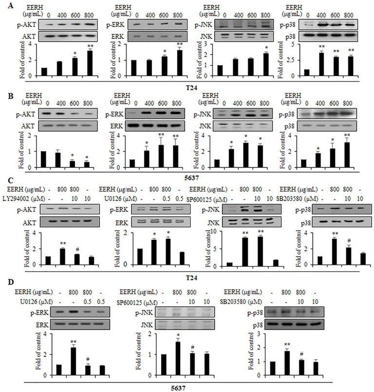
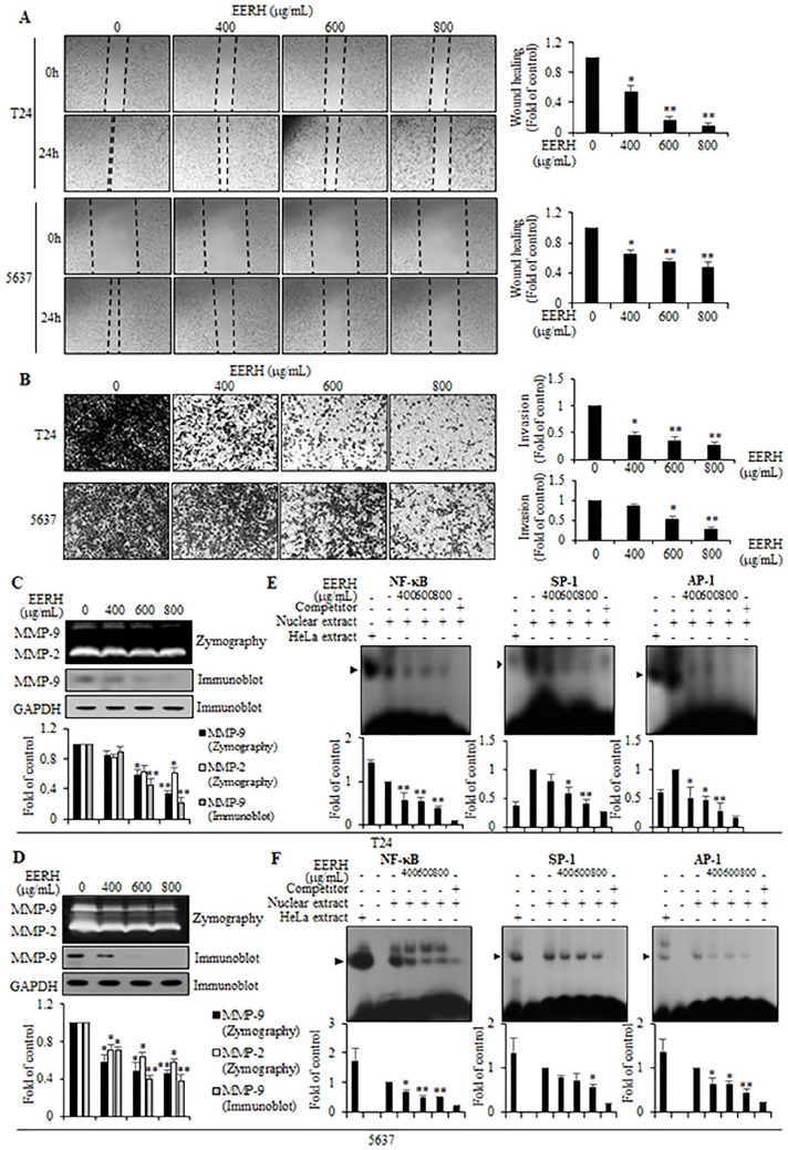
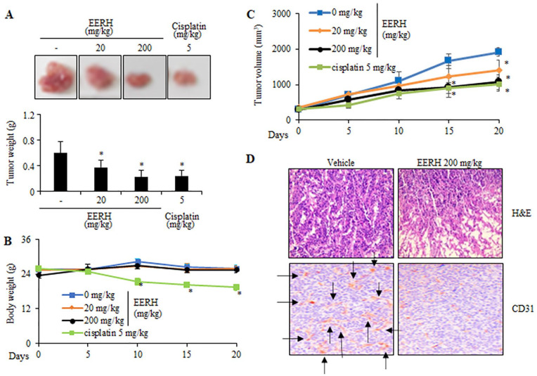
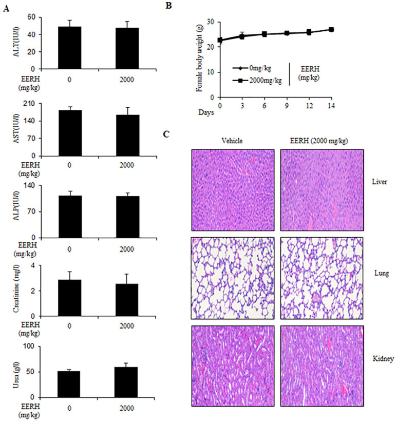
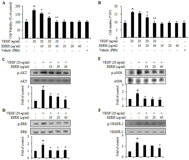
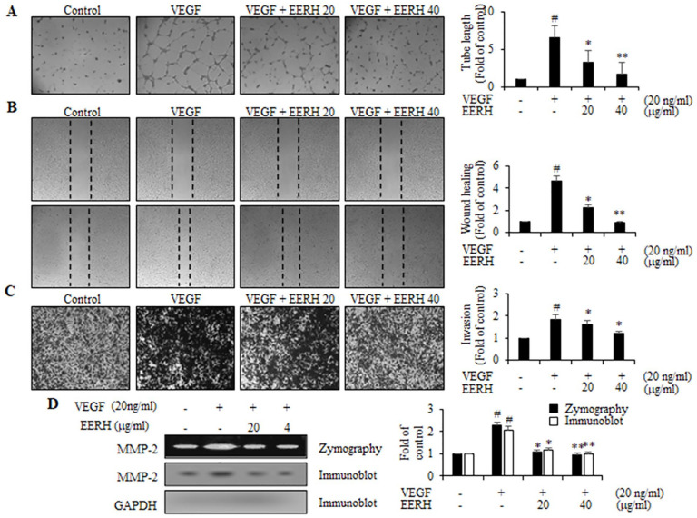
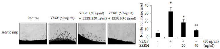
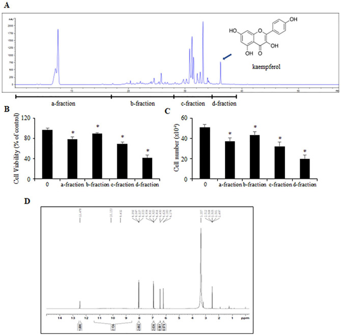
The edible Rosa hybrida (RH) petal is utilized in functional foods and cosmetics. Although the biological function of RH petal extract is known, mechanism of action studies involving tumor-associated angiogenesis have not yet been reported. Herein, we investigated the regulatory effect of the ethanol extract of RH petal (EERH) on tumor growth and tumor angiogenesis against bladder cancer. EERH treatment inhibited the bladder carcinoma T24 cell and 5637 cell proliferation because of G1-phase cell cycle arrest by inducing p21WAF1 expression and reducing cyclins/CDKs level. EERH regulated signaling pathways differently in both cells. EERH-stimulated suppression of T24 and 5637 cell migration and invasion was associated with the decline in transcription factor-mediated MMP-9 expression. EERH oral administration to xenograft mice reduced tumor growth. Furthermore, no obvious toxicity was observed in acute toxicity test. Decreased CD31 levels in EERH-treated tumor tissues led to examine the angiogenic response. EERH alleviated VEGF-stimulated tube formation and proliferation by downregulating the VEGFR2/eNOS/AKT/ERK1/2 cascade in HUVECs. EERH impeded migration and invasion of VEGF-induced HUVECs, which is attributed to the repressed MMP-2 expression. Suppression of neo-microvessel sprouting, induced by VEGF, was verified by treatment with EERH using the ex vivo aortic ring assay. Finally, kaempferol was identified as the main active compound of EERH. The present study demonstrated that EERH may aid the development of antitumor agents against bladder cancer.

Keywords: bladder cancer; bladder cancer cells; ethanol extract of Rosa hybrida; kaempferol; tumor angiogenesis; xenograft mice.


Conflict of interest statement

Declaration of Conflicting Interests: The author(s) declared no potential conflicts of interest with respect to the research, authorship, and/or publication of this article.

Figures

Figure 1.
Figure 1.
Effects of the EERH on cell proliferation and cell cycle in bladder cancer T24 and 5637 cells. T24 cells were incubated with various concentrations of EERH for 24 hours. (A) Viability of T24 and 5637 cells was measured using MTT assay. (B) The number of viable cells counted using a microscope. (C and E) Fluorescence-activated cell sorting (FACS) analysis of EERH-treated cells to determine the histogram of cell cycle pattern. (D and F) Percentages of cell population at each phase of cell cycle. The values are presented as the mean ± standard deviation (SD) of 3 independent experiments; *P < .05 and **P < .01 compared with the untreated control.
Figure 2.
Figure 2.
Effects of EERH on the cell cycle regulatory proteins. (A and B) Immunoblotting results for cyclin D1, cyclin E, cyclin-dependent kinase (CDK)−2, CDK4, p27KIP1, p21WAF1, and p53 for cells treated with indicated concentrations of EERH. Glyceraldehyde 3-phosphate dehydrogenase (GAPDH) was utilized as the internal control. (C and D) Cell lysates from T24 and 5637 cells treated with EERH for 24 hours were immunoprecipitated with antibodies against p21WAF1, CDK2, and CDK4 and then immunocomplexes were subjected to immunoblotting using cell lysates. Normal rabbit IgG and mouse control IgG were used as IgG control. The values are presented as the mean ±SD of 3 independent experiments; *P < .05 and **P < .01 compared with the untreated control.
Figure 3.
Figure 3.
Effects of EERH on the cell signaling pathway. (A and B) Immunoblotting for total and phosphorylated forms of AKT, ERK1/2, JNK, and p38MAPK in cells incubated with EERH for 24 hours. (C and D) Cells were pre-incubated with signaling inhibitors of SB203580 (10 μM), U0126 (0.5 μM), LY 294002 (10 μM), and SP600125 (10 μM) for 40 minutes, followed by EERH treatment for 24 hours. Immunoblotting was performed using cell lysates. Total forms were utilized as internal control. The values are presented as the mean ±SD of 3 independent experiments; *P < .05 and **P < .01 compared with the untreated control.
Figure 4.
Figure 4.
Effects of EERH on the migration, invasion, and transcription factors-controlled matrix metalloproteinase (MMP)-9 expression in bladder cancer cells. Cells were treated with indicated concentrations of EERH. (A) The distance between wounded areas was photographed (40× magnification) and visualized by inverted microscope. (B) Cells that invaded the bottom side of the membrane were stained and counted. (C and D) Activity and expression of MMP-9 was analyzed using zymography and immunoblotting. GAPDH was utilized as internal control. (E and F) Transcriptional activation of Sp-1, AP-1, and NF-κB was determined by electrophoretic mobility shift assay (EMSA) with radiolabeled oligonucleotide probes. All data are expressed as the means ± standard error (SE) of 3 experiments. **P < .05 and **P < .01 compared with the untreated control.
Figure 5.
Figure 5.
Anti-tumor efficacy of oral administration of EERH in the T24 bladder cancer xenograft mice. (A) Growth and weights of representative isolated tumors. (B) Body weights of mice with EERH treatment were compared with those of mice with cisplatin treatment. (C) Tumor volume was measured every 5 days. (D) Staining of growing tumor cells (H&E) and microvessel density (CD31) in tumor tissues. For (A-C), all data are expressed as the mean ± SE of 3 experiments. *P < .05 compared with the untreated control.
Figure 6.
Figure 6.
A single dose acute toxicity test of the mice administered with EERH on 14 days. Mice were orally administered with EERH (2000 mg/kg) for 14 days. (A) Analysis of biochemical test (AST, ALT, ALP, urea, and creatinine) between control and EERH-treated mice. (B) Changes in the body weights were observed in the EERH-treated mice. (C) H&E staining results of 3 main organs (kidney, liver, and lung) in mice orally injected EERH. For (A and B), values are presented as the mean ± SD of 3 independent experiments; *P < .05 compared with the control group.
Figure 7.
Figure 7.
EERH impeded the proliferation and VEGFR2-driven eNOS/AKT/ERK1/2 signaling in the VEGF-stimulated HUVECs. Cells were incubated with indicated concentrations of EERH for 40 minutes, prior to treatment with VEGF (20 ng/mL) for 24 hours. Cell proliferation was determined by both cell viability assay (A) and cell counting assay (B). (C-F) Cell lysates were prepared and phosphorylated level of VEGFR2, eNOS, AKT, and ERK1/2 was evaluated by immunoblot experiment. All data are shown as the means ± SE of 3 experiments. #P < .05 compared with control. *P < .05 and **P < .01 compared with VEGF treatment.
Figure 8.
Figure 8.
EERH suppressed the VEGF-stimulated angiogenic actions including colony tube formation, migration, invasion, and MMP-2 expression in HUVECs. After pre-treatment of cells with various concentrations of EERH for 40 minutes, cells were incubated with VEGF (20 ng/mL) for 24 hours. (A) The effect of EERH on colony tube formation assay using pre-coated Matrigel (scale bars = 200 µm). (B) Migratory potential of EERH was determined by wound-healing migration assay. (C) Transwell plate invasion assay was performed to examine the invasiveness of cells treated with EERH. (D) Gelatin zymographic assay and immunoblot experiment were employed to determine the activity and expression level of MMP-2. GAPDH was utilized as internal control. All data are obtained as the means ±SE of 3 experiments. #P < .05 compared with control. *P < .05 and **P < .01 compared with VEGF treatment.
Figure 9.
Figure 9.
Effects of EERH on the VEGF-induced neovessel sproung using an aortic ring ex vivo model. Representative images of the microvascular sprouting formed in the aortic ring assay between EERH-treated group and control group. A number of neovessel sprouting emerging from aortic ring in each group was photographed and counted. All data are represented as the means ± SE of 3 experiments. #P < .05 compared with control. *P < .05 and **P < .01 compared with VEGF treatment.
Figure 10.
Figure 10.
(A) HPLC chromatogram of EERH and chemical structure of active compound kaempferol. (B and C) MTT assay and cell counting analysis was employed to determine the anti-tumor effect in the sub-fractions of the EtOAc fraction from EERH. (D) 1H-NMR of kaempferol. The values are presented as the mean ± SD of 3 independent experiments; *P < .05 compared with the untreated control.

Similar articles

Cited by

References

    1. Bray F, Ferlay J, Soerjomataram I, Siegel RL, Torre LA, Jemal A. Global cancer statistics 2018: GLOBOCAN estimates of incidence and mortality worldwide for 36 cancers in 185 countries. CA Cancer J Clin. 2018;68:394-424. - PubMed
    1. Kamat AM, Hahn NM, Efstathiou JA, et al.. Bladder cancer. Lancet. 2016;388:2796-2810. - PubMed
    1. Smith AB, Deal AM, Woods ME, et al.. Muscle-invasive bladder cancer: evaluating treatment and survival in the National Cancer Data Base. BJU Int. 2014;114:719-726. - PubMed
    1. Schneider AK, Chevalier MF, Derré L. The multifaceted immune regulation of bladder cancer. Nat Rev Urol. 2019;16:613-630. - PubMed
    1. Black PC, Dinney CP. Bladder cancer angiogenesis and metastasis–translation from murine model to clinical trial. Cancer Metastasis Rev. 2007;26:623-634. - PubMed

Publication types

MeSH terms

Substances