. 2022 Jul 12:(185):10.3791/63476.
doi: 10.3791/63476.

Slicing the Embryonic Chicken Auditory Brainstem to Evaluate Tonotopic Gradients and Microcircuits

Affiliations

Slicing the Embryonic Chicken Auditory Brainstem to Evaluate Tonotopic Gradients and Microcircuits

Sandesh Mohan et al. J Vis Exp. .

Abstract

The chicken embryo is a widely accepted animal model to study the auditory brainstem, composed of highly specialized microcircuitry and neuronal topology differentially oriented along a tonotopic (i.e., frequency) axis. The tonotopic axis permits the segregated encoding of high-frequency sounds in the rostral-medial plane and low-frequency encoding in caudo-lateral regions. Traditionally, coronal brainstem slices of embryonic tissue permit the study of relative individual iso-frequency lamina. Although sufficient to investigate anatomical and physiological questions pertaining to individual iso-frequency regions, the study of tonotopic variation and its development across larger auditory brainstem areas is somewhat limited. This protocol reports brainstem slicing techniques from chicken embryos that encompass larger gradients of frequency regions in the lower auditory brainstem. The utilization of different slicing methods for chicken auditory brainstem tissue permits electrophysiological and anatomical experiments within one brainstem slice, where larger gradients of tonotopic properties and developmental trajectories are better preserved than coronal sections. Multiple slicing techniques allow for improved investigation of the diverse anatomical, biophysical, and tonotopic properties of auditory brainstem microcircuits.


Conflict of interest statement

DISCLOSURES:

All authors declare that the research was conducted without any commercial or financial interest and that they do not have any conflicts of interest.

Figures

Figure 1:
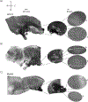
Figure 1:. Representative coronal serial sections of the brainstem.
(A–D) Left: slices from caudal to rostral axis, the auditory nuclei and connecting fibers marked with a white dashed circle. The middle insert is a larger view of the auditory region, where nuclei are shown within white dashed circles a: NM and b: NL. Arrows show auditory nerve afferent fibers, and arrowhead shows NM axon bifurcation in A,B. Arrow shows NA in C. Lateral white dashed circle shows SON in C,D. Right: satellite insert shows these nuclei at 60x objective: a: NM and b: NL. Abbreviations: NM = nucleus magnocellularis; NL = nucleus laminaris; NA = nucleus angularis; SON = superior olivary nucleus; LF = relatively low-frequency neurons; MF = medium-frequency neurons; HF = high-frequency neurons; D = dorsal; L = lateral; V = ventral.
Figure 2:
Figure 2:. Representative sagittal serial sections of the brainstem.
(A–C) Left: slices from lateral to medial axis with the auditory nuclei labeled in a white dashed circle. The middle insert shows the same auditory nuclei region in larger view, marked within white dashed circles. (A) White dashed circle in the center of slice highlights the SON; arrow showing auditory nerve fibers and arrowhead showing NA. A dark black spot at right-side tip of slice is an imaging artifact. Regions of the cerebellum can be seen dorsal to the auditory region in both slices A and B in left panel. (B) A sagittal slice whose orientation was changed to the coronal plane (during slicing). The auditory region was identified with blue dye (black arrow) and again sliced in the sagittal plane. (A–C) Middle insert NM and NL region marked under dashed white lines. Right: satellite view shows a: NM and b: NL observed in 60x objective magnification. LF and HF tonotopic gradient in auditory nuclei is shown along rostro-caudal axis. Arrows pointing to the dark area in (C) show heavily myelinated NM fibers running across the midline through the medial axis. The fibers connect either side of auditory nuclei. Abbreviations: NM = nucleus magnocellularis; NL = nucleus laminaris; NA = nucleus angularis; SON = superior olivary nucleus; LF = relatively low-frequency neurons; HF = high-frequency neurons; D = dorsal; V = ventral; R = rostral; C = caudal.
Figure 3:
Figure 3:. Electrophysiological recordings of neuronal response to somatic current injections (−100 pA to +200 pA, +10 pA increments, 100 ms duration) in current clamp mode.
Neurons were selected for recordings in the same slice but at extreme opposite regions of NM. (A,B) Representative neuronal responses in a single coronal slice indicating relative iso-frequency properties with subtle differences. Response properties represent two different MF neurons recorded from the most medial (A) and lateral (B) regions of NM in a coronal slice. (C,D) Representative neuronal recordings from a single sagittal slice. The recordings show a relatively LF NM response (C) and a HF NM response (D), highlighting the substantive differences in tonotopic gradient along within a single sagittal section. Abbreviations: NM = nucleus magnocellularis; LF = relatively low-frequency neurons; MF = mid-frequency neurons; HF = high-frequency neurons.
Figure 4:
Figure 4:. Representative horizontal serial sections of the brainstem.
(A,B) Left: slices along the dorsal to ventral axis, auditory nuclei are marked with white dashed circles. The 8th cranial nerve afferent fibers connect auditory nuclei marked with arrow. The middle insert is a larger view of auditory nuclei region with auditory nuclei marked under white dashed lines. A: NM and b: NL regions are shown. A clear topological movement of auditory nuclei can be seen in A,B. (A,B) Right: large satellite view showing a: NM and b: NL. Right insert shows auditory nuclei observed in 60x objective magnification and the curved topological axis from LF to HF along a caudo-lateral to rostral-medial axis. Abbreviations: NM = nucleus magnocellularis; NL = nucleus laminaris; LF = relatively low-frequency neurons; HF = high-frequency neurons; L = lateral; R = rostral; C = caudal.
Figure 5:
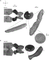
Figure 5:. Representative horizontal/transverse acute angular (45°) serial sections.
(A–C) Left: serial sections of the brainstem, auditory nuclei marked in white dashed circle. The middle insert is a larger view of the auditory region. (A) Middle insert shows the largest spread of NM and NL neurons in these slices. (B,C) Middle insert: auditory nuclei marked in white dashed lines show gradual topological change when compared to (A–C). Right: satellite insert showing auditory nuclei a: NM and b: NL in 60x objective magnification. The tonotopic axis from LF to HF regions in NM and NL rotates angularly from lateral to medial slices. Abbreviations: NM = nucleus magnocellularis; NL = nucleus laminaris; LF = relatively low-frequency neurons; HF = high-frequency neurons; V = ventral; R = rostral; D = dorsal; C = caudal.

Similar articles

References

    1. Rubel EW, Parks TN Organization and development of brain stem auditory nuclei of the chicken: tonotopic organization of n. magnocellularis and n. laminaris. Journal of Comparative Neurology 164 (4), 411–433 (1975). - PubMed
    1. Rubel EW et al. Organization and development of brain stem auditory nuclei of the chicken: ontogeny of n. magnocellularis and n. laminaris. Journal of Comparative Neurology 166 (4), 469–489 (1976). - PubMed
    1. Shao M et al. Spontaneous synaptic activity in chick vestibular nucleus neurons during the perinatal period. Neuroscience 127 (1), 81–90 (2004). - PubMed
    1. Fukui I, Ohmori H Tonotopic gradients of membrane and synaptic properties for neurons of the chicken nucleus magnocellularis. Journal of Neuroscience 24 (34), 7514–7523 (2004). - PMC - PubMed
    1. Sanchez JT, Seidl AH, Rubel EW, Barria A Preparation and culture of chicken auditory brainstem slices. Journal of Visualized Experiments (49), 2527 (2011). - PMC - PubMed

Publication types

LinkOut - more resources