. 2022 Aug 30;13(4):e0093522.
doi: 10.1128/mbio.00935-22. Epub 2022 Aug 1.

Structure and Function of BcpE2, the Most Promiscuous GH3-Family Glucose Scavenging Beta-Glucosidase

Affiliations

Structure and Function of BcpE2, the Most Promiscuous GH3-Family Glucose Scavenging Beta-Glucosidase

Benoit Deflandre et al. mBio. .

Abstract

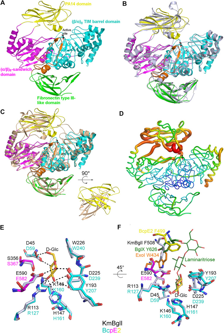
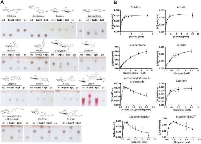
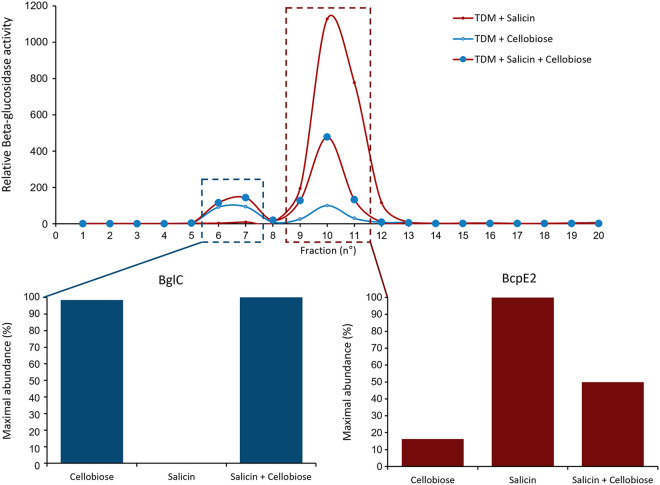
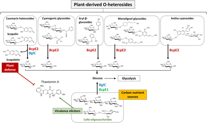
Cellulose being the most abundant polysaccharide on earth, beta-glucosidases hydrolyzing cello-oligosaccharides are key enzymes to fuel glycolysis in microorganisms developing on plant material. In Streptomyces scabiei, the causative agent of common scab in root and tuber crops, a genetic compensation phenomenon safeguards the loss of the gene encoding the cello-oligosaccharide hydrolase BglC by awakening the expression of alternative beta-glucosidases. Here, we revealed that the BglC compensating enzyme BcpE2 was the GH3-family beta-glucosidase that displayed the highest reported substrate promiscuity and was able to release the glucose moiety of all tested types of plant-derived heterosides (aryl β-glucosides, monolignol glucosides, cyanogenic glucosides, anthocyanosides, and coumarin heterosides). BcpE2 structure analysis highlighted a large cavity in the PA14 domain that covered the active site, and the high flexibility of this domain would allow proper adjustment of this cavity for disparate heterosides. The exceptional substrate promiscuity of BcpE2 provides microorganisms a versatile tool for scavenging glucose from plant-derived nutrients that widely vary in size and structure. Importantly, scopolin was the only substrate commonly hydrolyzed by both BglC and BcpE2, thereby generating the potent virulence inhibitor scopoletin. Next to fueling glycolysis, both enzymes would also fine-tune the strength of virulence. IMPORTANCE Plant decaying biomass is the most abundant provider of carbon sources for soil-dwelling microorganisms. To optimally evolve in such environmental niches, microorganisms possess an arsenal of hydrolytic enzymatic complexes to feed on the various types of polysaccharides, oligosaccharides, and monosaccharides. In this work, structural, enzymatic, and expression studies revealed the existence of a "swiss-army knife" enzyme, BcpE2, that was able to retrieve the glucose moiety of a multitude of plant-derived substrates that vary in size, structure, and origin. This enzyme would provide the microorganisms with a tool that would allow them to find nutrients from any type of plant-derived material.

Keywords: carbon metabolism; enzyme promiscuity; genetic compensation; glycosyl hydrolase; host-pathogen interaction; plant heterosides.


Conflict of interest statement

The authors declare no conflict of interest.

Figures

FIG 1
FIG 1
Overall fold and active site description of BcpE2 of S. scabiei. (A) Cartoon representation of the BcpE2 structure. The glycerol molecule in the active site (black sticks) results from the cryo-protectant solution used for freezing the crystal. (B) Superimposition of BcpE2 and KmBglI (light blue). (C) Superimposition of BcpE2 and DesR (light orange). Their PA14 is also shown with a 90° rotation to highlight the different orientations. (D) Ribbon representation of BcpE2. The ribbon radius was proportional to the mean B-factor value of the residues and a rainbow coloring scheme (blue low B factor value to red high B-factor value). (E) Superimposition of the catalytic site of BcpE2 (residues are colored by domain with the same coloring scheme as in (B) with KmBglI (gray) in complex with d-Glucose (d-Glc in gold)) in subsite −1. Hydrogen bonds with d-Glc are shown as black dashed lines. (F) Same as (E) but with a 45° rotation and the addition of ExoI (orange) and the BglX:Laminaritriose complex (green) superimposed. The aromatic residues of subsite +1 of KmBglI and BglX are also shown as sticks, as well as their likely equivalent in ExoI and BcpE2. The position of F499 in BcpE2 (yellow sticks) comes from the PA14 domain superimposed independently on the PA14 domain of KmBglI.
FIG 2
FIG 2
Substrate specificities of BcpE2 and BglC. (A) TLC plates revealed the release of glucose (glc) after incubation of a variety of substrates (5 mM) with BcpE2 or BglC (1 μM) compared to the intact substrate (standard [std]). The chemical structure is displayed above each substrate. Note that for scopolin, esculin, and 4-MUG the substrates are in the solvent front. (B) Nonlinear regressions of the kinetic analyses of BcpE2 toward seven substrates and one for BglC. The initial velocity (Vi, mM/min) was estimated by the rate of glucose released by the enzyme as a function of substrate concentrations (in mM). Individual values were entered into the GraphPad Prism software (9.2.0) which fitted the data to the Henri-Michaelis-Menten model by nonlinear regression. In the case of a decrease of the Vi at high substrate concentrations, the data were fitted to the Substrate Inhibition model. Error bars display the standard deviation values determined for the Vi by three replicates at each substrate concentration.
FIG 3
FIG 3
Induction of the respective production of BglC and BcpE2 by cellobiose and salicin. (Top) Relative beta-glucosidase activity in anion-exchange chromatography fractions obtained from the full protein extracts of S. scabiei cultured in TDM medium supplemented with salicin (red trait), cellobiose (blue trait), or both substrates (red trait with blue circles). (Bottom) Relative abundance of BglC in the first active peak (fractions 6 to 7) and of BcpE2 in the second active peak (fractions 9 to 11) was determined by targeted proteomics (LC-MRM [Liquid chromatography multiple reaction monitoring] after tryptic digestion of the protein fractions). In each culture condition, the relative protein abundance was reported to the maximal abundance measured for the given protein (see the Materials and methods section for the detailed protocol).
FIG 4
FIG 4
Active site flexibility of BcpE2. (A) Cartoon representation of the BcpE2 active site and the PA14 domain. Loops with missing amino acids are shown as dashed lines. Residues with a B factor of the Cα above 100 Å2 are in red and the others are in blue. The active site pocket is represented with a transparent surface with the d-glucose molecule in the (+1) subsite from the superimposed KmBglII structure as yellow sticks. (B) Same as in (A) but with a 90° rotation.
FIG 5
FIG 5
BcpE1 and BcpE2-mediated enzymatic compensation for the loss of BglC. BcpE1 (green) can compensate for the activity of BglC (blue) by generating glucose from the hydrolysis of cello-oligosaccharides cellobiose and cellotriose. BcpE2 (red) can also fuel glycolysis by removing glucose from multiple plant heterosides. In addition, BcpE2 can also compensate for the role of BglC in plant defense mechanism by displaying a substrate inhibition kinetic profile on scopolin, thereby generating the potent thaxtomin A production inhibitor scopoletin.

References

    1. Hodgson DA. 2000. Primary metabolism and its control in streptomycetes: a most unusual group of bacteria. Adv Microb Physiol 42:47–238. doi:10.1016/s0065-2911(00)42003-5. - DOI - PubMed
    1. Book AJ, Lewin GR, McDonald BR, Takasuka TE, Wendt-Pienkowski E, Doering DT, Suh S, Raffa KF, Fox BG, Currie CR. 2016. Evolution of high cellulolytic activity in symbiotic streptomyces through selection of expanded gene content and coordinated gene expression. PLoS Biol 14:e1002475. doi:10.1371/journal.pbio.1002475. - DOI - PMC - PubMed
    1. Lewin GR, Carlos C, Chevrette MG, Horn HA, McDonald BR, Stankey RJ, Fox BG, Currie CR. 2016. Evolution and ecology of actinobacteria and their bioenergy applications. Annu Rev Microbiol 70:235–254. doi:10.1146/annurev-micro-102215-095748. - DOI - PMC - PubMed
    1. Chater KF, Biró S, Lee KJ, Palmer T, Schrempf H. 2010. The complex extracellular biology of Streptomyces. FEMS Microbiol Rev 34:171–198. doi:10.1111/j.1574-6976.2009.00206.x. - DOI - PubMed
    1. Lakhundi S, Siddiqui R, Khan NA. 2015. Cellulose degradation: a therapeutic strategy in the improved treatment of Acanthamoeba infections. Parasit Vectors 8:23. doi:10.1186/s13071-015-0642-7. - DOI - PMC - PubMed

Publication types