. 2022 Aug 31;10(4):e0095022.
doi: 10.1128/spectrum.00950-22. Epub 2022 Aug 1.

Hibifolin, a Natural Sortase A Inhibitor, Attenuates the Pathogenicity of Staphylococcus aureus and Enhances the Antibacterial Activity of Cefotaxime

Affiliations

Hibifolin, a Natural Sortase A Inhibitor, Attenuates the Pathogenicity of Staphylococcus aureus and Enhances the Antibacterial Activity of Cefotaxime

Wu Song et al. Microbiol Spectr. .

Abstract

This study aimed to identify hibifolin as a sortase A (SrtA) inhibitor and to determine whether it could attenuate the virulence of methicillin-resistant Staphylococcus aureus (MRSA). We employed a fluorescence resonance energy transfer (FRET) assay to screen a library of natural molecules to identify compounds that inhibit SrtA activity. Fluorescence quenching assay and molecular docking were performed to verify the direct binding interaction between SrtA and hibifolin. The pneumonia model was established using C57BL/6J mice by MRAS nasal administration for evaluating the effect of hibifolin on the pathogenicity of MRSA. Herein, we found that hibifolin was able to inhibit SrtA activity with an IC50 of 31.20 μg/mL. Further assays showed that the capacity of adhesion of bacteria to the host cells and biofilm formation was decreased in hibifolin-treated USA300. Results obtained from fluorescence quenching assay and molecular docking indicated that hibifolin was capable of targeting SrtA protein directly. This interaction was further confirmed by the finding that the inhibition activities of hibifolin on mutant SrtA were substantially reduced after mutating the binding sites (TRP-194, ALA-104, THR-180, ARG-197, ASN-114). The in vivo study showed that hibifolin in combination with cefotaxime protected mice from USA300 infection-induced pneumonia, which was more potent than cefotaxime alone, and no significant cytotoxicity of hibifolin was observed. Taken together, we identified that hibifolin attenuated the pathogenicity of S. aureus by directly targeting SrtA, which may be utilized in the future as adjuvant therapy for S. aureus infections. IMPORTANCE We identified hibifolin as a sortase A (SrtA) inhibitor by screening the natural compounds library, which effectively inhibited the activity of SrtA with an IC50 value of 31.20 μg/mL. Hibifolin attenuated the pathogenic behavior of Staphylococcus aureus, including adhesion, invasion, and biofilm formation. Binding assays showed that hibifolin bound to SrtA protein directly. Hibifolin improved the survival of pneumonia induced by S. aureus USA300 in mice and alleviated the pathological damage. Moreover, hibifolin showed a synergistic antibacterial effect with cefotaxime in USA300-infected mice.

Keywords: cefotaxime; hibifolin; methicillin-resistant Staphylococcus aureus; pneumonia; sortase A.


Conflict of interest statement

The authors declare no conflict of interest.

Figures

FIG 1
FIG 1
The inhibitory effect of the hibifolin on SrtA activity. (A) FRET and CETSA were used to screen inhibitors from natural small molecule compounds. (B) The chemical structure of hibifolin. (C) The effect of hibifolin on SrtA activity with IC50 of 31.20 μg/mL. (D) Growth curves of S. aureus USA300 with or without hibifolin treatment (256 μg/mL). The viability of HepG2 (E), HEK-293T (F), and A549 (G) cells viability measured by MTT assay after being exposed to various concentrations of hibifolin (0 to 256 μg/mL).
FIG 2
FIG 2
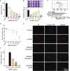
Effects of hibifolin on the sortase A-related virulence factors. (A) Multiple concentrations of hibifolin suppressed the adherence of S. aureus USA 300 to fibrinogen protein. (B) Biofilm formation is observed and quantified by staining with crystal violet. Hibifolin suppresses the S. aureus USA300 biofilm formation. (C) S. aureus surface protein (SpA) level is measured by flow cytometry using FITC-conjugated rabbit IgG. ΔsrtA and USA300 represent a positive-control group and a negative-control group, respectively. (D) Different concentrations of hibifolin-treated S. aureus significantly inhibited bacterial invasion of A549 cells. (E) Live/dead staining confirmed that hibifolin reduced damage to A549 cells by S. aureus, and consistent results were obtained from LDH release assays (F).
FIG 3
FIG 3
Determination of the binding sites for the hibifolin on SrtA. (A) Expression of different concentrations of hibifolin on SrtA expression by Western blot, which shows no significant effect. (B) The substrate-binding affinity of hibifolin was measured by SrtA fluorescence quenching. SrtA was treated with a 10-fold IC50 of hibifolin and then diluted the concentration. The group without hibifolin is assumed as 100% activity and the binding affinity is indicated by KA. (C) Reversible inhibitory effect of hibifolin on SrtA. (D) Molecular modeling predicted the interaction between hibifolin and SrtA. (E) Five mutants TRP-194, ALA-104, THR-180, ASN-114, and ARG-197 showed increasing resistance to hibifolin inhibition. Relative activities of recombinant SrtA were quantified via FRET assays.
FIG 4
FIG 4
Protective effect of hibifolin and cefotaxime combination against lethal pneumonia in MRSA-infected mice. (A) Flow chart of MRSA-induced mouse pneumonia infection model. (B) Survival rate of C57BL/6J mice treated with hibifolin (100 mg/kg/d) or combination (hibifolin [100 mg/kg/d] + cefotaxime [40 mg/kg/d]) which infected with a lethal dose of S. aureus USA300. (C) CFU number in lung tissues. (D) Gross pathological changes in the lungs were observed by HE staining. Scale bar, 1 cm and 100 μm, respectively. (E–G) The levels of inflammatory cytokines (IFN-γ; IL-6; TNF-α) in the alveolar lavage fluid of each group of mice were determined by ELISA. **, P < 0.01; ***, P < 0.001 compared with the USA300 group; #, P < 0.05, comparisons were considered statistically significant.
FIG 5
FIG 5
Hibifolin reduced the formation of biofilm of S. aureus and attenuated the adhesion and invasion to the host and the anchoring of surface proteins by inhibiting SrtA.

Similar articles

Cited by

References

    1. Tam K, Torres VJ. 2019. Staphylococcus aureus secreted toxins and extracellular enzymes. Microbiol Spectr 7:16. doi:10.1128/microbiolspec.GPP3-0039-2018. - DOI - PMC - PubMed
    1. Blair JM, Webber MA, Baylay AJ, Ogbolu DO, Piddock LJ. 2015. Molecular mechanisms of antibiotic resistance. Nat Rev Microbiol 13:42–51. doi:10.1038/nrmicro3380. - DOI - PubMed
    1. Bai Z, Chen M, Lin Q, Ye Y, Fan H, Wen K, Zeng J, Huang D, Mo W, Lei Y. 2021. Identification of methicillin-resistant Staphylococcus aureus from methicillin-sensitive Staphylococcus aureus and molecular characterization in Quanzhou. China Front Cell Dev Biol 9. - PMC - PubMed
    1. Tkaczyk C, Hamilton MM, Sadowska A, Shi Y, Chang CS, Chowdhury P, Buonapane R, Xiao X, Warrener P, Mediavilla J, Kreiswirth B, Suzich J, Stover CK, Sellman BR. 2016. Targeting alpha toxin and ClfA with a multimechanistic monoclonal-antibody-based approach for prophylaxis of serious Staphylococcus aureus disease. mBio 7:e00528-16. doi:10.1128/mBio.00528-16. - DOI - PMC - PubMed
    1. DiGiandomenico A, Sellman BR. 2015. Antibacterial monoclonal antibodies: the next generation? Curr Opin Microbiol 27:78–85. doi:10.1016/j.mib.2015.07.014. - DOI - PubMed

Publication types