Case Reports
. 2022 Dec 16;31(24):4121-4130.
doi: 10.1093/hmg/ddac156.

Newly identified disorder of copper metabolism caused by variants in CTR1, a high-affinity copper transporter

Affiliations
Case Reports

Newly identified disorder of copper metabolism caused by variants in CTR1, a high-affinity copper transporter

Spyros Batzios et al. Hum Mol Genet. .

Abstract

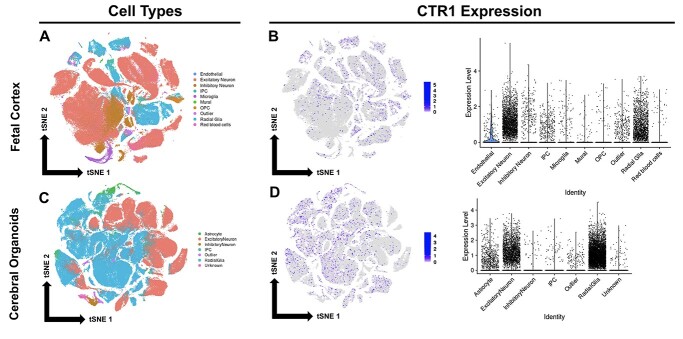
The high-affinity copper transporter CTR1 is encoded by CTR1 (SLC31A1), a gene locus for which no detailed genotype-phenotype correlations have previously been reported. We describe identical twin male infants homozygous for a novel missense variant NM_001859.4:c.284 G > A (p.Arg95His) in CTR1 with a distinctive autosomal recessive syndrome of infantile seizures and neurodegeneration, consistent with profound central nervous system copper deficiency. We used clinical, biochemical and molecular methods to delineate the first recognized examples of human CTR1 deficiency. These included clinical phenotyping, brain imaging, assays for copper, cytochrome c oxidase (CCO), and mitochondrial respiration, western blotting, cell transfection experiments, confocal and electron microscopy, protein structure modeling and fetal brain and cerebral organoid CTR1 transcriptome analyses. Comparison with two other critical mediators of cellular copper homeostasis, ATP7A and ATP7B, genes associated with Menkes disease and Wilson disease, respectively, revealed that expression of CTR1 was highest. Transcriptome analyses identified excitatory neurons and radial glia as brain cell types particularly enriched for copper transporter transcripts. We also assessed the effects of Copper Histidinate in the patients' cultured cells and in the patients, under a formal clinical protocol. Treatment normalized CCO activity and enhanced mitochondrial respiration in vitro, and was associated with modest clinical improvements. In combination with present and prior studies, these infants' clinical, biochemical and molecular phenotypes establish the impact of this novel variant on copper metabolism and cellular homeostasis and illuminate a crucial role for CTR1 in human brain development. CTR1 deficiency represents a newly defined inherited disorder of brain copper metabolism.


Figures

Figure 1
Figure 1
Molecular and clinical findings in twins with CTR1R95H. (A) DNA sequence analysis of CTR1 in the twins. Both parents are heterozygous for the R95H variant (data not shown). (B) Growth charts for Twins A and B according to World Health Organization standards for weight, length and head circumference percentiles from birth to 24 months. Blue arrows denote initiation of Copper Histidinate treatment; note increased velocity of weight gain associated with treatment. (C) Axial T2-weighted (left) and sagittal T1-weighted (right) brain MRI images at 2 years of age showing ventriculomegaly, and extensive cortical and cerebellar atrophy.
Figure 2
Figure 2
Characterization of CTR1. (A) Homology model of the human CTR1 homotrimer. The main chain is colored according to the sequence identity with the template structure (6m98.pdb), gold where identical (77%) and light blue where different. Arg 95 is shown as space-filling. At left is a view in the membrane plane and, on the right, a view from the intracellular side obtained by 90° rotation. Copper and zinc ions are shown as green and purple spheres, respectively. Only crystals grown in conditions containing zinc acetate diffracted X-rays well and allowed the CTR1 structure determination on which this homology model is based (13). CTR1 is not a known transporter of zinc, however. (B) Confocal imaging of transfected HEK293T cells, revealing normal plasma membrane localization of CTR1R95H. (C) Expression of endogenous CTR1 in primary dermal fibroblasts derived from the affected siblings and from healthy controls. Cell lysates were analyzed by western blotting with the indicated antibodies. A representative blot from among several performed is shown (top), together with a bar graph depicting densitometric analysis of CTR1 expression normalized to beta-actin (P = 0.0185). Error bars = SEM. (D) Representative electron microscopy images of control and patient fibroblasts. Patient fibroblasts exhibit markedly dilated endoplasmic reticulum (yellow arrows).
Figure 3
Figure 3
Treatment with copper histidinate (CuHis) enhances mitochondrial function in CTR1R95H fibroblasts. (A) Reduced fibrobast copper content in the patients’ cultured fibroblasts compared with normal control cells. Error bars = SD. (B) CCO activity measured spectrophotometrically by loss of ferrocytochrome c following addition of fresh fibroblast lysates normalized to total protein. Results of three independent experiments are shown, with data from two control, both patients’ and CuHis-treated both patients’ cells pooled. The results indicate reduced activity in the patients’ cells (P < 0.0001), and increased activity with 50 μM CuHis treatment. (C,D) Mitochondrial function in primary dermal fibroblasts as measured by a Seahorse XF Analyzer. Results from a representative experiment from among several are depicted as a scatter plot with OCR plotted against time (top). At the time points indicated by dotted lines, the cells were exposed to oligomycin (Olig), carbonyl cyanide-4-(trifluoromethoxy)phenyl-hydrazone (FCCP) and a combination of rotenone and antimycin A (R&A). Mean results from three independent experiments are depicted, with data from affected sibling fibroblasts pooled. The following are plotted: basal respiration (BR, P = 0.0001, P = 0.0299 after treatment), ATP-linked respiration (ATP, P = 0.0012, P = 0.0419 after treatment), maximal respiration (MR, P < 0.0001, P < 0.0001 after treatment), spare capacity (SC, P < 0.0001, P < 0.0001 after treatment), proton leak (PL, P = 0.7513, P = 0.9884 after treatment) and non-mitochondrial oxygen consumption (NM, P = 0.0050, P = 0.9122 after treatment). Error bars = SE
Figure 4
Figure 4
Representation of CTR1 in fetal brain cortex and CO transcriptomes. (A) t-distributed stochastic neighbor embedding (t-SNE) plot generated from fetal brain cortex single cell RNA sequencing (scRNA-seq) data. Each point on the tSNE represents a single cell (189 409 cells shown). Cells are color-coded to specify cell type. (B) CTR1 expression in fetal brain cortex. Left, tSNE feature plot. Right, violin plots grouped by cell cluster identity. (C) t-SNE plot generated from COs single cell RNA sequencing (scRNA-seq) data. Cells are color-coded to specify cell type. (D) CTR1 expression in COs. Left, tSNE feature plot. Right, violin plots grouped by cell cluster identity.
Figure 5
Figure 5
CTR1 expression exceeds copper exporters ATP7A and ATP7B in COs. (A) Bulk expression of CTR1, ATP7A and ATP7B in day 93 and 140 COs. (B) Single cell transcriptomic analysis of CTR1, ATP7A and ATP7B in COs generated from a publicly available dataset of 235 121 single cells from 37 COs cultured across a developmental time window spanning 3–10 weeks (19,20). See text for further details.
Figure 6
Figure 6
Models of systemic and central nervous system copper uptake. The left panel shows a working model of gastrointestinal copper uptake in which CTRl is primarily active at the basolateral membranes of enterocytes. CTR1-mediated copper absorption could also occur at the luminal (apical) aspect, however, a number of alternative copper uptake mechanisms have been described. The latter would explain how the infants described here (and mouse models) with defective CTR1 maintain normal blood copper levels. The copper chaperone ATOX1 ferries Cu to the Cu exporter, ATP7A, defective in Menkes disease, which normally mediates copper passage from enterocytes into the blood. The middle panel shows a model of choroid plexus-mediated Cu uptake to the brain in which CTR1 is also localized predominantly at the basolateral aspect of polarized choroid plexus epithelia, reflecting the intense expression documented in this tissue. The well-fenestrated capillaries of the choroid plexuses enable passive Cu exodus from blood to the basolateral epithelial surfaces where CTR1 is presumed to mediate uptake to the epithelial cells. This enables delivery of Cu to ATP7A at the opposite pole, via the cytosolic copper chaperone ATOX1, for delivery to the cerebrospinal fluid (CSF). This model is highly consistent with prior human (15) and murine (24) studies that document copper entry to the developing brain as primarily choroid plexus-mediated. The location and orientation of the Wilson disease gene product, ATP7B, at the basolateral aspect of choroid plexus epithelia are consistent with the toxic brain copper accumulation that occurs in that illness, when untreated (29). The right panel shows a model of copper transport at the blood–brain barrier that we propose is less important based on prior evidence. Tight junctions are present in brain capillary endothelial cells in contrast to choroid plexus and GI tract capillary endothelia, which are well-fenestrated and facilitate passive Cu transport.

References

    1. Festa, R.A. and Thiele, D.J. (2011) Copper: an essential metal in biology. Curr. Biol., 21, R877–R883. - PMC - PubMed
    1. Inesi, G. (2017) Molecular features of copper binding proteins involved in copper homeostasis. IUBMB Life, 69, 211–217. - PubMed
    1. Zhou, B. and Gitschier, J. (1997) hCTR1: a human gene for copper uptake identified by complementation in yeast. Proc. Natl. Acad. Sci. U. S. A., 94, 7481–7486. - PMC - PubMed
    1. Tsang, T., Posimo, J.M., Gudiel, A.A., Cicchini, M., Feldser, D.M. and Brady, D.C. (2020) Copper is an essential regulator of the autophagic kinases ULK1/2 to drive lung adenocarcinoma. Nat. Cell Biol., 22, 412–424. - PMC - PubMed
    1. Ge, E., Bush, A., Casini, A., Cobine, P., Cross, J., DeNicola, G., Dou, Q.P., Franz, K.J., Gohil, V.M., Gupta, S. et al. (2022) Connecting copper and cancer: from transition metal signaling to metalloplasia. Nat. Rev. Cancer, 22, 102–113. - PMC - PubMed

Publication types