. 2022 Aug 1;18(8):e1010331.
doi: 10.1371/journal.pgen.1010331. eCollection 2022 Aug.

PrrA modulates Mycobacterium tuberculosis response to multiple environmental cues and is critically regulated by serine/threonine protein kinases

Affiliations

PrrA modulates Mycobacterium tuberculosis response to multiple environmental cues and is critically regulated by serine/threonine protein kinases

David Giacalone et al. PLoS Genet. .

Abstract

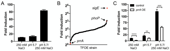
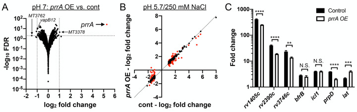
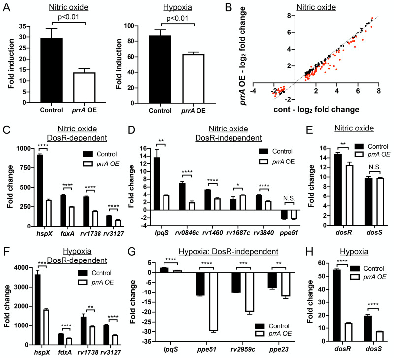
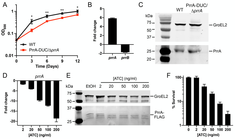
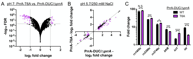
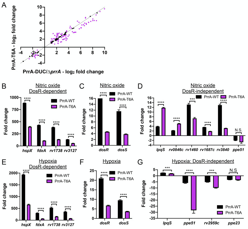
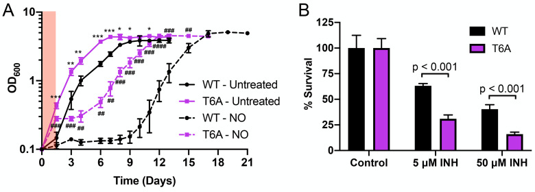
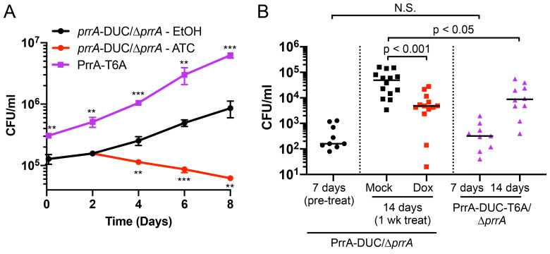
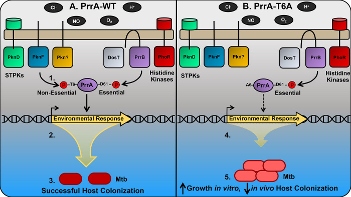
The ability of Mycobacterium tuberculosis (Mtb) to adapt to its surrounding environment is critical for the bacterium to successfully colonize its host. Transcriptional changes are a vital mechanism by which Mtb responds to key environmental signals experienced, such as pH, chloride (Cl-), nitric oxide (NO), and hypoxia. However, much remains unknown regarding how Mtb coordinates its response to the disparate signals seen during infection. Utilizing a transcription factor (TF) overexpression plasmid library in combination with a pH/Cl--responsive luciferase reporter, we identified the essential TF, PrrA, part of the PrrAB two-component system, as a TF involved in modulation of Mtb response to pH and Cl-. Further studies revealed that PrrA also affected Mtb response to NO and hypoxia, with prrA overexpression dampening induction of NO and hypoxia-responsive genes. PrrA is phosphorylated not just by its cognate sensor histidine kinase PrrB, but also by serine/threonine protein kinases (STPKs) at a second distinct site. Strikingly, a STPK-phosphoablative PrrA variant was significantly dampened in its response to NO versus wild type Mtb, disrupted in its ability to adaptively enter a non-replicative state upon extended NO exposure, and attenuated for in vivo colonization. Together, our results reveal PrrA as an important regulator of Mtb response to multiple environmental signals, and uncover a critical role of STPK regulation of PrrA in its function.


Conflict of interest statement

The authors have declared that no competing interests exist.

Figures

Fig 1
Fig 1. A rv2390c’::luciferase reporter transcription factor overexpression screen identifies PrrA as a TF that regulates Mtb response to pH and Cl-.
(A) Mtb carrying a chromosomal rv2390c’::luciferase reporter was grown in 7H9, pH 7.0 ± 250 mM NaCl, or 7H9, pH 5.7 ± pH 250 mM NaCl for 9 days before light output (relative light units, RLU) and OD600 were measured. Fold induction compares RLU/OD600 in each condition to RLU/OD600 in the control pH 7.0 condition. Data are shown as means ± SD from three experiments. (B) A library of inducible TF overexpression plasmids (P1’::TF-FLAG-tetON) in the background of a rv2390c’::luciferase reporter Mtb strain was screened for their response to 250 mM NaCl. TF overexpression was induced with 200 ng/ml ATC 1 day prior to exposure to 7H9, pH 7.0 ± 250 mM NaCl media for 9 days, in the continued presence of ATC. RLU/OD600 was measured and fold induction calculated as in (A) for each strain. Empty vector plasmid controls were included for comparison. (C) prrA overexpression represses Mtb response to acidic pH and high [Cl-]. Mtb(P1’::prrA-FLAG-tetON, rv2390c’::luciferase) was grown in 7H9, pH 7.0 ± 250 mM NaCl, or 7H9, pH 5.7 ± pH 250 mM NaCl for 9 days, with 0.1% ethanol (EtOH) as a carrier control (“control”) or 200 ng/ml ATC (“prrA OE”) added 6 days post-assay start. RLU/OD600 was measured at the end of the assay and fold induction calculated as in (A). Data are shown as means ± SD from three experiments. p-values were obtained with an unpaired t-test. ** p<0.01, *** p<0.001, **** p<0.0001.
Fig 2
Fig 2. Perturbation of PrrA globally alters Mtb response to pH and Cl-.
(A) prrA overexpression does not significantly alter Mtb transcriptional profile in standard pH 7.0 growth conditions. Mtb(P1’::prrA-FLAG-tetON, rv2390c’::luciferase) was grown in 7H9, pH 7.0 media and treated with 0.1% EtOH or 200 ng/ml ATC for 6 hours before RNA was extracted for RNA sequencing analysis. Log2-fold change compares gene expression in the ATC (“prrA OE”) versus EtOH (“cont”) treatment sets. (B and C) prrA overexpression alters Mtb response to acidic pH and high [Cl-]. Mtb(P1’::prrA-FLAG-tetON, rv2390c’::luciferase) was grown in 7H9, pH 7.0 media and treated with 0.1% EtOH or 200 ng/ml ATC for 2 hours, before exposure to 7H9, pH 7.0 or 7H9, pH 5.7 + 250 mM NaCl for 4 hours in the continued presence of EtOH or ATC as appropriate. RNA was extracted for RNA sequencing analysis (B) or qRT-PCR (C). In (B), log2-fold change compares gene expression in the 7H9, pH 5.7 + 250 mM NaCl condition versus the 7H9, pH 7 control condition for each of the EtOH (“cont”) or ATC (“prrA OE”) treatment sets. Genes marked in red had a log2-fold change difference ≥0.25 between the ATC and EtOH treatment sets (p<0.05, FDR<0.01 in both sets, with log2-fold change ≥1 in the EtOH set). In (C), fold change compares gene expression in the pH 5.7/250 mM NaCl versus the control pH 7.0 condition for each of the EtOH (“control”) or ATC (“prrA OE”) treatment sets. sigA was used as the control gene, and data are shown as means ± SD from 3 technical replicates. p-values were obtained with an unpaired t-test. N.S. not significant, ** p<0.01, *** p<0.001, **** p<0.0001.
Fig 3
Fig 3. Perturbation of PrrA globally alters Mtb response to NO and hypoxia.
(A) prrA overexpression dampens hspX’::GFP reporter response to NO and hypoxia. Mtb(P606’::prrA-FLAG-tetON, hspX’::GFP) was grown in 7H9, pH 7.0 media and treated with 0.1% ETOH (“control”) or 200 ng/ml ATC (“prrA OE”) for 1 day before exposure to 100 μM DETA NONOate for an additional 1 day (left panel), or to 1% oxygen for 2 days (right panel). EtOH or ATC was maintained as appropriate throughout the exposure. Reporter GFP signal was analyzed by flow cytometry, and fold induction is in comparison to control conditions (no added DETA NONOate and atmospheric O2 respectively). Data are shown as means ± SD from 3 experiments. p-values were obtained with an unpaired t-test. (B-E) Overexpression of prrA globally modulates Mtb response to NO. Log-phase Mtb(P1’::prrA-FLAG-tetON, rv2390c’::luciferase) was grown in 7H9, pH 7.0 media and treated with 0.1% EtOH or 200 ng/ml ATC for 2 hours, before exposure to 7H9, pH 7.0 ± 100 μM DETA NONOate for 4 hours in the continued presence of EtOH or ATC as appropriate, and samples extracted for RNA sequencing analysis (B) or qRT-PCR (C-E). In (B), log2-fold change compares gene expression in the 7H9, pH 7 + 100 μM DETA NONOate condition versus the 7H9, pH 7 control condition for each of the EtOH (“cont”) or ATC (“prrA OE”) treatment sets. Genes marked in red had a log2-fold change difference ≥0.25 between the ATC and EtOH treatment sets (p<0.05, FDR<0.01 in both sets, with log2-fold change ≥1 in the EtOH set). In (C-E), fold change compares gene expression in the 7H9, pH 7 + 100 μM DETA-NONOate condition versus the 7H9, pH 7 control condition for each of the EtOH (“control”) or ATC (“prrA OE”) treatment sets. sigA was used as the control gene, and data are shown as means ± SD from 3 technical replicates. p-values were obtained with an unpaired t-test. N.S. not significant, * p<0.05, ** p<0.01, **** p<0.0001. (F-H) prrA overexpression inhibits induction of multiple hypoxia-responsive genes. qRT-PCR of Mtb(P1’::prrA-FLAG-tetON, rv2390c’::luciferase) in 7H9 pH 7.0 media treated with 0.1% EtOH (“control”) or 200 ng/ml ATC (“prrA OE”) for 2 hours under aerated conditions before exposure to 1% oxygen for an additional 4 hours, in the continued presence of EtOH or ATC as appropriate. Fold change compares the 1% oxygen condition at 4 hours to the aerated 0 hour time point. sigA was used as the control gene, and data are shown as means ± SD from 3 technical replicates. p-values were obtained with an unpaired t-test. ** p<0.01, *** p<0.001, **** p<0.0001.
Fig 4
Fig 4. Characterization of dual inducible prrA gene repression/PrrA protein degradation system.
(A) PrrA-DUC/ΔprrA Mtb grows more slowly than WT Mtb. WT or PrrA-DUC/ΔprrA Mtb were grown in 7H9, pH 7.0 media and growth tracked over time. Data are shown as means ± SD from three experiments. p-values were obtained with an unpaired t-test, comparing the strains at each time point. * p<0.05, ** p<0.01. (B) PrrA-DUC/ΔprrA Mtb has elevated prrA transcript levels compared to WT Mtb. qRT-PCR of WT or PrrA-DUC/ΔprrA Mtb grown in 7H9, pH 7.0 for four hours. Fold change compares the PrrA-DUC/ΔprrA strain to WT. sigA was used as the control gene, and data are shown as means ± SD from three technical replicates. (C) Elevated PrrA levels is detected by western blot in PrrA-DUC/ΔprrA Mtb. WT or PrrA-DUC/ΔprrA Mtb were grown in 7H9, pH 7.0 media for nine days. Cultures were normalized to the lowest OD600 and lysates analyzed by western blot. Membranes were blotted with either anti-GroEL2 antibody as a loading control (top panel) or an anti-PrrA antibody (bottom panel). Blot is representative of 3 experiments. (D and E) ATC concentration-dependent repression of prrA expression in PrrA-DUC/ΔprrA Mtb. (D) shows qRT-PCR data of prrA expression in PrrA-DUC/ΔprrA Mtb grown in 7H9, pH 7.0 media and treated with 0.1% EtOH or indicated concentrations of ATC for six hours. Fold change compares each ATC-treated condition to the EtOH control. sigA was used as the control gene, and data are shown as means ± SD from three technical replicates. (E) shows western blot analysis of PrrA-DUC/ΔprrA Mtb grown in 7H9, pH 7.0 media and treated with 0.1% EtOH or indicated concentrations of ATC for two days. Cultures were normalized to the lowest OD600 before lysate preparation. Membranes were blotted with either an anti-GroEL2 antibody as a loading control (top panel) or an anti-FLAG antibody (bottom panel). Blot is representative of 2–3 experiments. (F) Viability of PrrA-DUC/ΔprrA Mtb is affected in an ATC-concentration dependent manner. PrrA-DUC/ΔprrA Mtb was treated with 0.1% EtOH (0 ng/ml ATC) or indicated concentrations of ATC for two days. Cultures were normalized to the lowest OD600 and plated for colony forming units (CFUs). CFUs observed in the control 0 ng/ml ATC concentration was set at 100% survival. Data are shown as means ± SD from three experiments.
Fig 5
Fig 5. STPK phosphorylation of PrrA is important for Mtb response to pH and Cl-.
(A) Blocking STPK phosphorylation of PrrA significantly alters Mtb transcriptional profile in standard 7H9, pH 7.0 growth conditions. PrrA-DUC/ΔprrA and PrrA-T6A-DUC/ΔprrA Mtb were grown in 7H9, pH 7.0 media for four hours before RNA was extracted for RNA sequencing analysis. Log2-fold change compares gene expression in the PrrA-T6A-DUC/ΔprrA (“PrrA-T6A”) versus PrrA-DUC/ΔprrA strain (p<0.05, FDR<0.01). (B and C) A STPK-phosphoablative PrrA variant alters Mtb response to acidic pH and high [Cl-]. PrrA-DUC/ΔprrA and PrrA-T6A-DUC/ΔprrA strains were grown in 7H9, pH 7.0 or 7H9, pH 5.7 + 250 mM NaCl for four hours, and RNA extracted for RNA sequencing analysis (B) or qRT-PCR (C). In (B), log2-fold change compares gene expression in the 7H9, pH 5.7 + 250 mM NaCl condition versus the 7H9, pH 7 control condition for each of the PrrA-DUC/ΔprrA or PrrA-T6A-DUC/ΔprrA strains. Genes marked in purple had a log2-fold change difference ≥0.25 between the PrrA-T6A-DUC/ΔprrA and PrrA-DUC/ΔprrA strains (p<0.05, FDR<0.01 in both sets, with log2-fold change ≥1 in the PrrA-DUC/ΔprrA set). In (C), fold change compares the 7H9, pH 5.7 + 250 mM NaCl condition to the control 7H9, pH 7.0 condition for each strain. sigA was used as the control gene, and data are shown as means ± SD from 3 technical replicates. p-values were obtained with an unpaired t-test. N.S. not significant, * p<0.05, *** p<0.001, **** p<0.0001.
Fig 6
Fig 6. STPK phosphorylation of PrrA is critical for Mtb response to NO and hypoxia.
(A-D) A STPK-phosphoablative PrrA variant strongly affects Mtb response to NO. PrrA-DUC/ΔprrA and PrrA-T6A-DUC/ΔprrA (“PrrA-T6A”) Mtb were grown in 7H9, pH 7.0 ± 100 μM DETA NONOate for four hours. RNA was extracted for RNA sequencing analysis (A) or qRT-pCR (B-D). In (A), log2-fold change compares gene expression in the 7H9, pH 7.0 + 100 μM DETA NONOate condition versus the 7H9, pH 7.0 control condition. Genes marked in purple had a log2-fold change difference ≥0.25 between the PrrA-T6A-DUC/ΔprrA and PrrA-DUC/ΔprrA strains (p<0.05, FDR<0.01 in both sets, with log2-fold change ≥1 in the PrrA-DUC/ΔprrA set). In (B-D), fold change compares the 7H9, pH 7 + 100 μM DETA NONOate condition versus the 7H9, pH 7 control condition. sigA was used as the control gene, and data are shown as means ± SD from 3 technical replicates. p-values were obtained with an unpaired t-test. N.S. not significant, *** p<0.001, **** p<0.0001. (E-G) The PrrA-T6A variant strain modulates Mtb response to hypoxia. qRT-PCR of PrrA-DUC/ΔprrA (“PrrA-WT”) and PrrA-T6A-DUC/ΔprrA (“PrrA-T6A”) were grown under aerated conditions before exposure to 1% oxygen for four hours. Fold change compares the 1% oxygen condition at 4 hours to the aerated 0 hour time point. sigA was used as the control gene, and data are shown as means ± SD from 3 technical replicates. p-values were obtained with an unpaired t-test. N.S. not significant, *** p<0.001, **** p<0.0001.
Fig 7
Fig 7. STPK phosphorylation of PrrA is critical for Mtb entry into an adaptive state of growth arrest upon extended NO exposure.
(A) Blocking STPK phosphorylation of PrrA affects the ability of Mtb to enter a state of growth arrest upon extended NO exposure. PrrA-DUC/ΔprrA (“WT”) and PrrA-T6A-DUC/ΔprrA (“T6A”) were treated with 4 doses of 100 μM DETA NONOate over 30 hours (red shaded region indicates period of treatment) and growth tracked over time. Data are shown as means ± SD from 3 experiments. p-values were obtained with an unpaired t-test, comparing PrrA-T6A-DUC/ΔprrA to PrrA-DUC/ΔprrA within each condition. * symbols indicate p-values for the untreated conditions. # symbols indicate p-values for the NO conditions. * p<0.05, ** p<0.01, *** p<0.001, **** p<0.0001. (B) The PrrA-T6A variant is more sensitive to isoniazid. Log-phase PrrA-DUC/ΔprrA ("WT") and PrrA-T6A-DUC/ΔprrA (“T6A”) Mtb were treated with isoniazid (INH) for 24 hours. Percent survival compares the number of CFUs from each INH treatment to the untreated control condition for each strain. Data are shown as means ± SD from three experiments. p-values were obtained with an unpaired t-test.
Fig 8
Fig 8. PrrA is critical for successful host colonization by Mtb.
(A) prrA perturbation affects Mtb colonization of primary macrophages. Murine bone marrow-derived macrophages were infected with PrrA-DUC/ΔprrA or PrrA-T6A-DUC/ΔprrA (“PrrA-T6A”) Mtb. 0.1% EtOH or 200 ng/ml ATC was added 2 days post-infection for the PrrA-DUC/ΔprrA strain, with media replenished every 2 days for all strains. Data are shown as means ± SD from 3 wells. p-values were obtained with an unpaired t-test, comparing ATC-treated PrrA-DUC/ΔprrA or PrrA-T6A-DUC/ΔprrA to EtOH-treated PrrA-DUC/ΔprrA. ** p<0.01, *** p<0.001. (B) prrA perturbation inhibits Mtb colonization of mice. C3HeB/FeJ mice were infected with PrrA-DUC/ΔprrA or PrrA-T6A-DUC/ΔprrA Mtb. PrrA-DUC/ΔprrA Mtb infected mice were provided drinking water supplemented with 5% sucrose (“mock”) or 5% sucrose + 1 mg/ml doxycycline (“dox”) one-week post-infection for an additional week (two weeks total). Mice were sacrificed and lungs homogenized and plated for bacterial load at indicated time points. Each data point represents an individual mouse. Horizontal lines depict the median. P-values were obtained with a Mann-Whitney statistical test. N.S. not significant.
Fig 9
Fig 9. Schematic model of environmental signal integration via PrrA in Mtb.
A schematic model illustrating signal integration and outcome in the presence of WT PrrA (A) versus a STPK-phosphoablative PrrA-T6A variant (B) is shown. 1. In addition to the cognate HK PrrB, other HKs are predicted to interact with PrrA [64], while several STPKs are predicted to phosphorylate PrrA [29, 48]. Phosphorylation of PrrA by STPKs is non-essential for Mtb viability in rich broth. In contrast, HK-mediated phosphorylation of PrrA is essential for Mtb viability in rich broth, even though PrrB is non-essential. 2. PrrA contributes to the control of Mtb transcriptional response to acidic pH, Cl-, nitric oxide (NO), and hypoxia (low O2), with PrrA function regulated by both STPK- and HK-mediated phosphorylation. 3. Appropriate environmental response via WT PrrA results in adaptation, proper Mtb growth control, and successful host colonization. 4. When STPK-mediated phosphorylation of PrrA is prevented at the T6 residue (PrrA-T6A variant), appropriate transcriptional response to pH, Cl-, NO, and hypoxia is prevented (predominantly dampened). 5. The disrupted transcriptional response consequently results in failure of Mtb to adapt to its local environment, with the failure to respond to growth-inhibiting cues increasing Mtb growth in vitro and attenuating the bacterium’s ability to colonize the host in vivo.

Similar articles

Cited by

References

    1. Rohde KH, Abramovitch RB, Russell DG. Mycobacterium tuberculosis invasion of macrophages: linking bacterial gene expression to environmental cues. Cell Host Microbe. 2007;2(5):352–64. doi: 10.1016/j.chom.2007.09.006 - DOI - PubMed
    1. Voskuil MI, Schnappinger D, Visconti KC, Harrell MI, Dolganov GM, Sherman DR, et al.. Inhibition of respiration by nitric oxide induces a Mycobacterium tuberculosis dormancy program. J Exp Med. 2003;198(5):705–13. doi: 10.1084/jem.20030205 - DOI - PMC - PubMed
    1. Tan S, Sukumar N, Abramovitch RB, Parish T, Russell DG. Mycobacterium tuberculosis responds to chloride and pH as synergistic cues to the immune status of its host cell. PLoS Pathog. 2013;9(4):e1003282. doi: 10.1371/journal.ppat.1003282 - DOI - PMC - PubMed
    1. Sherman DR, Voskuil M, Schnappinger D, Liao R, Harrell MI, Schoolnik GK. Regulation of the Mycobacterium tuberculosis hypoxic response gene encoding alpha -crystallin. Proc Natl Acad Sci USA. 2001;98(13):7534–9. doi: 10.1073/pnas.121172498 - DOI - PMC - PubMed
    1. Park HD, Guinn KM, Harrell MI, Liao R, Voskuil MI, Tompa M, et al.. Rv3133c/dosR is a transcription factor that mediates the hypoxic response of Mycobacterium tuberculosis. Mol Microbiol. 2003;48(3):833–43. doi: 10.1046/j.1365-2958.2003.03474.x - DOI - PMC - PubMed

Publication types

MeSH terms