. 2022 Aug 1;18(8):e1010693.
doi: 10.1371/journal.ppat.1010693. eCollection 2022 Aug.

Capsule type defines the capability of Klebsiella pneumoniae in evading Kupffer cell capture in the liver

Affiliations

Capsule type defines the capability of Klebsiella pneumoniae in evading Kupffer cell capture in the liver

Xueting Huang et al. PLoS Pathog. .

Abstract

Polysaccharide capsule is the main virulence factor of K. pneumoniae, a major pathogen of bloodstream infections in humans. While more than 80 capsular serotypes have been identified in K. pneumoniae, only several serotypes are frequently identified in invasive infections. It is documented that the capsule enhances bacterial resistance to phagocytosis, antimicrobial peptides and complement deposition under in vitro conditions. However, the precise role of the capsule in the process of K. pneumoniae bloodstream infections remains to be elucidated. Here we show that the capsule promotes K. pneumoniae survival in the bloodstream by protecting bacteria from being captured by liver resident macrophage Kupffer cells (KCs). Our real-time in vivo imaging revealed that blood-borne acapsular K. pneumoniae mutant is rapidly captured and killed by KCs in the liver sinusoids of mice, whereas, to various extents, encapsulated strains bypass the anti-bacterial machinery in a serotype-dependent manner. Using capsule switched strains, we show that certain high-virulence (HV) capsular serotypes completely block KC's capture, whereas the low-virulence (LV) counterparts confer partial protection against KC's capture. Moreover, KC's capture of the LV K. pneumoniae could be in vivo neutralized by free capsular polysaccharides of homologous but not heterologous serotypes, indicating that KCs specifically recognize the LV capsules. Finally, immunization with inactivated K. pneumoniae enables KCs to capture the HV K. pneumoniae. Together, our findings have uncovered that KCs are the major target cells of K. pneumoniae capsule to promote bacterial survival and virulence, which can be reversed by vaccination.


Conflict of interest statement

The authors have declared that no competing interests exist.

Figures

Fig 1
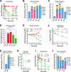
Fig 1. Dramatic variation in the virulence of K. pneumoniae isolates.
Virulence levels of K. pneumoniae isolates were assessed in CD1 mice by intraperitoneal (i.p.) infection with 106 CFU of each isolate. A. Correlation between capsule type and mean survival time of mice. Mean survival time of mice infected by each high- (HV, red dot) or low-virulent (LV, green dot) isolates in day is presented as a filled circle and grouped in a single column based on capsule type. Capsule type and total isolates tested for each type are indicated at the bottom and top of each group, respectively. UT, untypable. n = 3. B, C. Virulence traits of the HV and LV isolates. Survival rate (B) and bacteremia level (C) of representative HV (K1, K2 and K20) and LV (K3, K47 and K64) strains were determined. Strains are denoted by their capsule types: K1 (TH9885), K2 (ATCC43816), K3 (ATCC13883), K20 (TH12908), K47 (TH12846) and K64 (ATCC35657). n = 5–6. The mice died immediately after bleeding at 12 h in (C) were considered as being survived at this time point. The data were pooled from two independent experiments and presented as mean value ± standard deviation (SD). D, E. Mean survival time (D) and bacteremia level (E) of the mice at 12 and 24 hr post infection with HV and LV strains. n = 3. Each dot represents the mean value of three mice infected with a single strain. Dash line indicates the detection limit. Unpaired t test (D) and two-way ANOVA with Sidak’s multiple comparisons test (E) were performed. ****, P < 0.0001.
Fig 2
Fig 2. The role of capsule type in K. pneumoniae virulence.
A. Schematic diagram of the capsule type replacement strategy in ATCC43816 (capsule type K2). The cps gene cluster of recipient strain (red region) was first deleted to produce an unencapsulated (Δcps) mutant before the cps gene amplicon (green region) of donor strain were transformed into the Δcps strain to yield capsule replacement strain. B, C. Virulence traits of K2 derivatives. CD1 mice were i.p. infected with 106 CFU of acapsular mutant of ATCC43816 (Δcps) or isogenic derivative carrying the cps gene cluster of capsule type K1 from strain TH12908 (K2K1), K2 from ATCC43816 (K2K2), K3 from ATCC13883 (K2K3), and K23 from TH12852 (K2K23) to determine survival rate (B) and bacteremia level (C). Dash line denotes the detection limit. n = 6. D, E. Quantification of capsule production (D) and mucoviscosity (E) of the recipient strain (K2), acapsular mutant (Δcps), capsule switched variants (hybrids) and respective cps donor strains (donor). The capsular polysaccharide (CPS) was quantified by the uronic acid method and presented as the amount of uronic acid per OD600. Mucoviscosity status was evaluated by the sedimentation assay and shown as the OD600 ratio of the supernatant and culture. The data were pooled from two independent experiments and presented as mean ± SD, and statistically tested with one-way ANOVA with Tukey’s multiple comparisons. ns, not significant; **, P < 0.01; ***, P <0.001.
Fig 3
Fig 3. Capsule type-dependent capture of circulating K. pneumoniae in the liver.
A-D. Superior protection of HV capsule types against hepatic clearance of K. pneumoniae in the early phase of septic infection. ATCC43816 (K2) derivatives in the first 30 min of septic infection. CD1 mice were intravenously (i.v.) infected with 5 × 10⁶ CFU of isogenic acapsular mutant (Δcps), and capsule switched HV (K2K1 and K2K2) or LV (K2K3 and K2K23) derivatives. CFU counts in the blood at various time points (A) and the major organs at 5 min (B) and 30 min (C), were determined; bacterial killing in the first 30 min was estimated by comparing the combined CFU detected in the blood and five organs with the corresponding inoculum (D). n = 6. E. The impact of the capsule and capsule types on bacterial survival/killing in the first 6 hr of septic infection. CD1 mice were infected with the capsular mutant (Δcps), capsule switched HV derivative (K2K2) or LV derivative (K2K23) as in (A); the CFU counts obtained from the blood and five organs of each mouse are combined and presented as a single value at each time point. n = 6. F-H. The contribution of LV capsule type to bacterial survival. CD1 mice were i.v. infected with 5 × 10⁸ CFU of acapsular (Δcps) or LV (K2K23) derivative of ATCC43816; CFU counts obtained with the blood (F) and major organs (G) samples; mouse survival observed for 7 days post infection (H). n = 6. I, J. K2 capsule-mediated protection of K3 and K7 strains against hepatic capture. LV strains K3 (TH12849) and K7 (TH12880) were compared with their progenitors K2 capsule-switched derivatives (K3K2 and K7K2) in CFU counts in the blood (I) and major organs (J) as in (A) and (B), respectively. n = 4. The data were pooled from two independent experiments in CD1 mice and presented as mean ± SD, except for data of (H), which were from one experiment. One-way ANOVA with Tukey’s multiple comparisons test (D), two-way ANOVA with Tukey’s multiple comparisons test (E, F and I) and log-rank test (H) were performed. **, P < 0.01; ***, P < 0.001; ****, P < 0.0001.
Fig 4
Fig 4. Dispensable role of the spleen in the clearance of blood-borne K. pneumoniae in the early phase of septic infection.
A. Bacterial kinetics in the blood circulation of asplenic mice. The spleens of CD1 mice were surgically removed (splenectomy, SPX) 7 days before i.v. infection with 5 × 10⁶ CFU of ATCC43816 (K2) or isogenic acapsular mutant (Δcps) and capsule switched LV strain (K2K23). CFU counts in the blood were enumerated on agar plates. Mice with sham operation (SHM) were used as a negative control. Dash line denotes the detection limit. n = 4. B. Bacterial distribution in the major organs of asplenic mice. CFU counts in the blood and major organs of asplenic mice were compared with those of sham operation control 30 min post i.v. infection as in (A). n = 4. The data were from one experiment and presented as mean ± SD.
Fig 5
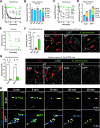
Fig 5. Capsule type-dependent capture of K. pneumoniae by KCs.
A, B. Importance of macrophages in clearing the LV derivative (K2K23) of ATCC43816. CD1 mice were pretreated with PBS, ISO (isotype control), CLL (depleting macrophage), 1A8 antibody (depleting neutrophil) or Gr1 antibody (depleting neutrophil and inflammatory monocyte) before being i.v. infected with 5 × 10⁶ CFU of K2K23 to determine bacteria in the bloodstream (A) and organs (B). n = 6. C, D. Essentiality of KCs in capturing acapsular and LV variants of ATCC43816. Clec4f-DTR mice were pretreated with (+DT) or without (-DT) diphtheria toxin and i.v. infected with 5 × 10⁶ CFU of the acapsular (Δcps) and LV derivative (K2K23) of ATCC43816 to determine bacteria in the bloodstream (C) and organs (D). Dash line denotes the detection limit. n = 6. E. The importance of KCs in the host survival against infection of LV K. pneumoniae. Clec4f-DTR mice were pretreated with (+DT) or without (-DT) diphtheria toxin and i.v. infected with 2 × 10⁷ CFU of K2K23. n = 6. F. Intravital microscopy (IVM) detection of K. pneumoniae capture by KCs in the liver sinusoids. CD1 mice were pretreated with antibodies to fluorescently labelling KC (red), liver sinusoidal endothelial cells (LSEC) (cyan), i.v infected with 5 × 10⁷ CFU of isogenic HV (K2), LV (K2K23) or Δcps K. pneumoniae (green), and imaged for bacterium-KC interactions in the liver sinusoids approximately 10 min post infection. Immobilized bacteria on KCs and occasionally vascular wall were quantified in 6 random fields of images (right) and presented as bacteria per field of view (FOV) (left). The process of bacterial interaction with KCs is manifested in S1 Movie. Scale bar, 10 μm. n = 2. G. The loss of K. pneumoniae capture in the liver in KC-deficient mice. Clsec4f-DTR mice were pretreated with (+DT) or without (-DT) diphtheria toxin before being used to detect KC capture of Δcps and K2K23 by IVM as in (F). H. Phagocytosis of Δcps and K2K23 by KCs. The phagocytosis was assessed by i.v. infection of mice with bacteria double labeled with FITC and pH-sensitive pHrodo dyes; extracellular (green) and intracellular (red) localization of bacteria bound by KCs (blue) were tracked by IVM within 2–30 min after infection as in (F). The internalized bacteria changed the color appearance from green (FITC), yellow (FITC + pHrodo) to red (pHrodo) after pHrodo was activated in the low pH environment where FITC lost fluorescence. Six random fields of representative images at 30 min were used to quantify the extracellular and intracellular bacteria. The percentage of intracellular bacteria out of the total immobilized bacteria per field is indicated in the images (30 min). Scale bar, 10 μm. The data of (A-E) were pooled from two independent experiments and presented as mean ± SD. Two-way ANOVA with Tukey’s (A and C) or Sidak’s (G) multiple comparisons test, log-rank test (E) and ordinary one-way ANOVA with Tukey’s multiple comparisons test (F) were performed. **, P < 0.01; ***, P < 0.001; ****, P < 0.0001.
Fig 6
Fig 6. Receptor-dependent recognition of K. pneumoniae in the liver.
A. Dose dependent inhibition of free K23 CPS against the clearance of K2K23 from the bloodstream. CD1 mice were intravenously inoculated with PBS or various amounts of purified CPS from K2K23 2 min before i.v. infection with 5 × 10⁶ CFU of K2K23 and determine blood bacteria at 5 time points in the first 30 min of infection. n = 3. B. Dose dependent inhibition of free K23 CPS against hepatic capture of K2K23 in CD1 mice treated as in (A). n = 3. C, D. Capsule type-specific blocking of free CPS against the clearance of LV variants. CD1 mice were pretreated with PBS or 800 μg free CPS of types K3, K23 or KL108 before infection with the strain of homologous (C) or heterologous (D) capsule type and enumeration of blood bacteria at 30 min. Capsule type of free CPS and strain inoculated are indicated at the bottom of each column. The strains used are specified in S1 Table. n = 3. E, F. Role of the complement system and scavenger receptors in the clearance of blood-borne K2K23. The involvement of the complement system in the clearance of LV K. pneumoniae was assessed by comparing C57BL/6 (WT) mice to those deficient in complement protein C3 (C3-/-) or major C3 receptor CRIg on KCs (CRIg-/-) after i.v. infection with 5 × 10⁶ CFU of K2K23 to determine bacteria in the bloodstream in the first 30 min (E). The contribution of scavenger receptors in the clearance of LV K. pneumoniae was evaluated by comparing CD1 mice with those pretreated with i.v. inoculation of 400 μg poly(I), a pan-scavenger receptor inhibitor, 2 min before infection with K2K23 and enumeration of bacteria in the bloodstream as in (E). Mice treated with 400 μg poly(C) were used as a negative control. n = 5. G, H. Role of the complement system and scavenger receptors in the clearance of blood-borne acapsular K. pneumoniae. Same as in (E) and (F) except for using the Δcps strain in i.v. infection. Dash line denotes the detection limit. The data are presented as mean ± SD. Two-way ANOVA with Tukey’s multiple comparisons (A, E, F, G and H) and one-way ANOVA test (C and D) were performed. ***, P < 0.001; ****, P < 0.0001.
Fig 7
Fig 7. Variation in virulence among type K47 strains.
A. Virulence of capsule type-K47 HV and LV strains. CD1 mice were i.p. infected with 106 CFU of high-virulence TH12845 (K47-H) or low-virulence strain TH12846 (K47-L). Mortality was assessed in a 7-day period. n = 6. B-D. Difference of HV and LV K47 strains in resisting hepatic capture. CD1 mice were i.v. infected with 5 × 10⁶ CFU of K47-H and K47-L to determine bacteria in the bloodstream (B) and major organs at 5 min (C) and 30 min (D). n = 4. E. Capsule production of K47-H and K47-L. K47 CPS was quantified as in Fig 2D. F. Schematic illustration of sequence difference in the K47 cps locus between K47-H and K47-L. The DNA sequences between the galF and wbaP genes of the K47 cps locus of K47-H and K47-L are drawn to show the two sequence differences between the two strains. The coding sequences are identified with gene names. The 776-bp insertion sequence upstream of wzi in K47-H is marked with a yellow line; the C-G SNP at the 2144th nucleotide of the wzc coding region in K47-L indicated with a vertical dashed line between the two sequences; putative promoter and JUMPstart sequences marked with black arrow and indigo rectangle, respectively. G. Capsule production of K47 variants was quantified as in (E). H. Effects of the IS and wzc SNP on the virulence of K47 variants. Mice were infected with the K47 cps variants of ATCC43816 to determine survival rates as in (A). n = 5. I-K. Virulence traits of the K47 cps variants. Bacteremia kinetics (I) and bacterial load in major organs at 5 min (J) and 30 min (K) were determined in mice i.v. infected as in (B). All data are presented as mean ± SD. Log-rank test (A and G), two-way ANOVA with Tukey’s multiple comparisons test (B and I) and one-way ANOVA with Tukey’s multiple comparisons test (L) were performed. **, P < 0.01; ***, P < 0.001; ****, P < 0.0001.
Fig 8
Fig 8. Vaccination to boost capture of HV K. pneumoniae by KCs.
A. Anti-K. pneumoniae IgG titer in vaccinated mice. CD1 mice were immunized twice, one week apart, by subcutaneous injection of 108 formaldehyde inactivated ATCC43816). Sera were collected from untreated (naïve) or immunized mice by retro-orbital bleeding two weeks after the second immunization and antibody titers against ATCC43816 were evaluated by ELISA. n = 3. B, C. Immunoprotection against HV K. pneumoniae infection. CD1 mice were immunized as in (A), and i.p. infected with different lethal doses (104, 105, or 106 CFU) of ATCC43816 to determine survival (B) and bacteremia levels (C). n = 5. D, E. Impact of immunization on the early clearance of HV K. pneumoniae by the liver. Bacteremia kinetics (D) and bacterial distribution (E) in the blood and five major organs of naïve or immunized mice at 30 min post i.v. infection with 106 CFU of ATCC43816 are presented. n = 3. F. Vaccine-activated pathogen capture by Kupffer cells. Pathogen capture by KCs in the liver sinusoids of naïve and vaccinated mice was assessed by IVM post i.v. infection with 5 × 10⁷ CFU of ATCC43816 as in Fig 5F. Six random fields of IVM images were used to quantify KC-associated bacteria per field of view (FOV) (left panel). Representative IVM images (right panel) are shown at the right panel. The process of bacterial capture is demonstrated in supplemental S4 Movie. Scale bar, 10 μm. n = 2. G. Immune serum-mediated clearance of HV K. pneumoniae. Naïve CD1 mice were i.v. infected with 106 CFU of ATCC43816 pre-incubated with 25% natural or immune serum for 10 minutes at room temperature to determine bacteremia kinetics in the first 30 min. n = 5. Dash line denotes the detection limit. All data are presented as mean ± SD. Unpaired t test (A and F), log-rank test (B) and two-way ANOVA with Tukey’s multiple comparisons test (D and G) were performed. **, P < 0.01; ****, P < 0.0001.

Similar articles

Cited by

References

    1. Paczosa MK, Mecsas J. Klebsiella pneumoniae: going on the offense with a strong defense. Microbiol Mol Biol Rev. 2016;80(3):629–61. doi: 10.1128/MMBR.00078-15 ; PubMed Central PMCID: PMC4981674. - DOI - PMC - PubMed
    1. Russo TA, Marr CM. Hypervirulent Klebsiella pneumoniae. Clin Microbiol Rev. 2019;32(3). doi: 10.1128/CMR.00001-19 ; PubMed Central PMCID: PMC6589860. - DOI - PMC - PubMed
    1. Cheng HY, Chen YS, Wu CY, Chang HY, Lai YC, Peng HL. RmpA regulation of capsular polysaccharide biosynthesis in Klebsiella pneumoniae CG43. J Bacteriol. 2010;192(12):3144–58. doi: 10.1128/JB.00031-10 ; PubMed Central PMCID: PMC2901686. - DOI - PMC - PubMed
    1. Fang CT, Chuang YP, Shun CT, Chang SC, Wang JT. A novel virulence gene in Klebsiella pneumoniae strains causing primary liver abscess and septic metastatic complications. J Exp Med. 2004;199(5):697–705. doi: 10.1084/jem.20030857 ; PubMed Central PMCID: PMC2213305. - DOI - PMC - PubMed
    1. Russo TA, Olson R, Fang CT, Stoesser N, Miller M, MacDonald U, et al.. Identification of biomarkers for differentiation of hypervirulent Klebsiella pneumoniae from Classical K. pneumoniae. J Clin Microbiol. 2018;56(9):e00776–18. Epub 2018/06/22. doi: 10.1128/JCM.00776-18 ; PubMed Central PMCID: PMC6113484. - DOI - PMC - PubMed

Publication types

Substances