. 2022 Aug 9;119(32):e2204779119.
doi: 10.1073/pnas.2204779119. Epub 2022 Aug 1.

Chemical interference with DSIF complex formation lowers synthesis of mutant huntingtin gene products and curtails mutant phenotypes

Affiliations

Chemical interference with DSIF complex formation lowers synthesis of mutant huntingtin gene products and curtails mutant phenotypes

Ning Deng et al. Proc Natl Acad Sci U S A. .

Abstract

Earlier work has shown that siRNA-mediated reduction of the SUPT4H or SUPT5H proteins, which interact to form the DSIF complex and facilitate transcript elongation by RNA polymerase II (RNAPII), can decrease expression of mutant gene alleles containing nucleotide repeat expansions differentially. Using luminescence and fluorescence assays, we identified chemical compounds that interfere with the SUPT4H-SUPT5H interaction and then investigated their effects on synthesis of mRNA and protein encoded by mutant alleles containing repeat expansions in the huntingtin gene (HTT), which causes the inherited neurodegenerative disorder, Huntington's Disease (HD). Here we report that such chemical interference can differentially affect expression of HTT mutant alleles, and that a prototypical chemical, 6-azauridine (6-AZA), that targets the SUPT4H-SUPT5H interaction can modify the biological response to mutant HTT gene expression. Selective and dose-dependent effects of 6-AZA on expression of HTT alleles containing nucleotide repeat expansions were seen in multiple types of cells cultured in vitro, and in a Drosophila melanogaster animal model for HD. Lowering of mutant HD protein and mitigation of the Drosophila "rough eye" phenotype associated with degeneration of photoreceptor neurons in vivo were observed. Our findings indicate that chemical interference with DSIF complex formation can decrease biochemical and phenotypic effects of nucleotide repeat expansions.

Keywords: DSIF; Huntington’s disease; SUPT4H; Spt4; nucleotide repeats.


Conflict of interest statement

The authors declare no competing interest.

Figures
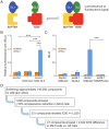
Fig. 1.
Fig. 1.
Interaction between SUPT4H and NGN domain of SUPT5H. (A) Schematic diagram depicting the interaction between SUPT4H and NGN to yield a luminescence signal by bringing together fragments of the split-Gaussia luciferase (Gluc). (B) Bar graph showing relative luminescence generated by the indicated constructs in the presence of the expression-inducing ligand RSL1 (RheoSwitch ligand 1) or a noninducing control element (DMSO). (C) Relative luminescence units (RLU) generated by cotransfection mediated complementation between Gluc reporter fragments fused to SUPT4H (4H-GL1). Controls were fusion proteins containing a leucine zipper motif capable of forming homodimers but not able to interact with SUPT4H or NGN (Zip-GL1 and Zip-GL2), or NGN (NGN-GL2). The NGN(SF)-GL2 construct contains a 214 Ser-to-Phe substitution in NGN known to preclude NGN interaction with SUPT4H. The values shown in panels B and C are the mean ± SD (n = 4; ***P < 0.001, two-tailed paired Student’s t test). (D) Screening procedures for identification of small molecules that interfere with the luminescence segment generated by interaction between SUPT4H and SUPT5H-NGN. Z’-factor = 0.83.
Fig. 2.
Fig. 2.
Effect of 6-azauridine (6-AZA) and 6-AZA-TA. (A) Comparison of 6-AZA and related compounds on luminescence generated by the SUPT4H-SUPT5H-NGN interaction. M2- 8 cells were incubated with compounds having chemical structures as shown, and luminescence signals determined for each sample were quantified using procedures described in Materials and Methods. The signal from lysates of untreated cells (i.e., relative luminescence units; RLU) was set as 1. Mean values ± SDs are indicated. (n = 3) (B) Western blot of immunoprecipitation analysis of SUPT4H interaction with SUPT5H-NGN. M2-8 cells were treated with 6-AZA at indicated concentrations. Flag-tagged NGN-GL2 protein was collected by immunoprecipitation using anti-Flag magnetic beads. The precipitates were then probed and quantitated using anti-Gluc antibody as indicated in Materials and Methods to detect SUPT4H-GL1 (arrowhead) that had coprecipitated with NGN-GL2. Band intensity measurements determined for SUPT4H-GL1 were normalized against NGN-GL2 measurements and the intensity of the SUPT4H-GLI band relative to NGN-GL2 is indicated by %. The normalized value for the untreated sample was set as 100%. (C) Analysis of endogenous SUPT4H interaction with SUPT5H. ST HdhQ111/Q111 cells were treated with 10 μM 6-AZA for 24 h. Protein lysates were collected and subjected to immunoprecipitation using anti-SUPT4H antibody. Precipitates were then probed by anti-SUPT4H and anti-SUPT5H antibody respectively. SUPT5H band intensity was measured and normalized to that of SUPT4H. The normalized value for the untreated sample was set as 100%. (n = 3).
Fig. 3.
Fig. 3.
Effects of 6-AZA on protein production by mutant or normal HTT alleles. (A) Effects in GM14044 human lymphoblastoid cells, a mutant HTT allele containing 750 CAG repeats and a normal allele containing 19 CAG repeat cells, treated with indicated concentrations of 6-AZA were subjected to Western blot analysis (left). Relative protein abundance was calibrated by scanning and normalized against total protein (TP) and the normalized values were plotted (right). Proteins in untreated samples were set as 1. (B) HTT, SUPT4H, SUPT5H, and TBP proteins in ST HdhQ111/Q111 and ST HdhQ7/Q7 cells treated with 6-AZA were detected by Western blotting (left). Values were normalized against α-tubulin and plotted (right). The value obtained for each protein from untreated sample was set as 1. (C) Measurement of cell number in cells treated with Q7/Q7 and Q111/Q111with 6-AZA concentrations shown in B. (D) Effect of the indicated concentrations of uridine on relative number of ST HdhQ111/Q111 cells incubated with 10 μM 6-AZA (left) or lacking 6-AZA (right) are shown. The viability of cells grown in the absence of either agent was set to 100%. (E) ST HdhQ111/Q111 cells treated with 6-AZA as described in D were analyzed by Western blotting and values were normalized against those obtained for total protein (TP). Samples collected from 6-AZA treated cells were shown a line above the panel. Quantification of the effects on mHTT protein in 6-AZA treated vs. untreated cells shown at the right panel. The relative occurrence of mHTT protein value in cells not exposed to either 6-AZA or uridine was set as 1. Mean values and ± SDs are indicated. (F) Effects of 6-AZA on MSN-GM23225 cells. Protein lysates were collected and analyzed by Western blotting (left) for the abundance of mHTT, wHTT, TBP, and α-tubulin relative to total protein (TP) and compared to the value obtained for cells lacking 6-AZA treatment, which was set to 1 (right). HTT ratio represents the calculated value of mHTT vs. wHTT. Error bars represent the SD. (n = 3; ns: no significant, *P < 0.05, **P < 0.01, ***P < 0.001, two-tailed paired Student’s t test).
Fig. 4.
Fig. 4.
6-AZA-mediated modification of effects of mutant HTT on oxidative stress gene expression in MSN neurons. (A) GSTT1 and GSTT2 gene expression was analyzed by qRT-PCR with SnRNA U6 as a loading control in GM23225-, GMC#1-, and GMC#2-differentiated MSN GABAergic neurons with or without 6-AZA treatment. The GSTT1 and GSTT2 mRNA levels relative to untreated GM23225 are shown. All the values shown are the mean ± SD (B) Experimental outline for cell viability determination in MSN GABAergic neurons. Mature MSN GABAergic neurons derived from GM23225 (HD subject), or GMC#1 and GMC#2 (HD correction subject) were treated with 1 μM 6-AZA for 13 d. 100 μM H2O2 was added and cultures were incubated for an additional 24 h. Cell viability was determined by Trypsin blue assay. Values for samples not exposed to H2O2 or 6-AZA were set as 100%. (n = 3), **P < 0.01, ***P < 0.001, in two-tailed paired Student’s t test.
Fig. 5.
Fig. 5.
Effect of 6-AZA and its prodrug on rough eye phenotype associated with expression of mutant HTT in Drosophila. (A) Representative images of compound eye of Gmr > HTT-97Q/+ flies developed from larvae cultured in agar containing 100 μM of 6-AZA or its prodrug 6-AZA-TA (indicated as +). Gmr/+ flies served as a control (top panel). Percentage of flies showing rough eye phenotype after culture of larvae in agar containing Gmr > HTT-97Q/+, 10 or 100 μM 6-AZA or 6-AZA-TA. Ten flies were randomly picked from each experimental group and the number of flies showing the rough eye phenotype was determined by examination under a microscope. The values shown in this panel are the mean ± SD for three independent experiments (n = 10, N = 3, *P < 0.05, **P < 0.01, two-tailed paired Student’s t test). (B) 100 eggs of w1118 strain were cultured in a vial containing 10 mL of media supplemented with 10 or 100 μM of 6-AZA or 6-AZA-TA. Posteclosion flies were counted in each vial and the number of flies collected in untreated samples was set as 100%. (n = 100, N = 3, Error bars represent the SD, two-tailed paired Student’s t test). (C) Mutant HTT protein was assessed by Western blotting using the same set of HD flies as described in A. α-tubulin was included as a loading control. The relative levels of mutant HTT in treated vs. untreated HD flies after normalization are shown. (n = 3; **P < 0.01, two-tailed paired Student’s t test).

Similar articles

Cited by

References

    1. Paulson H., Repeat expansion diseases. Handb. Clin. Neurol. 147, 105–123 (2018). - PMC - PubMed
    1. Rodriguez C. M., Todd P. K., New pathologic mechanisms in nucleotide repeat expansion disorders. Neurobiol. Dis. 130, 104515 (2019). - PMC - PubMed
    1. Malik I., Kelley C. P., Wang E. T., Todd P. K., Molecular mechanisms underlying nucleotide repeat expansion disorders. Nat. Rev. Mol. Cell Biol. (2021). - PMC - PubMed
    1. Wada T., et al. , DSIF, a novel transcription elongation factor that regulates RNA polymerase II processivity, is composed of human Spt4 and Spt5 homologs. Genes Dev. 12, 343–356 (1998). - PMC - PubMed
    1. Yamaguchi Y., et al. , Structure and function of the human transcription elongation factor DSIF. J. Biol. Chem. 274, 8085–8092 (1999). - PubMed

Publication types

MeSH terms

LinkOut - more resources