. 2022 Sep:349:796-811.
doi: 10.1016/j.jconrel.2022.07.040. Epub 2022 Aug 3.

CD44 receptor targeted nanoparticles augment immunity against tuberculosis in mice

Affiliations

CD44 receptor targeted nanoparticles augment immunity against tuberculosis in mice

Vipul K Singh et al. J Control Release. 2022 Sep.

Abstract

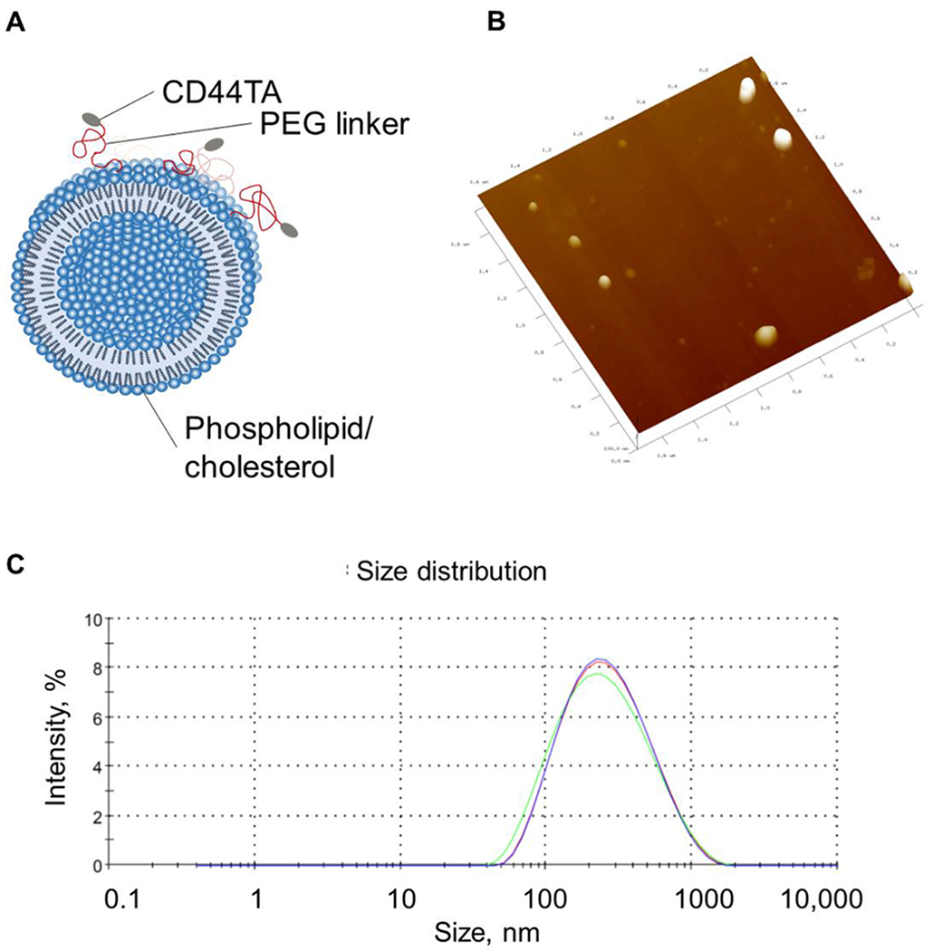
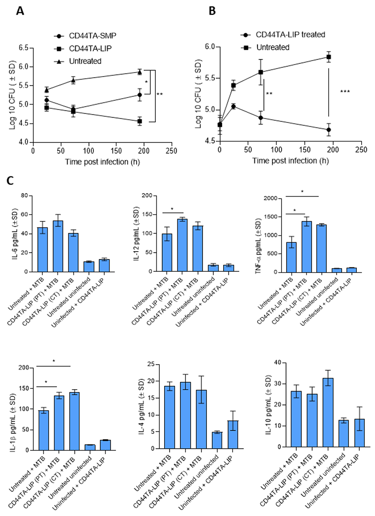
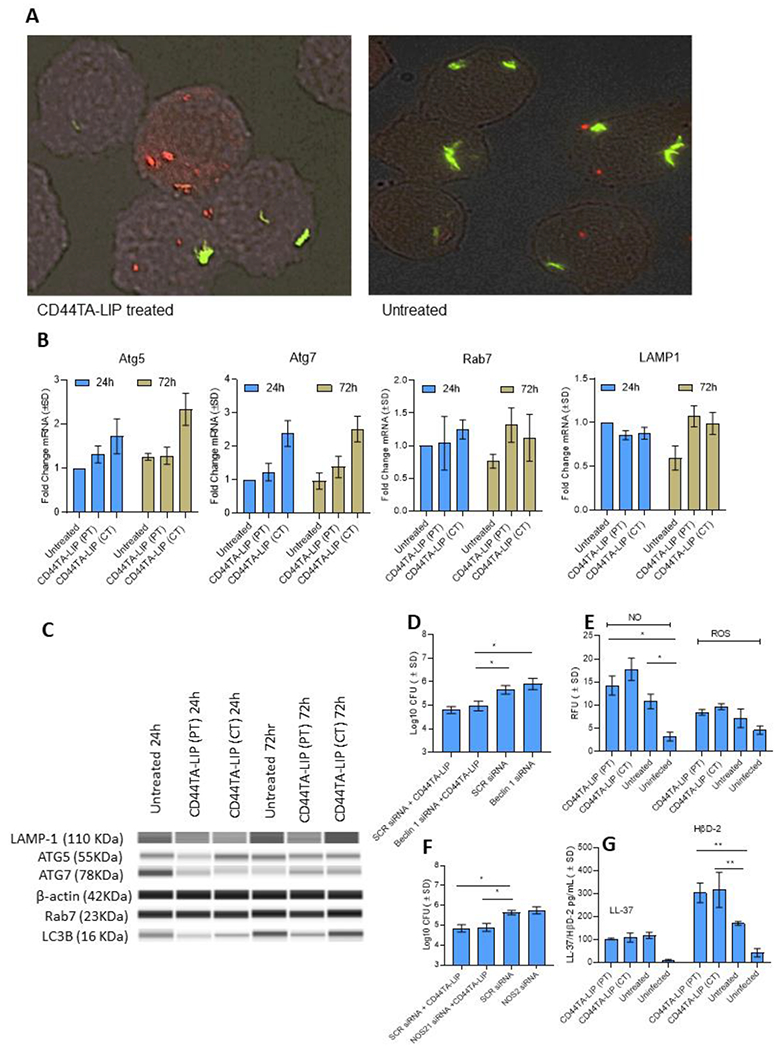
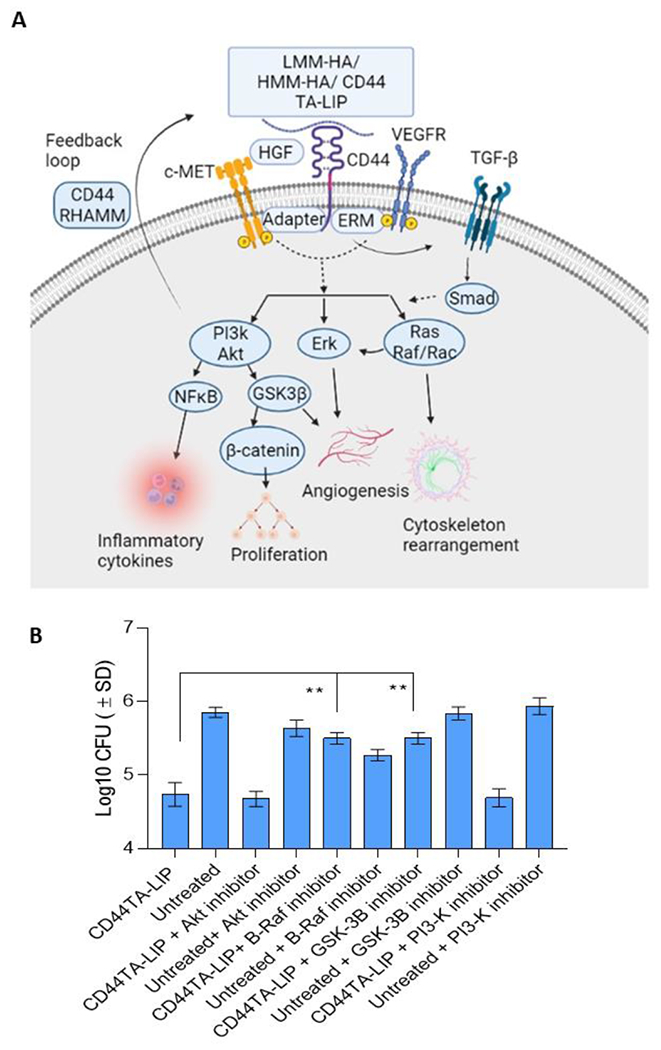
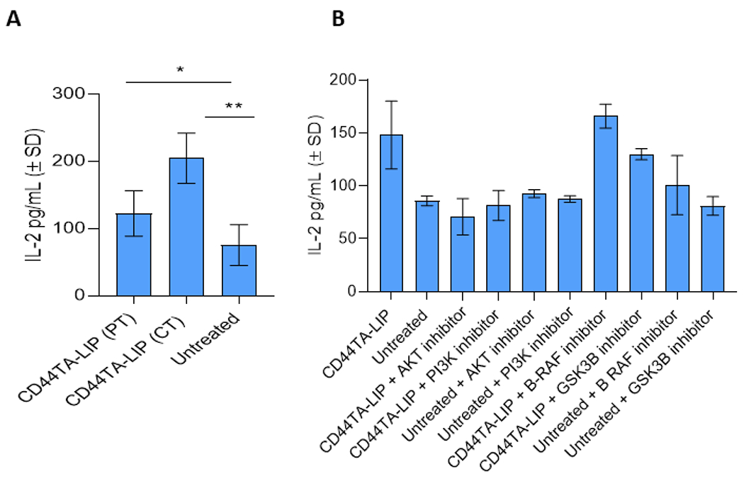
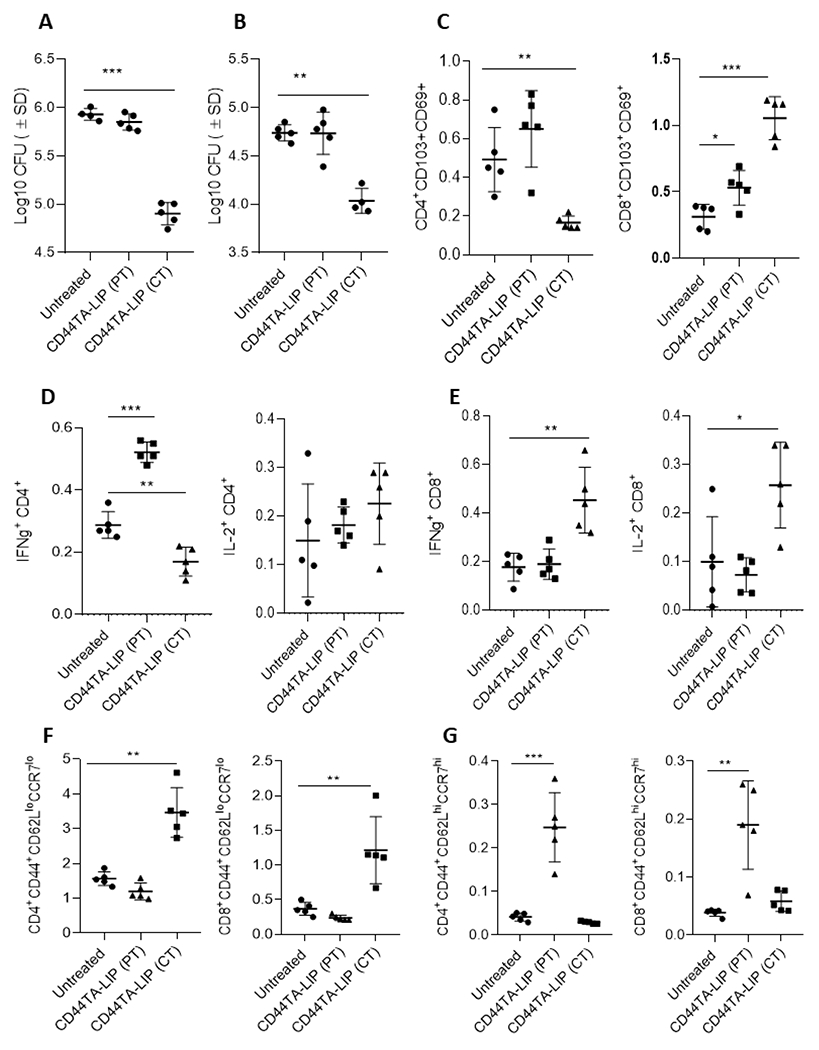
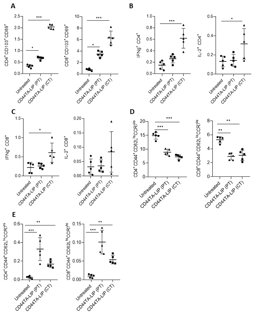
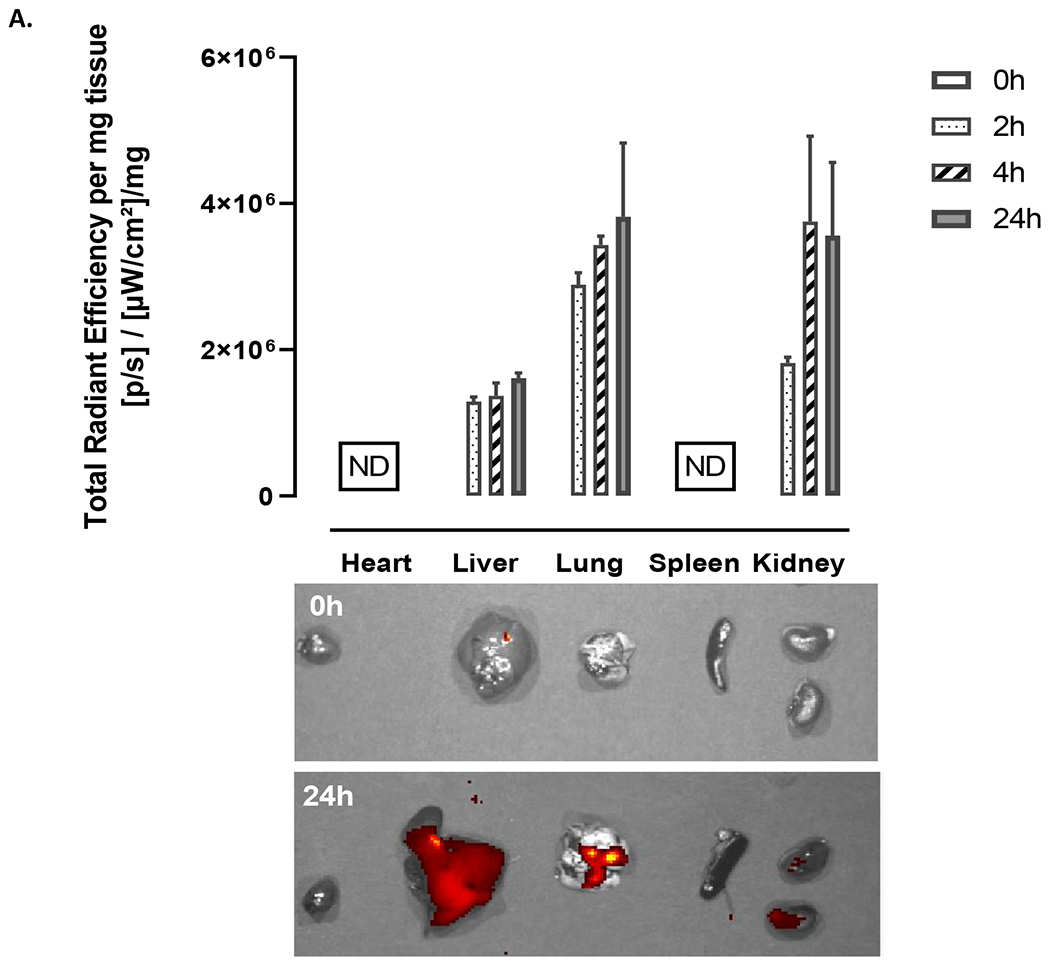
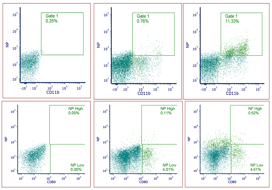
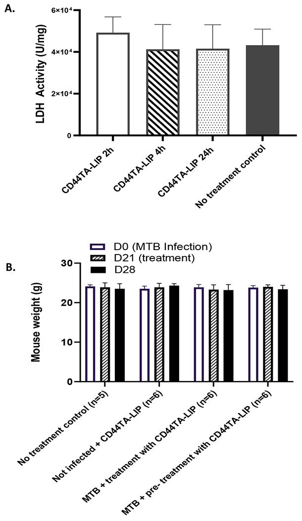
We describe a role of CD44-mediated signaling during host-defense against tuberculosis (TB) using a mouse model of TB and studies in M. tuberculosis (Mtb) infected human macrophage (MФ). Liposomes targeting CD44 using thioaptamers (CD44TA-LIP) were designed and tested as new vaccines to boost host immunity in TB. CD44TA-LIP enhanced killing of Mtb in human MФ, which correlated with an increased production of pro-inflammatory cytokines IL-1β, TNF-α and IL-12. CD44TA-LIP activated MФ showed an enhanced MHC-II dependent antigen presentation to CD4 T-cells. Inhibition of cellular proliferation and cytoskeleton rearrangement pathways downstream of CD44 signaling abrogated CD44TA-LIP-induced antimicrobial effects. Blockade of inflammatory pathways also reduced antigen presentation by MФ and activation of CD4 T cells. Mtb infected MФ treated with CD44TA-LIP exhibited increased nitric oxide and HβD2 defensin peptide production. Among Mtb infected mice with increased lung and spleen loads of organisms, intranasal administration of CD44TA-LIP led to a ten-fold reduction of colony forming units of Mtb and elevated IFN-γ + CD4, effector, central and resident memory T cells. Biodistribution studies demonstrated that CD44TA-LIP preferentially accumulated in the lungs and were associated with CD11b + cells. CD44TA-LIP treated mice showed no weight loss or increased liver LDH levels. This study highlights the importance of CD44-mediated signaling in host-defense during TB and the therapeutic potential of CD44TA-LIP.

Keywords: CD4 T cells; CD44; Host-defense; Host-directed therapy; Immunity; Liposomes; Macrophages; Mycobacterium tuberculosis; Thioaptamers; Tuberculosis.


Conflict of interest statement

Declaration of Competing Interest None.

Figures

Fig. 1:
Fig. 1:. Design (A) and characterization (B-D) of CD44TA-LIP.
A) Schematics of LIP composition and conjugation with CD44-TA. (B) Atomic force microscopy (AFM) image shows CD44TA-LIP of ~200 nm, confirmed by dynamic light scattering measurement (C) which showed the diameter of 204.9 ± 6.3 nm.
Figure 2:
Figure 2:. Effect of CD44TA-LIP treatment on antimicrobial activity and cytokine response of human MΦ during Mtb infection.
Mtb burden in CD44TA-LIP treated (Panel A: Concurrent treatment; Panel B: pre-treatment) vs untreated human MΦ over seven days of infection as measured by CFU assay. During concurrent treatment MΦ were treated with CD44TA-LIP at the beginning of infection (4 h after the infection) whereas during pre-treatment MΦ were treated 24 h prior to infection. B. Secreted levels of pro- and anti-inflammatory cytokines by CD44TA-LIP treated, untreated and uninfected human MΦ at 24 h post-infection/treatment. CD44TA-LIP concentration used: 10 μg/106 MΦ. PT: pre-treatment; CT: concurrent treatment. pg = picograms; MOI used for the experiments=1. Number of MΦ used per replicate: 1x10⁶. h= h. Representative data shown from three independent experiments carried out in duplicate. Bars and error bars represent mean and SD, respectively. *p≤0.05, **p≤0.005, ***p≤0.0005, ****p≤0.0001.
Figure 3:
Figure 3:. Effect of CD44TA-LIP treatment on autophagy, reactive species and antimicrobial peptide production by human MΦ during Mtb infection.
A: Localization of Mtb (green) phagosomes and autophagy related LC3B+ protein (red) within CD44TA-LIP treated vs untreated MΦ after 24 h of infection. Phagosomes were labeled using GFP Mtb and LC3B was labeled using specific antibody. Images were acquired at 60x magnification with an inverted fluorescent microscope using MΦ of a single random healthy donor and were analyzed using confocal microscopy and NIKON NIS element software. Transcriptional (B) and translational (C) level expression of autophagy and phagosome maturation associated genes Atg5, Atg7, Rab7 and Lamp in CD44TA-LIP treated and untreated MΦ at 24 and 72 h post Mtb infection. Mtb burden (at 72 h post infection) in CD44TA-LIP treated (Concurrent treatment) and untreated human MΦ after siRNA mediated knockdown of beclin1 (D) and NOS2 (F) as measured by CFU assay. Production of ROS/ NO (E) and antimicrobial peptides LL-37/HβD2 (G) by CD44TA-LIP treated and untreated MΦ at 24 h post-Mtb infection, as measured through fluorescent probes, and ELISA assays respectively, using a fluorimeter/plate reader. CD44TA-LIP concentration used: 10 μg/106 MΦ. PT: pre-treatment; CT: concurrent treatment. pg = picograms; MOI used for the experiments = 1. Number of MΦ used per replicate: 1x10⁶. h= h. Representative data shown from three independent experiments carried out in duplicate. Bars and error bars represent mean and SD, respectively. *p≤0.05, **p≤0.005, ***p≤0.0005, ****p≤0.0001.
Figure 4:
Figure 4:. CD44 signal transduction pathways and effect of specific pharmacological inhibitors of its downstream effectors on antimicrobial effect of CD44TA-LIP on survival of Mtb in human MΦ.
A: Ligand binding to cell surface receptor CD44 triggers a complex formation between CD44 and co-receptors such as c-Met, VEGFR, and TGF-β receptors leading to activation of downstream effectors such as Akt, PI3K, PP2A, ERK1/2, and Ras/Raf/Rac. By inducing these signaling events and downstream effectors, CD44 signaling can modulate various cellular processes including proliferation, invasion, cytoskeleton rearrangement and inflammation. B: Effect of pharmacological inhibitors of Akt, PI3K, GSK3β and B-Raf on Mtb burden in CD44TA-LIP treated (Concurrent treatment) vs untreated human MΦ at 72 h post infection as measured by CFU assay. CD44TA-LIP concentration used: 10 μg/106 MΦ. Respective concentration used for Akt, PI3K, GSK3β and B-Raf inhibitors/106 MΦ= 10 nM, 50 nM, 5 nM and 100 nM. Pharmacological inhibitors of Akt, PI3K, GSK3β and B-Raf were added at the beginning of infection and removed after 24 h post infection. pg= picograms; MOI used for the experiments = 1. Representative data shown from three independent experiments carried out in duplicate. Bars and error bars represent mean and SD, respectively. *p≤0.05, **p≤0.005, ***p≤0.0005, ****p≤0.0001.
Figure 5:
Figure 5:. Effect of CD44 signaling on antigen presentation capabilities of CD44TA-LIP treated vs untreated human MΦ during Mtb infection.
A: Secreted levels of IL-2 by Mtb Ag85B-specific T cells (F9A6) during co-culture with CD44TA-LIP treated and untreated MΦ at 72 h post-infection. B: Effect of pharmacological inhibitors of Akt, PI3K, GSK3β and B-Raf on antigen presentation capabilities of CD44TA-LIP treated (Concurrent treatment) vs untreated human MΦ at 72 h post infection as measured by CFU assay. CD44TA-LIP concentration used: 10 μg/106 MΦ. Respective concentration used for Akt, PI3K, GSK3β and B-Raf inhibitors /106 MΦ= 10 nM, 50 nM, 5 nM and 100 nM. Pharmacological inhibitors of Akt, PI3K, GSK3β and B-Raf were added at the beginning of infection and removed after 24 h post infection. pg= picograms; MOI used for the experiments = 1. Representative data shown from three independent experiments carried out in duplicate. Bars and error bars represent mean and SD, respectively. *p≤0.05, **p≤0.005, ***p≤0.0005, ****p≤0.0001.
Figure 6:
Figure 6:. Effect of CD44TA-LIP on bacterial burden and various T cell populations in vivo during experimental murine tuberculosis.
Mtb CFU in Lungs (A) and spleens (B) of mice treated intranasally with CD44TA-LIP. Immune cell analysis was done by flow cytometry. Frequencies of resident memory CD4 and CD8 T cells (CD103+CD69+) in lungs (C) of untreated, and CD44TA-LIP treated mice. Percentages of IFN-γ and IL-2 expressing CD4 (D) and CD8 (E) T cells in lungs of untreated, and CD44TA-LIP treated mice. Frequencies of effector memory (F) and central memory (G) CD4 and CD8 T cells in lungs of untreated, and CD44TA-LIP treated mice. Treatment with CD44TA-LIP prior to infection is depicted as CD44TA-LIP (PT) whereas treatment with CD44TA-LIP after infection is depicted as CD44TA-LIP (CT) in all panels. Flow panels show the % of live leukocytes. CD44+CD62LloCCR7lo= Effector memory population; CD44+CD62LhiCCR7hi= Central memory population. *p < 0.05, **p < 0.001, ***p < 0.0001, ***p < 0.0001; t-test.
Figure 7:
Figure 7:. Effect of CD44TA-LIP treatment on various T cell populations of mice during tuberculosis infection.
Frequencies of resident memory CD4 and CD8 T cells (CD103+CD69+) in spleens (A) of untreated, and CD44TA-LIP treated mice. Percentages of IFN-γ and IL-2 expressing CD4 (B) and CD8 (C) T cells in spleens of untreated, and CD44TA-LIP treated mice. Frequencies of effector memory (D) and central memory (E) CD4 and CD8 T cells in spleens of untreated, and CD44TA-LIP treated mice. Treatment with CD44TA-LIP prior to infection is depicted as CD44TA-LIP (PT) whereas treatment with CD44TA-LIP after infection is depicted as CD44TA-LIP (CT) in all panels. CD44+CD62LloCCR7lo= Effector memory population; CD44+CD62LhiCCR7hi= Central memory population. *p < 0.05, **p < 0.001, ***p < 0.0001, ***p < 0.0001; t-test.
Figure 8:
Figure 8:. Organ distribution of CD44TA-LIP in lung inflammation model.
CD44TA-LIP were labeled with Cy 5.5. At pre-determined times, animals were sacrificed and IVIS images were taken at 0 (prior to administration), 2, 4 and 24h. (A) The signal (total radiant efficiency) was assessed using IVIS Spectrum Imaging System (Perkin Elmer, MA, USA) at excitation and emissions wavelengths of 675nm and 720nm for Cy 5.5 fluorescence intensity detection. The data was quantified using Living Image® Software and normalized to the organ weight.
Figure 9:
Figure 9:
Flow cytometry analysis of CD11b (A, B, C) and CD80 (D, E. F) positive cell populations associated with CD44TA-LIP (NP on the scatters). No treatment control (A and D); 2h (B and E) and 24h (C and D). CD44TA-LIP were Cy 5.5 labeled and the samples were stained with CD11b-Pacific Blue (117322, Biolegend) and CD80-PE (104708, Biolegend). Samples were analyzed using a Becton-Dickenson FACS Fortessa (5 laser 17 channel) using Diva 9.1. Gates were based on single and unstained controls.
Figure 10:
Figure 10:
Safety of CD44TA-LIP: (A) LDH levels in the liver 2, 4, and 24h following CD44TA-LIP administration. (B) Weights of the mice in therapy experiments. No changes were observed up to 7 days following CD44TA-LIP administration.

Similar articles

Cited by

References

    1. Chakaya J, Khan M, Ntoumi F, Aklillu E, Fatima R, Mwaba P, Kapata N, Mfinanga S, Hasnain SE, Katoto PDMC, et al. Global Tuberculosis Report 2020 – Reflections on the Global TB burden, treatment and prevention efforts. Int J Infect Dis (2021) doi:10.1016/j.ijid.2021.02.107 - DOI - PMC - PubMed
    1. Seung KJ, Keshavjee S, Rich ML. Multidrug-Resistant Tuberculosis and Extensively Drug-Resistant Tuberculosis. Cold Spring Harb Perspect Med (2015) 5:a017863. doi:10.1101/cshperspect.a017863 - DOI - PMC - PubMed
    1. Bates M, Marais BJ, Zumla A. Tuberculosis Comorbidity with Communicable and Noncommunicable Diseases. Cold Spring Harb Perspect Med (2015) 5:a017889. doi:10.1101/cshperspect.a017889 - DOI - PMC - PubMed
    1. Palucci I, Delogu G. Host Directed Therapies for Tuberculosis: Futures Strategies for an Ancient Disease. Chemotherapy (2018) 63:172–180. doi:10.1159/000490478 - DOI - PubMed
    1. O’Garra A, Redford PS, McNab FW, Bloom CI, Wilkinson RJ, Berry MPR. The Immune Response in Tuberculosis. Annu Rev Immunol (2013) 31:475–527. doi:10.1146/annurev-immunol-032712-095939 - DOI - PubMed

Publication types