

Review
doi: 10.1101/cshperspect.a041020.
Caspase Activation and Inhibition
Affiliations
- PMID: 35914782
- PMCID: PMC9341465
- DOI: 10.1101/cshperspect.a041020
Item in Clipboard
Review
Caspase Activation and Inhibition
Cold Spring Harb Perspect Biol.
.
No abstract available
Figures
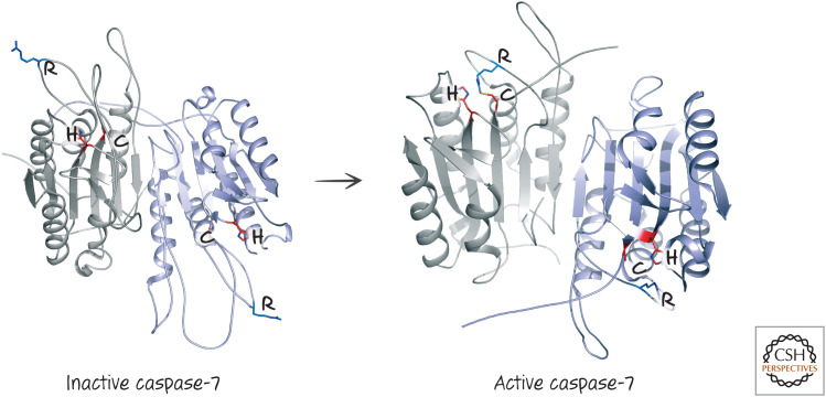
The structures of inactive and active caspase-7. The arginine (R) in the specificity loop is indicated, as is the cysteine–histidine (C–H) catalytic dyad. (Left, PDB 1K86 [Chai et al. 2001]; right, PDB 1GOF [Reidl et al. 2001a].)
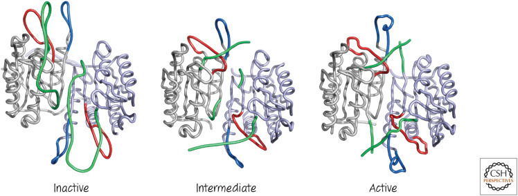
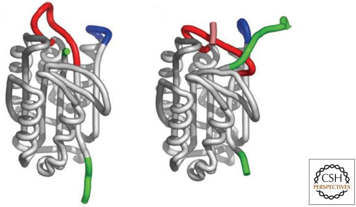
Close-up view of caspase-7 activation. Three loops mediate caspase-7 active site assembly: the specificity loop (red), a neighboring loop that draws the specificity loop into position (blue), and the linker between the large and small subunits (green). Structures are the inactive zymogen (left), the inhibitor-bound enzyme (center), and the active enzyme (right). (Left, PDB 1K86 [Chai et al. 2001]; center, PDB 1F1J [Wei et al. 2000]; right, PDB 1GOF [Reidl et al. 2001a].)
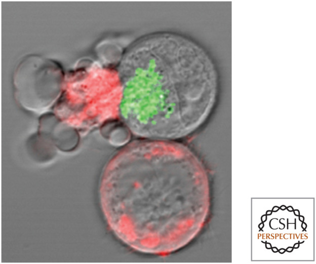
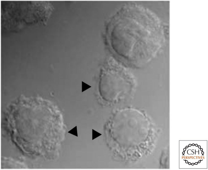
Cytotoxic lymphocyte killing a target (cell). The target cells are stained red, and the cell at left is undergoing apoptosis. Green: cytotoxic granules in the cytotoxic lymphocyte. (Reproduced from Bleakley 2008, ©2008 with permission from Lippincott, Williams & Wilkins. Image kindly provided by Dr. I.S. Goping, University of Alberta.)
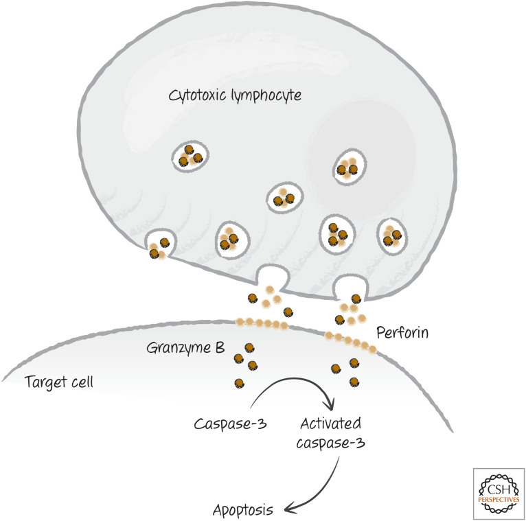
Simplified scheme of cytotoxic granule killing.
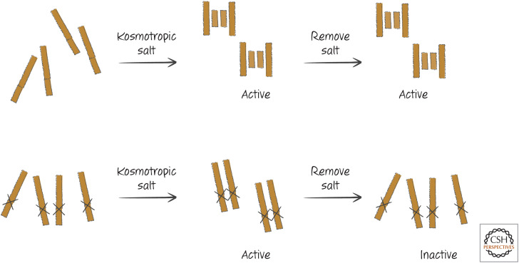
Initiator caspases can be activated by induced proximity. Kosmotropic salts, which cause protein aggregation, activate initiator caspases that remain active after the salt is removed. If the cleavage sites between the protease subunits are made uncleavable by mutation, the salts can still activate the enzyme, but the enzyme becomes inactive when the salts are removed.
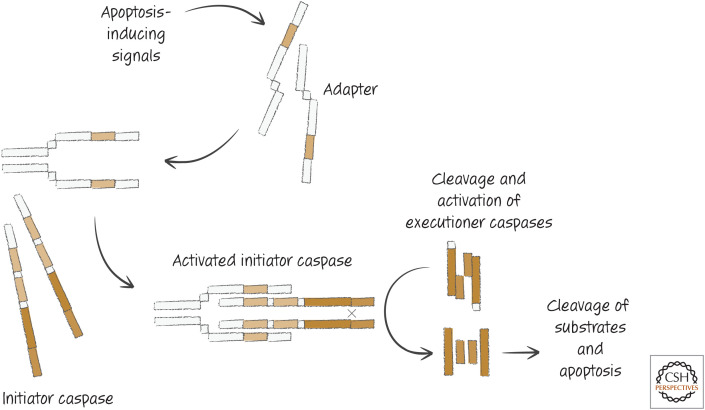
General apoptotic pathways.
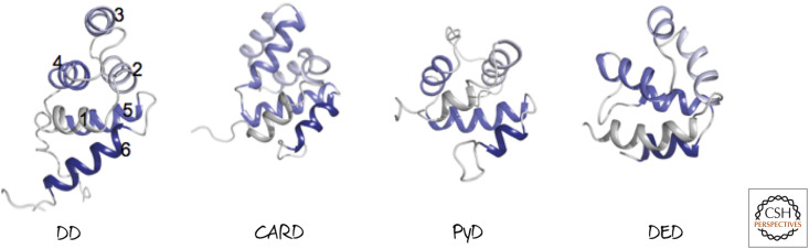
Representative death folds. Although different death folds do not have sequence similarity, they are structurally related. Each has six globular helical bundles, shown here colored gray to dark purple from the amino to carboxyl termini.2 (Left to right, PDB 1DDF [Huang et al. 1996]; PDB 1CWW [Day et al. 1999]; PDB 1PN5 [Hiller et al. 2003]; PDB 1A1Z [Eberstadt et al. 1998].)
The transition from inactive to active caspase-9. When caspase-9 is dimerized, the specificity-determining loop (red) is brought into position by a loop (blue) from the other monomer. Cleavage of the region between the protease subunits (green) is not necessary for activation. Only one site in the dimer can form at a time: One is inactive (left) and the other is active (right). (Reproduced with permission from Fuentes-Prior and Salvesen 2004, ©The Biochemical Society.)
Pyroptosis. Cells die in a caspase-1-dependent manner, indicated by arrows. (Reprinted by permission from Macmillan Publishers Ltd.: Fernandes-Alnemri et al. 2007, ©2007.)
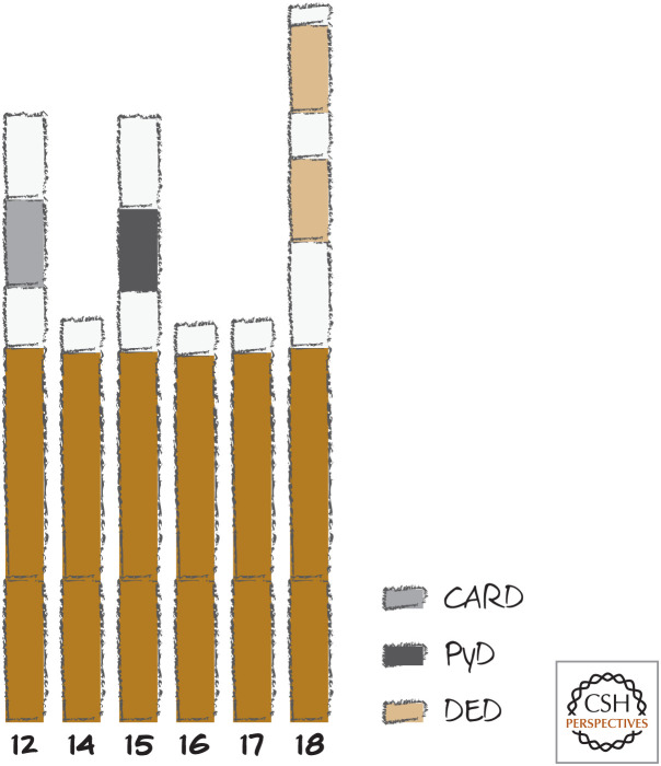
Caspase-12 and beyond in mammals. Lengths shown are arbitrary. Caspases-15, -17, and -18 are not found in humans, and caspase-12 is frequently not found in humans. Caspases-11 (mouse) and -13 (cow) are homologs of caspase-5 and caspase-3, respectively, and are not shown.
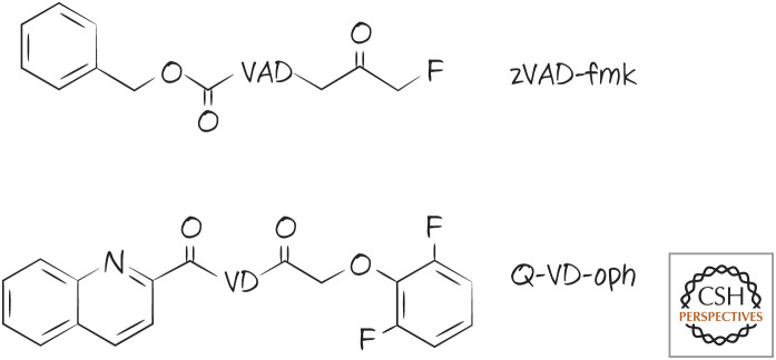
Caspase inhibitors. The peptide portion is shown with a single-letter code.
Caspase-3 bound to the inhibitor zVAD-fmk. (PDB 1CP3 [Mittl et al. 1997].)
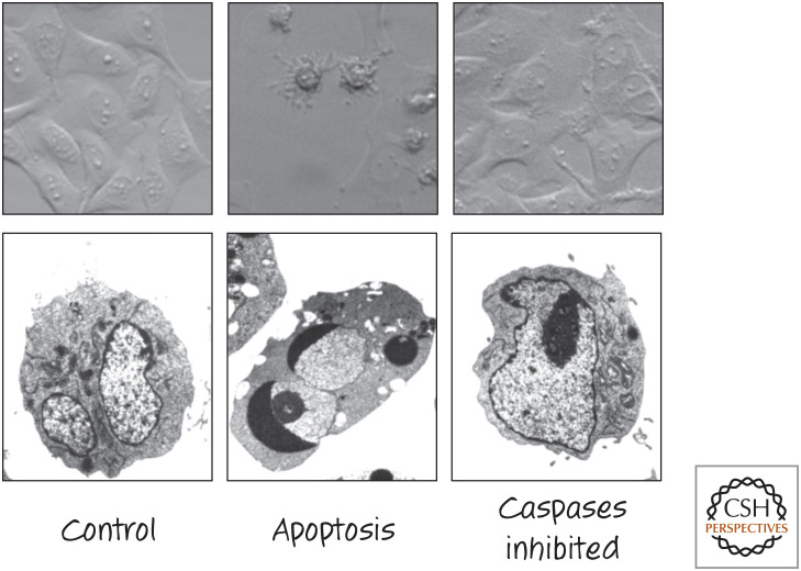
Caspase inhibitors block the morphological features of apoptosis. Appearance of different cell types treated to induce apoptosis in the presence or absence of the caspase inhibitor zVAD-fmk. (Upper) Bright-field images of fibroblasts. (Lower) Electron micrographs of tumor cells. (Images courtesy of Dr. Nigel Waterhouse, Mater Medical Research Institute, Brisbane, Australia.)
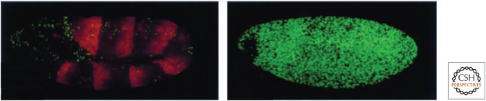
Loss of DIAP1 causes apoptosis. Extensive cell death, staining green, is not seen in a wild-type embryo (left), but is observed in a DIAP1 mutant embryo (right). (Reprinted from Wang et al. 1999, ©1999 with permission from Elsevier.)
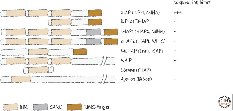
Some IAPs.
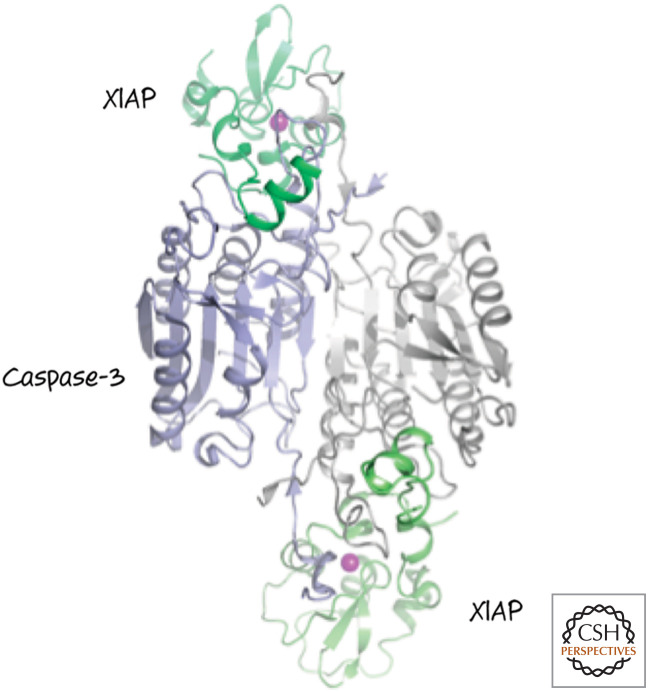
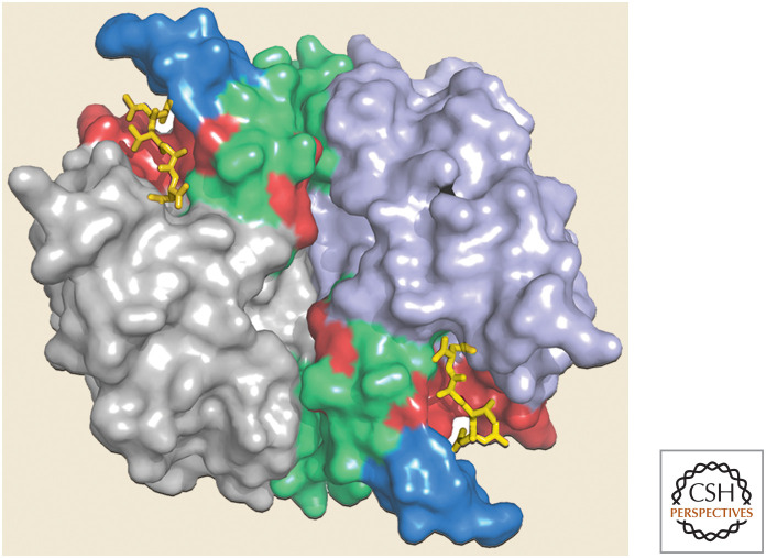
XIAP binds to the active sites in caspase-3. Only the BIR2 domain in XIAP (green) is shown. (Purple circles indicate zinc atoms that coordinate the folding of XIAP.) (PDB 1I3O [Riedl et al. 2001b].)
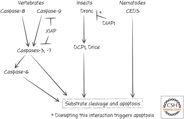
Caspases and IAPs in apoptosis.
References
FIGURE CREDITS
-
- Bleakley RC. 2008. Cytotoxic T lymphocytes. In Fundamental immunology, 6th ed. (ed. Paul WE.), p. 1079. Lippincott, Williams & Wilkins, Philadelphia.
-
- Fernandes-Alnemri T, Wu J, Yu J-W, Datta P, Miller B, Jankowski W, Rosenberg S, Zhang J, Alnemri ES. 2007. The pyroptosome: a supramolecular assembly of ASC dimers mediating inflammatory cell death via caspase-1 activation. Cell Death Differ 14: 1590–1604. 10.1038/sj.cdd.4402194 - DOI - PMC - PubMed
Publication types
MeSH terms
Substances
LinkOut - more resources
Full Text Sources