. 2022 Aug 2;99(5):e500-e511.
doi: 10.1212/WNL.0000000000200750. Epub 2022 May 27.

Automated Detection of Speech Timing Alterations in Autopsy-Confirmed Nonfluent/Agrammatic Variant Primary Progressive Aphasia

Affiliations

Automated Detection of Speech Timing Alterations in Autopsy-Confirmed Nonfluent/Agrammatic Variant Primary Progressive Aphasia

Adolfo M García et al. Neurology. .

Abstract

Background and objectives: Motor speech function, including speech timing, is a key domain for diagnosing nonfluent/agrammatic variant primary progressive aphasia (nfvPPA). Yet, standard assessments use subjective, specialist-dependent evaluations, undermining reliability and scalability. Moreover, few studies have examined relevant anatomo-clinical alterations in patients with pathologically confirmed diagnoses. This study overcomes such caveats using automated speech timing analyses in a unique cohort of autopsy-proven cases.

Methods: In a cross-sectional study, we administered an overt reading task and quantified articulation rate, mean syllable and pause duration, and syllable and pause duration variability. Neuroanatomical disruptions were assessed using cortical thickness and white matter (WM) atrophy analysis.

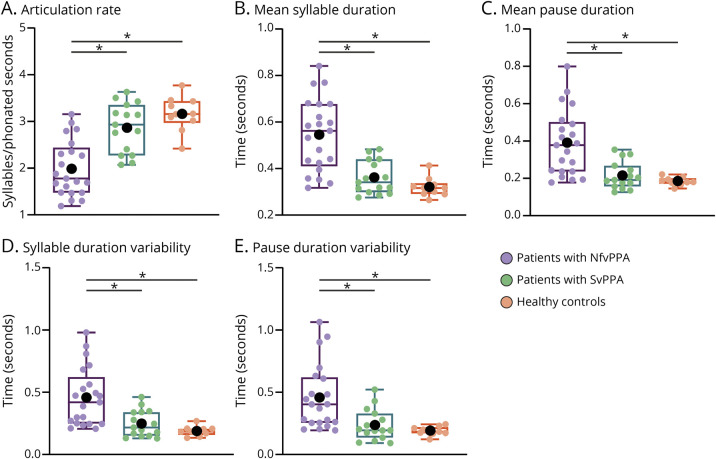
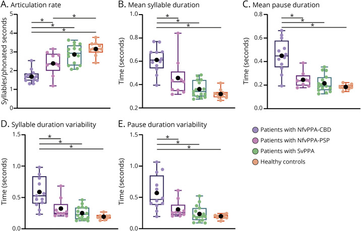
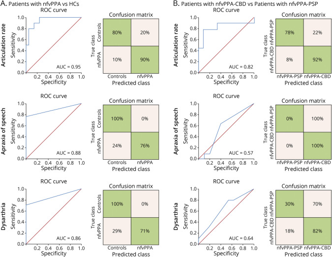
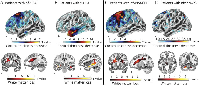
Results: We evaluated 22 persons with nfvPPA (mean age: 67.3 years; 13 female patients) and confirmed underlying 4-repeat tauopathy, 15 persons with semantic variant primary progressive aphasia (svPPA; mean age: 66.5 years; 8 female patients), and 10 healthy controls (HCs; 70 years; 5 female patients). All 5 speech timing measures revealed alterations in persons with nfvPPA relative to both the HC and svPPA groups, controlling for dementia severity. The articulation rate robustly discriminated individuals with nfvPPA from HCs (area under the ROC curve [AUC] = 0.95), outperforming specialist-dependent perceptual measures of dysarthria and apraxia of speech severity. Patients with nfvPPA exhibited structural abnormalities in left precentral and middle frontal as well as bilateral superior frontal regions, including their underlying WM. The articulation rate correlated with atrophy of the left pars opercularis and supplementary/presupplementary motor areas. Secondary analyses showed that, controlling for dementia severity, all measures yielded greater deficits in patients with nfvPPA and corticobasal degeneration (nfvPPA-CBD, n = 12) than in those with progressive supranuclear palsy pathology (nfvPPA-PSP, n = 10). The articulation rate robustly discriminated between individuals in each subgroup (AUC = 0.82). More widespread cortical thinning was observed for the nfvPPA-CBD than the nfvPPA-PSP group across frontal regions.

Discussion: Automated speech timing analyses can capture specific markers of nfvPPA while potentially discriminating between patients with different tauopathies. Thanks to its objectivity and scalability; this approach could support standard speech assessments.

Classification of evidence: This study provides Class III evidence that automated speech analysis can accurately differentiate patients with nonfluent PPA from normal controls and patients with semantic variant PPA.


Figures

Figure 1
Figure 1. Results for the 3-Group Setting (All Patients With nfvPPA Together)
All 5 speech timing measures revealed significant impairments in patients with nfvPPA relative to both healthy controls and patients with svPPA. No differences were observed between the latter 2 groups. In the box plot, middle horizontal lines show each group's median, with lower and upper lines representing the 25th and 75th percentiles, respectively. The whiskers represent the smallest and largest values in the distribution. Colored dots indicate each participant's individual value. Black dots represent the group's mean. *Denotes significant differences at p < 0.05. All statistics were calculated after outlier removal (namely, a single nfvPPA-PSP participant). NfvPPA = nonfluent variant primary progressive aphasia; svPPA = semantic variant primary progressive aphasia.
Figure 2
Figure 2. Results of the 4-Group Analysis (Separating Patients With nfvPPA-CBD From nfvPPA-PSP)
All 5 speech timing measures captured significant impairments in patients with nfvPPA-CBD compared with both healthy controls and patients with svPPA. By contrast, only the articulation rate was impaired in the patients with nfvPPA-PSP. Deficits in all measures were significantly greater for patients with nfvPPA-CBD than nfvPPA-PSP. In the box plot, middle horizontal lines show each group's median, with lower and upper lines representing the 25th and 75th percentiles, respectively. The whiskers represent the smallest and largest values in the distribution. Colored dots indicate each participant's individual value. Black dots represent the group's mean. *Denote significant differences at p < 0.05. All statistics were calculated after outlier removal (namely, a single participant with nfvPPA-PSP). NfvPPA-CBD = nonfluent variant primary progressive aphasia with corticobasal degeneration pathology; nfvPPA-PSP = nonfluent variant primary progressive aphasia with progressive supranuclear palsy pathology; svPPA = semantic variant primary progressive aphasia.
Figure 3
Figure 3. Binary Logistic Regression Results
(A) Classification between patients with nfvPPA and HCs was significant for the automated measure of the articulation rate (top inset) as well as the perceptual measures of apraxia of speech severity (middle inset) and dysarthria severity (bottom inset), with maximal AUC score for the former. (B) Classification between patients with nfvPPA-CBD and nfvPPA-PSP was significant for the automated measure of the articulation rate (top inset) but not for the perceptual measures of apraxia of speech severity (middle inset) and dysarthria severity (bottom inset), the former yielding a robust AUC value. AUC = area under the ROC curve; HCs = healthy controls; nfvPPA = nonfluent variant primary progressive aphasia; nfvPPA-CBD = nonfluent variant primary progressive aphasia with corticobasal degeneration pathology; nfvPPA-PSP = nonfluent variant primary progressive aphasia with progressive supranuclear palsy pathology; ROC = receiving operating characteristic; svPPA = semantic variant primary progressive aphasia.
Figure 4
Figure 4. Brain Structural Abnormality Patterns of Each Patient Group
The combined nfvPPA group (A) and the svPPA group (B), as well as subgroups with nfvPPA-CBD (C) and nfvPPA-PSP (D), were compared with HCs to estimate their patterns of cortical thickness (top insets) and white matter volume (bottom insets) using surface-based and voxel-based morphometry, respectively. For better visualization of differences and similarities across groups, t-map values are reported at an uncorrected p < 0.001 threshold. NfvPPA = nonfluent variant primary progressive aphasia; nfvPPA-CBD = nonfluent variant primary progressive aphasia with corticobasal degeneration pathology; nfvPPA-PSP = nonfluent variant primary progressive aphasia with progressive supranuclear palsy pathology; svPPA = semantic variant primary progressive aphasia.

Comment in

Similar articles

Cited by

References

    1. Josephs KA, Duffy JR. Apraxia of speech and nonfluent aphasia: a new clinical marker for corticobasal degeneration and progressive supranuclear palsy. Curr Opin Neurol. 2008;21(6):688-692. - PubMed
    1. Peterson KA, Patterson K, Rowe JB. Language impairment in progressive supranuclear palsy and corticobasal syndrome. J Neurol. 2021;268(3):796-809. - PMC - PubMed
    1. Marshall CR, Hardy CJD, Volkmer A, et al. . Primary progressive aphasia: a clinical approach. J Neurol. 2018;265(6):1474-1490. - PMC - PubMed
    1. Spinelli EG, Mandelli ML, Miller ZA, et al. . Typical and atypical pathology in primary progressive aphasia variants. Ann Neurol. 2017;81(3):430-443. - PMC - PubMed
    1. Santos-Santos MA, Mandelli ML, Binney RJ, et al. . Features of patients with nonfluent/agrammatic primary progressive aphasia with underlying progressive supranuclear palsy pathology or corticobasal degeneration. JAMA Neurol. 2016;73(6):733-742. - PMC - PubMed

Publication types