. 2022 Aug 1;12(1):13162.
doi: 10.1038/s41598-022-17310-z.

Profiling of gene expression in the brain associated with anxiety-related behaviors in the chronic phase following cranial irradiation

Affiliations

Profiling of gene expression in the brain associated with anxiety-related behaviors in the chronic phase following cranial irradiation

Hae-June Lee et al. Sci Rep. .

Abstract

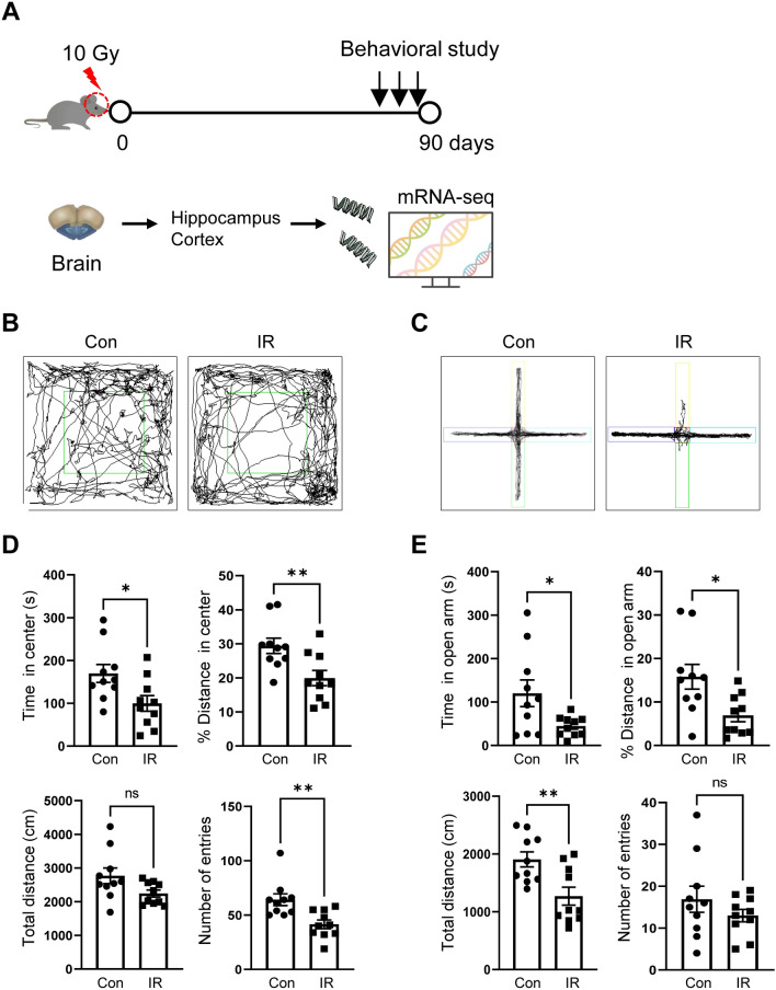
Although the brain is exposed to cranial irradiation in many clinical contexts, including malignant brain tumor therapy, such exposure can cause delayed neuropsychiatric disorders in the chronic phase. However, how specific molecular mechanisms are associated with irradiation-induced behavioral dysfunction, especially anxiety-like behaviors, is unclear. In the present study, we evaluated anxiety-like behaviors in adult C57BL/6 mice using the open-field (OF) and elevated plus maze (EPM) tests 3 months following single cranial irradiation (10 Gy). Additionally, by using RNA sequencing (RNA-seq), we analyzed gene expression profiles in the cortex and hippocampus of the adult brain to demonstrate the molecular mechanisms of radiation-induced brain dysfunction. In the OF and EPM tests, mice treated with radiation exhibited increased anxiety-like behaviors in the chronic phase. Gene expression analysis by RNA-seq revealed 89 and 106 differentially expressed genes in the cortex and hippocampus, respectively, following cranial irradiation. Subsequently, ClueGO and STRING analyses clustered these genes in pathways related to protein kinase activity, circadian behavior, and cell differentiation. Based on our expression analysis, we suggest that behavioral dysfunction following cranial irradiation is associated with altered expression of Cdkn1a, Ciart, Fos, Hspa5, Hspb1 and Klf10. These novel findings may provide potential genetic targets to investigate for the development of radioprotective agents.


Conflict of interest statement

The authors declare no competing interests.

Figures

Figure 1
Figure 1
Anxiety-related behavior during the chronic phase following cranial irradiation. (A) Schematic diagram of our experimental procedure. (B) Representative tracking of mouse movement during the open-field (OF) test. (C) Representative tracking of mouse movement during the elevated plus maze (EPM) test. (D) The time spent in the center, % distance traveled from the center, total distance traveled, and number of entries in the OF apparatus were assessed in sham-irradiated (Con) and 10 Gy-irradiated (IR) mice. (E) The time spent in the open arm, % distance traveled in the open arm, total distance traveled, and number of entries in the EPM test were assessed in the Con and IR groups. Data are expressed as the mean ± SE (n = 10 per group). *p < 0.05 and **p < 0.01 vs. the Con group.
Figure 2
Figure 2
Gene ontology (GO) analysis of RNA-seq screening data. (A) Venn diagram of differentially expressed genes (DEGs) in the cortex and hippocampus between sham-irradiated (Con) and 10 Gy-irradiated (IR) mice. (B,C) ClueGO network analysis of enriched categories in the cortex and/or hippocampus after cranial irradiation. GO terms describing molecular interactions among targets are represented as nodes, and node size represents the term's enrichment significance.
Figure 3
Figure 3
STRING network and enrichment analyses. (A) STRING analysis of 181 differentially expressed genes (DEGs) obtained upon RNA-seq analysis of the cortex and hippocampus between sham-irradiated (Con) and 10 Gy-irradiated (IR) mice. (B) The top 10 Gene Ontology (GO) biological processes that were functionally enriched in genes in the network were selected in order of highest strength with an FDR < 0.05.
Figure 4
Figure 4
Changes in 13 selected differentially expressed genes (DEGs) in the cortex and hippocampus of mouse brains after cranial irradiation. The bar graphs show the relative expression levels of upregulated (A) and downregulated (B) genes from RNA-seq data collected from both the cortex and hippocampus. Data are expressed as the mean ± SE (n = 5 per group). *p < 0.05, **p < 0.01, and ***p < 0.001 vs. the Con group.

Similar articles

Cited by

References

    1. Laperriere, N., Zuraw, L., Cairncross, G. & Cancer Care Ontario Practice Guidelines Initiative Neuro-Oncology Disease Site, G. Radiotherapy for newly diagnosed malignant glioma in adults: a systematic review. Radiother Oncol64, 259–273, doi:10.1016/s0167-8140(02)00078-6 (2002). - PubMed
    1. Gzell C, Back M, Wheeler H, Bailey D, Foote M. Radiotherapy in glioblastoma: The past, the present and the future. Clin. Oncol. (R Coll. Radiol.) 2017;29:15–25. doi: 10.1016/j.clon.2016.09.015. - DOI - PubMed
    1. Greene-Schloesser D, Moore E, Robbins ME. Molecular pathways: radiation-induced cognitive impairment. Clin. Cancer Res. 2013;19:2294–2300. doi: 10.1158/1078-0432.CCR-11-2903. - DOI - PMC - PubMed
    1. Janda M, et al. Quality of life among patients with a brain tumor and their carers. J. Psychosom. Res. 2007;63:617–623. doi: 10.1016/j.jpsychores.2007.06.018. - DOI - PubMed
    1. Tibbs MD, et al. Longitudinal analysis of depression and anxiety symptoms as independent predictors of neurocognitive function in primary brain tumor patients. Int. J. Radiat. Oncol. Biol. Phys. 2020;108:1229–1239. doi: 10.1016/j.ijrobp.2020.07.002. - DOI - PMC - PubMed

Publication types