. 2022 Aug;29(8):774-780.
doi: 10.1038/s41594-022-00807-6. Epub 2022 Aug 1.

Mechanism of client selection by the protein quality-control factor UBE2O

Affiliations

Mechanism of client selection by the protein quality-control factor UBE2O

Matthew C J Yip et al. Nat Struct Mol Biol. 2022 Aug.

Abstract

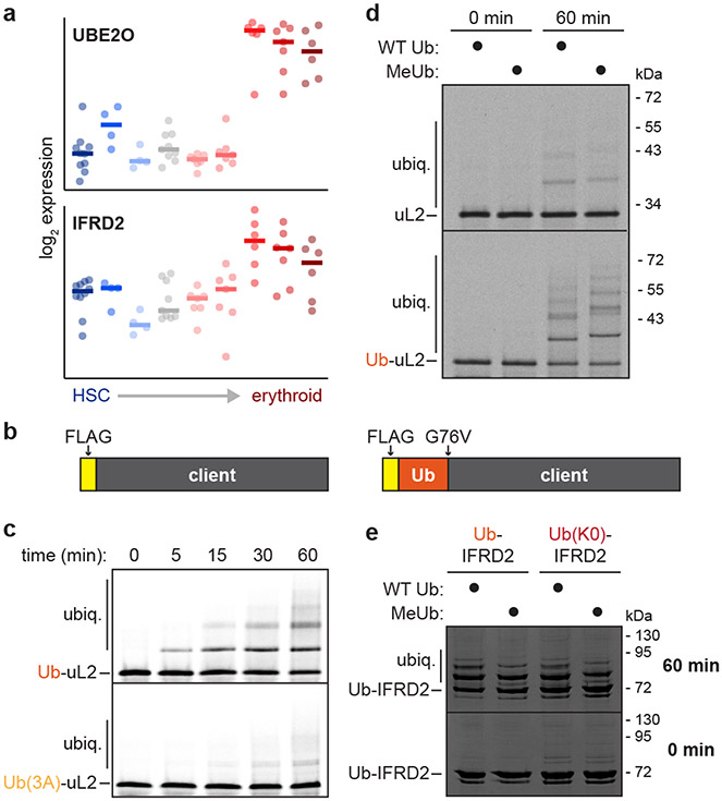
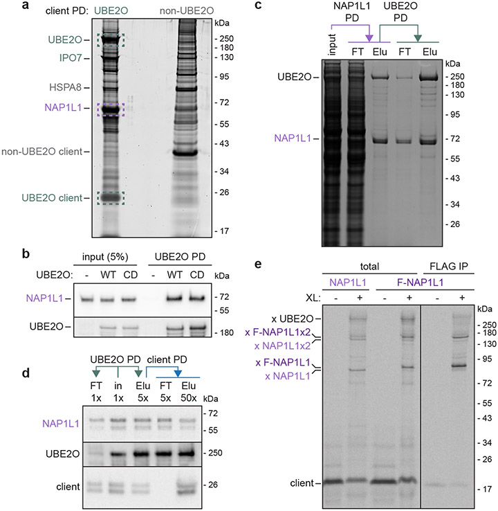
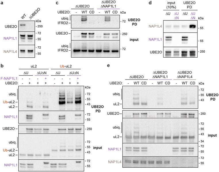
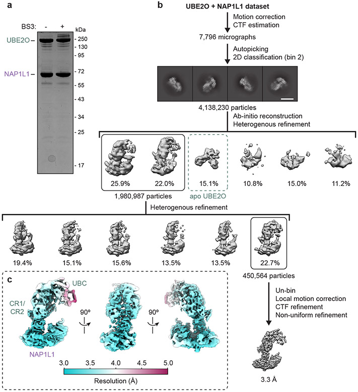
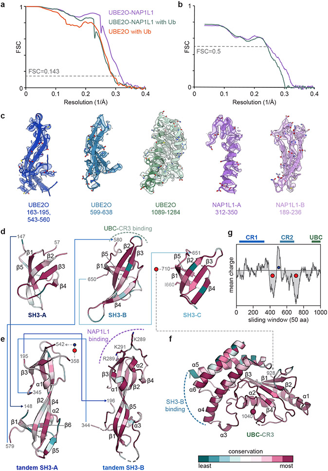
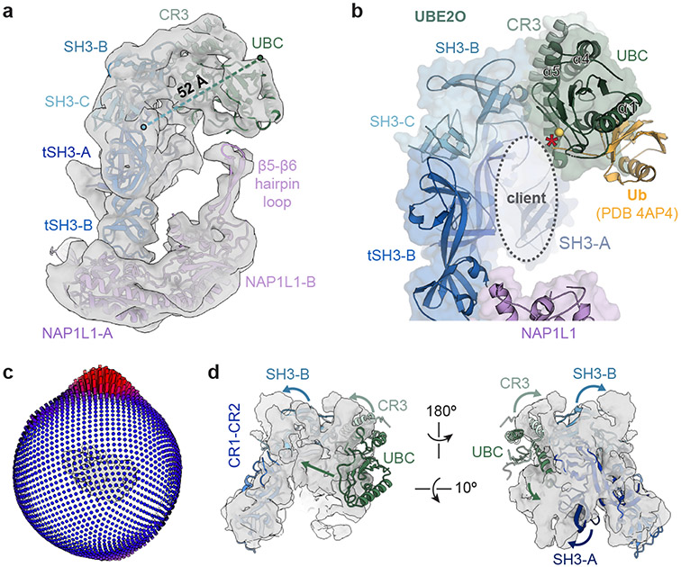
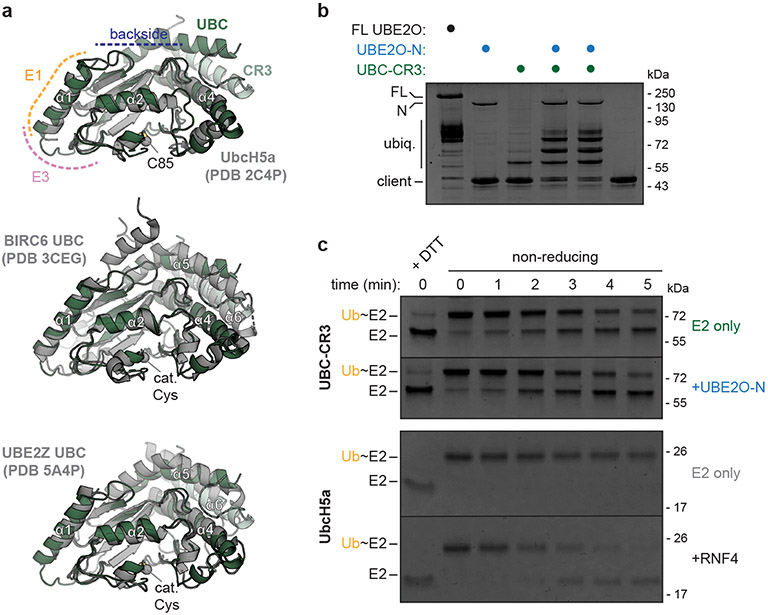
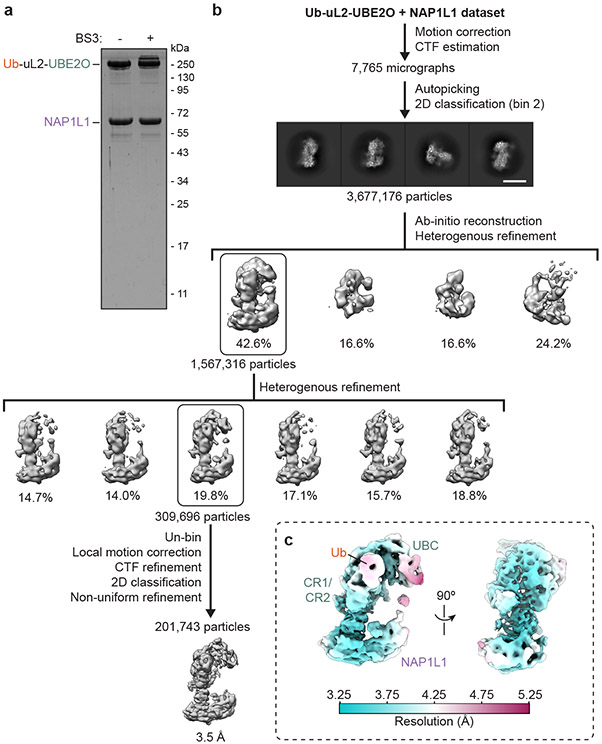
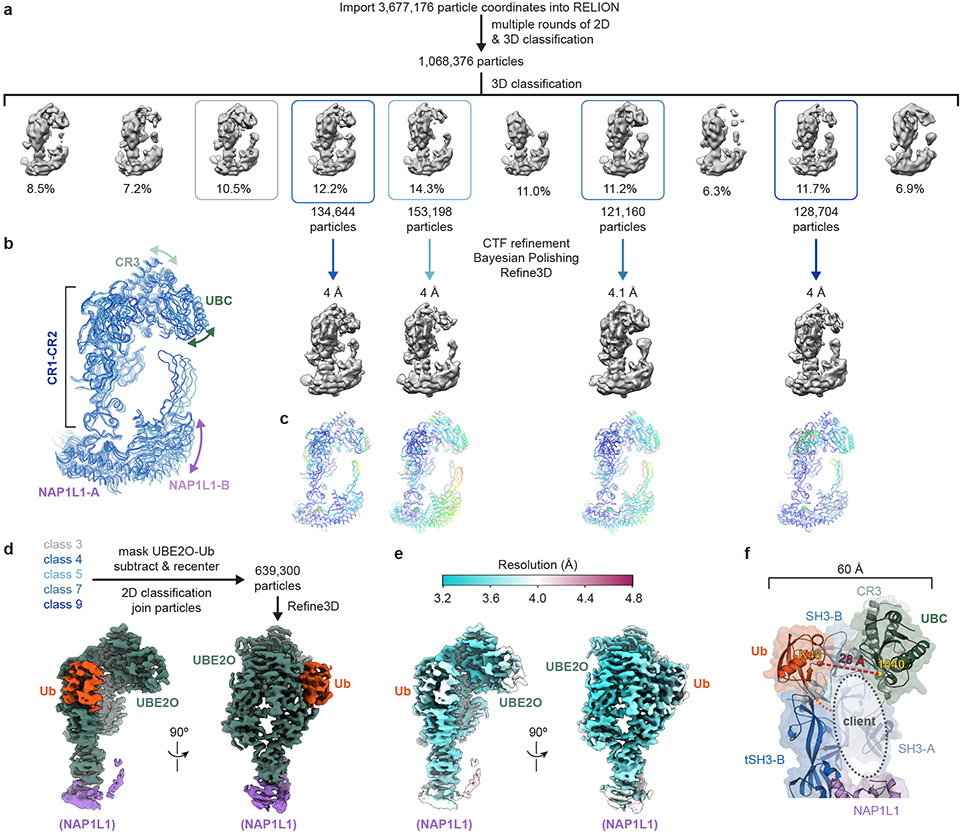
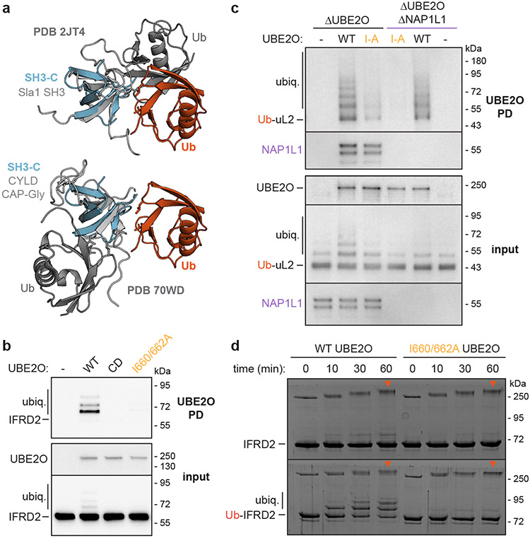
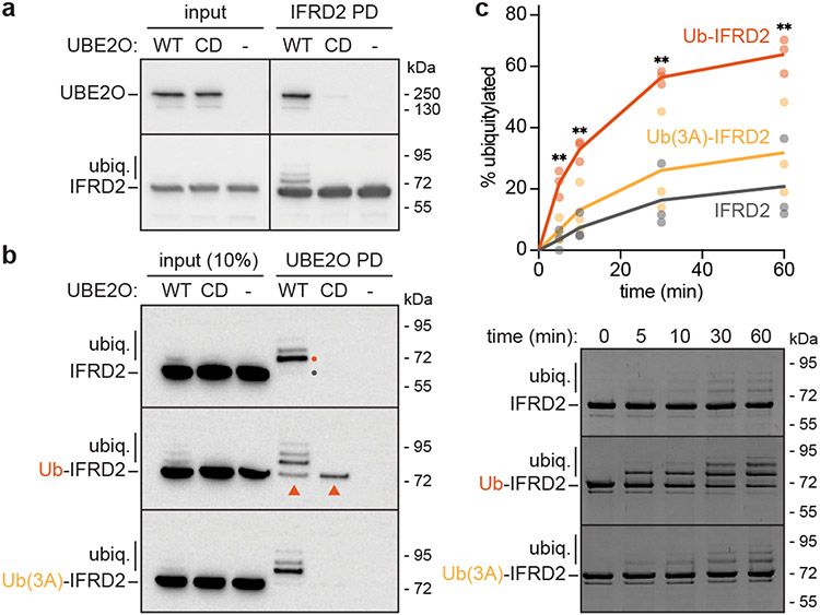
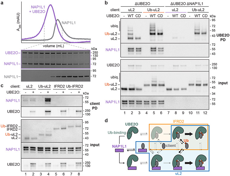
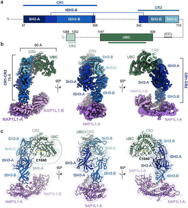
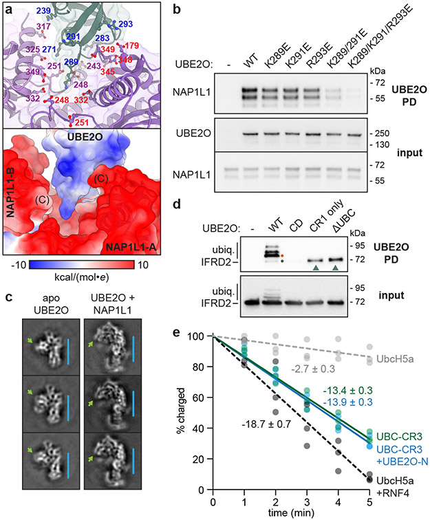
The E2/E3 enzyme UBE2O ubiquitylates diverse clients to mediate important processes, including targeting unassembled 'orphan' proteins for quality control and clearing ribosomes during erythropoiesis. How quality-control factors, such as UBE2O, select clients on the basis of heterogeneous features is largely unknown. Here, we show that UBE2O client selection is regulated by ubiquitin binding and a cofactor, NAP1L1. Attaching a single ubiquitin onto a client enhances UBE2O binding and multi-mono-ubiquitylation. UBE2O also repurposes the histone chaperone NAP1L1 as an adapter to recruit a subset of clients. Cryo-EM structures of human UBE2O in complex with NAP1L1 reveal a malleable client recruitment interface that is autoinhibited by the intrinsically reactive UBC domain. Adding a ubiquitylated client identifies a distinct ubiquitin-binding SH3-like domain required for client selection. Our findings reveal how multivalency and a feed-forward mechanism drive the selection of protein quality-control clients.


Figures

Extended Data Fig. 1 ∣
Extended Data Fig. 1 ∣. UBE2O has a ubiquitin-binding domain.
a, Expression (log2) values of UBE2O or IFRD2 in human hematopoietic cells at different stages of differentiation from hematopoietic stem cells (HSC) through the erythroid lineage,. Data (left to right) are from HSC (CD133+ CD34dim), HSC (CD38− CD34+), common myeloid progenitor (CMP) cells, megakaryocyte-erythroid progenitor (MEP) cells, and the following erythroid cell types: CD34+ CD71+ GlyA−, CD34− CD71+ GlyA−, CD34− CD71+ GlyA+, CD34− CD71lo GlyA+, CD34− CD71− GlyA+. b, Scheme of IFRD2 and ubiquitin-fused IFRD2 (Ub-IFRD2) variants analyzed in Fig. 1. c, SDS-PAGE and phosphorimaging of in vitro ubiquitylation timecourses, representative of 3 replicates, of radiolabeled uL2 fused to wildtype ubiquitin (Ub-uL2) or to ubiquitin with a mutated hydrophobic patch [Ub(3A)-uL2], which were synthesized in the PURE translation system and then incubated with 500 nM UBE2O, 75 nM E1, 10 μM ubiquitin, and an energy regenerating system. Note: ubiquitylation of Ub-uL2 is more efficient than that of Ub(3A)-uL2. d, Autoradiography of in vitro ubiquitylation reactions of uL2 (top) and Ub-uL2 (bottom) as in c, with wildtype ubiquitin (WT Ub) or methylated ubiquitin (MeUb) incapable of polyubiquitin chain formation at the indicated timepoints. e, Coomassie staining of in vitro ubiquitylation reactions as in Fig. 1c of IFRD2 conjugated either to wildtype ubiquitin (Ub-IFRD2) or to ubiquitin in which all lysines are mutated to arginines [Ub(K0)-IFRD2] with WT Ub or MeUb. Note: the degree of client ubiquitylation in reactions shown in d and e does not change substantially with Ub(K0) or MeUb.
Extended Data Fig. 2 ∣
Extended Data Fig. 2 ∣. NAP1L1 interacts with UBE2O and UBE2O clients.
a, SDS-PAGE and SYPRO Ruby staining of pulldowns (PD) of a FLAG-tagged UBE2O client (uL14) or a FLAG-tagged non-UBE2O client (uS3) synthesized in a mammalian in vitro translation system. Labels indicate abundant bands which were excised and identified by mass spectrometry. Teal dotted boxes, UBE2O and UBE2O client. Purple dotted box indicates stoichiometric recovery of NAP1L1 with the UBE2O client but not the non-UBE2O client. b, UBE2O knockout Flp-In 293 T-REx cells co-expressing FLAG-tagged NAP1L1 without or with wildtype (WT) or catalytically dead (CD) Strep-tagged UBE2O were lysed (input), subjected to UBE2O PD, and analyzed by SDS-PAGE and immunoblotting, representative of >3 replicates. Note: NAP1L1 is not ubiquitylated and interacts equally well with WT and CD UBE2O. c, SDS-PAGE and Coomassie staining of sequential PDs of FLAG-tagged NAP1L1 and Strep-tagged UBE2O showing stoichiometric complex purification, representative of 2 replicates. FT, flow-through; Elu, elution. d, Cells co-expressing CD Strep-tagged UBE2O and a FLAG-tagged UBE2O client (uL14) were lysed and subjected to sequential UBE2O and client PDs. Input (in), FT, and Elu samples at the indicated relative concentrations were analyzed by SDS-PAGE and immunoblotting, representative of 2 replicates, suggesting recovery of ternary complexes of UBE2O, NAP1L1, and uL14,. e, The radiolabeled UBE2O client uL14 was synthesized in vitro with either 1 μM untagged or FLAG-tagged NAP1L1 (F-NAP1L1), subjected to chemical crosslinking with 250 μM BMH as indicated, and analyzed directly (total) or after denaturing anti-FLAG immunoprecipitations (IP) by SDS-PAGE and autoradiography, representative of 2 replicates. Client crosslinks (x target) to UBE2O, NAP1L1, F-NAP1L1, and NAP1L1 or F-NAP1L1 dimers (x2) are indicated.
Extended Data Fig. 3 ∣
Extended Data Fig. 3 ∣. NAP1L1 recruits a subset of UBE2O clients.
a, Immunoblotting for UBE2O and NAP1 paralogs in wildtype (WT) or UBE2O knockout (ΔUBE2O) Flp-In 293 T-Rex cells. b, Pulldown (PD) of WT Strep-tagged UBE2O co-expressed with HA-tagged uL2 or Ub-uL2 in UBE2O knockout (ΔU) or UBE2O/NAP1L1 double knockout (ΔUΔN) Flp-In 293 T-Rex cells without or with induced re-expression of FLAG-tagged NAP1L1 (F-NAP1L1) incorporated into the Flp-In locus. F-NAP1L1 successfully rescues the interaction between uL2 and UBE2O which is impaired by knocking out NAP1L1, representative of 3 replicates. c, PD of WT or catalytically dead (CD) UBE2O co-expressed with FLAG-tagged IFRD2 in ΔUBE2O or ΔUBE2O ΔNAP1L1 Flp-In 293 T-Rex cells, showing that knocking out NAP1L1 does not strongly impair the interaction between IFRD2 and UBE2O, representative of 2 replicates. d, UBE2O purifications from ΔU or ΔUΔN cells showing NAP1L1 and NAP1L4 association. e, PD of WT or CD UBE2O co-expressed with FLAG-tagged uL2 in ΔUBE2O, ΔUBE2O ΔNAP1L1, or UBE2O/NAP1L4 double knockout (ΔUBE2O ΔNAP1L4) Flp-In 293 T-Rex cells. Note: knocking out NAP1L1 but not NAP1L4 impairs uL2 association with UBE2O, representative of 3 replicates.
Extended Data Fig. 4 ∣
Extended Data Fig. 4 ∣. Cryo-EM data processing of UBE2O in complex with NAP1L1.
a, SDS-PAGE and Coomassie staining of purified UBE2O-NAP1L1 complex without and with crosslinking with 250 μM BS3 used for cryo-EM analysis, representative of 2 independent preparations. b, Summary of processing strategy for the UBE2O + NAP1L1 dataset. Teal dotted box denotes class of apo UBE2O. Scale bar, 10 nm. c, Cryo-EM map of UBE2O-NAP1L1 colored by local resolution.
Extended Data Fig. 5 ∣
Extended Data Fig. 5 ∣. Cryo-EM maps and models.
a, Fourier shell correlation (FSC) coefficient vs. resolution (Å−1) curves of the indicated maps. Resolution was estimated at FSC=0.143 (gray dotted line). b, Model vs. map FSC curves for structures as in a. c, Segmented EM map densities of the sharpened UBE2O-NAP1L1 map contoured at 8.6σ with corresponding atomic model. d-f, Secondary structure designations of the d, SH3-like domains, e, tandem SH3 domains, and f, UBC-CR3 of UBE2O colored by sequence conservation. Position of the catalytic cysteine is shown as a sphere. Connectivity between domains is indicated by lines with arrowheads and residue numbering. Dotted lines and labeled residues indicate interfaces involved in intra- and inter-molecular interactions. g, Net charge (calculated as +1 for K/R, +0.5 for H, −1 for D/E) over sliding windows of 50 amino acids across the UBE2O sequence. Positions of CR1, CR2, and the beginning of UBC are indicated above. Red and blue circles indicate acidic and basic stretches, respectively.
Extended Data Fig. 6 ∣
Extended Data Fig. 6 ∣. Contributions to UBE2O client binding.
a, Model of UBE2O-NAP1L1 docked into a downsampled and unsharpened cryo-EM map contoured at 4.9σ showing the position of the β5-β6 hairpin loop of NAP1L1-B and the linear distance (dotted line) between the C-terminal end of SH3-C and the N-terminal end of the UBC domain on UBE2O, which are connected by residues 711-927. These features may influence client engagement. b, Position of UBC-charged ubiquitin (Ub, light orange) in the UBE2O-NAP1L1 complex relative to the putative client binding cavity, based on superposition of the UBE2O UBC domain with ubiquitin-charged UbcH5a (PDB 4AP4). Yellow sphere, position of C1040. Red asterisk denotes active site for client ubiquitylation. c, Angular distribution of a cryo-EM map of apo UBE2O. Preferential orientation was not significantly improved despite combining three datasets of unmodified apo UBE2O and of PEGylated apo UBE2O without and with a stage tilt of 35°. d, Rigid body docking of the model of UBE2O in the UBE2O-NAP1L1 complex (Fig. 3c) into a cryo-EM map of apo UBE2O. Arrows indicate putative movements of the indicated domains to account for conformational differences.
Extended Data Fig. 7 ∣
Extended Data Fig. 7 ∣. Analysis of UBE2O E2 activity.
a, Superposition of UBC-CR3 of UBE2O (green) with the UBC domain (gray) of UbcH5a (PDB 2C4P, top), BIRC6 (PDB 3CEG, middle), or UBE2Z (PDB 5A4P, bottom). The E1 binding site (orange), the canonical RING E3 binding site (yellow), and the backside of the UBC domain (blue) are indicated relative to UbcH5a (top). Catalytic cysteines are shown as spheres. Note the additional helices (α5 and α6) present in UBC-CR3 of UBE2O and the UBC domains of BIRC6 and UBE2Z. b, In vitro ubiquitylation assays of a recombinant UBE2O client (Cmd1-DDX56) with full-length (FL) UBE2O, the CR1 and CR2 regions of UBE2O (UBE2O-N), and/or UBC-CR3 of UBE2O as indicated. The non-endogenous Cmd1-DDX56 client was analyzed due to its efficient ubiquitylation. Note: effective ubiquitylation is achieved with FL UBE2O or with both UBC-CR3 and UBEO-N, but not with either domain individually, representative of 2 replicates. c, Ubiquitin discharging assays from UBE2O UBC-CR3 (top) or UbcH5a (bottom) onto free lysine without or with a corresponding E3 activity (UBE2O-N or RNF4, respectively), representative of 3 independent experiments quantified in Fig. 4e. Ub~E2, ubiquitin-charged E2.
Extended Data Fig. 8 ∣
Extended Data Fig. 8 ∣. Cryo-EM data processing of UBE2O-NAP1L1 bound to Ub-uL2.
a, SDS-PAGE and Coomassie staining of purified Ub-uL2-UBE2O-NAP1L1 complex without and with crosslinking with 250 μM BS3 used for cryo-EM analysis, representative of 2 independent preparations. Note: Ub-uL2 is fused to UBE2O by a long linker but remains associated with the complex if this linker is cleaved after purification. b, Summary of processing strategy for the Ub-uL2-UBE2O + NAP1L1 dataset. Scale bar, 10 nm. c, Cryo-EM map of UBE2O-NAP1L1 bound to Ub-uL2 colored by local resolution.
Extended Data Fig. 9 ∣
Extended Data Fig. 9 ∣. Analysis of UBE2O-NAP1L1 with Ub-uL2.
a, Summary of processing strategy to examine conformational heterogeneity of UBE2O-NAP1L1 bound to ubiquitin. b, Superposition of models in which individual UBE2O and NAP1L1 domains were fitted to cryo-EM maps of the indicated classes (blue boxes) showing conformational heterogeneity in the relative positions and orientations of NAP1L1-B, UBC, and CR3 around the putative client binding interface. c, Models as in b, fitted to the indicated cryo-EM maps and superposed with the consensus model of the Ub-UBE2O-NAP1L1 complex (gray; Fig. 5b) colored by RMSD (blue – lowest, red – highest, normalized to each model). d, Processing strategy to generate subtracted particles of ubiquitin-bound UBE2O for focused refinement. e, Cryo-EM map resulting from focused refinement of ubiquitin-bound UBE2O as in d, colored by local resolution. f, Position of ubiquitin (orange) bound to SH3-C of UBE2O. Lysine at position 48 (K48) of ubiquitin is shown together with the distance (red) to the catalytic cysteine at position 1040 (yellow) of UBE2O. The C-terminal end of ubiquitin, which is modeled up to residue 73 is indicated by the orange dotted line leading to the putative client binding cavity.
Extended Data Fig. 10 ∣
Extended Data Fig. 10 ∣. SH3-C of UBE2O binds ubiquitin.
a, Superposition of ubiquitin (Ub; orange) bound to SH3-C of UBE2O (light blue) with the SH3 domain of Sla1 (top; PDB 2JT4) or the CAP-Gly SH3-like domain of CYLD (bottom; PDB 70WD) (light gray) bound to ubiquitin (dark gray). Note: although the SH3 and SH3-like domains are well-aligned, they bind ubiquitin through different interfaces. b, Pulldown (PD) of wildtype (WT), catalytically dead (CD), or I660A/I662A Strep-tagged UBE2O co-expressed with FLAG-tagged IFRD2, representative of 2 replicates. ubiq., ubiquitylated client. c, PD of WT or I660A/I662 (I-A) Strep-tagged UBE2O co-expressed with FLAG-tagged ubiquitin-fused uL2 (Ub-uL2) in UBE2O knockout (ΔUBE2O) or UBE2O/NAP1L1 double knockout (ΔUBE2O ΔNAP1L1) cells, representative of 2 replicates. Note: I-A UBE2O loses ubiquitin-enhanced client binding activity. d, In vitro ubiquitylation timecourses of IFRD2 or ubiquitin-fused IFRD2 (Ub-IFRD2) with WT or I660A/I662A UBE2O, representative of 2 replicates. Orange arrowheads, autoubiquitylated UBE2O.
Fig. 1 ∣
Fig. 1 ∣. UBE2O preferentially binds and modifies ubiquitin-conjugated clients.
a, SDS-PAGE and immunoblotting of input and pulldown (PD) samples of FLAG-tagged IFRD2 co-expressed with wildtype (WT) or catalytically dead (CD) Strep-tagged UBE2O, or a transfection control (−), in HEK293T cells reveal a specific interaction between IFRD2 and WT but not CD UBE2O, representative of 3 replicates. ubiq., ubiquitylated IFRD2. b, UBE2O PD with unmodified IFRD2 (top) or with IFRD2 fused to G76V (Ub-IFRD2) or L8A/I44A/V70A/G76V ubiquitin (Ub(3A)-IFRD2) in UBE2O knockout HEK293T cells, representative of 3 replicates. Gray dot, unmodified IFRD2; orange dot, mono-ubiquitylated IFRD2; orange triangles, Ub-IFRD2 that pulls down with UBE2O. c, In vitro ubiquitylation timecourses of 2 μM of the indicated IFRD2 variant with 500 nM UBE2O, 75 nM E1, 10 μM ubiquitin, and an energy regenerating system. The average (line) percentage of ubiquitylated IFRD2 variant for 3 replicates (dots) is plotted versus reaction time (top). **, p<0.01 (p=0.0043 at 5 min, p=0.0014 at 15 min, p=0.0028 at 30 min, p=0.0071 at 60 min), unpaired two-tailed t-test between Ub-IFRD2 and IFRD2. SDS-PAGE and Coomassie staining of one replicate is shown below.
Fig. 2 ∣
Fig. 2 ∣. NAP1L1 is a UBE2O cofactor that regulates client selection.
a, Size exclusion chromatography trace (top) and SDS-PAGE and Coomassie staining (bottom) of purified NAP1L1 without (gray) or with (purple) UBE2O, representative of 2 replicates. b, Pulldowns (PD) of wildtype (WT) or catalytically dead (CD) Strep-tagged UBE2O co-expressed with the large ribosomal subunit protein uL2 without or with a ubiquitin fusion (Ub-uL2) in UBE2O knockout (ΔUBE2O) or UBE2O/NAP1L1 double-knockout (ΔUBE2O ΔNAP1L1) cells, representative of 2 replicates. c, PD of the indicated FLAG-tagged client expressed in ΔUBE2O cells without or with Strep-tagged UBE2O to detect endogenous NAP1L1 (purple) association, representative of 3 replicates. d, Model for UBE2O client selection mediated by an unannotated ubiquitin (Ub)-binding site and NAP1L1 as substrate adaptors. Opaque complexes are detected biochemically in our study. Outlined boxes indicate complexes observed with IFRD2 (light orange) or uL2 (blue).
Fig. 3 ∣
Fig. 3 ∣. Cryo-EM structure of UBE2O in complex with NAP1L1.
a, Linear domain organization of UBE2O. SH3-like domains (SH3-A, SH3-B, SH3-C), tandem SH3-like domains (tSH3-A, tSH3-B), the UBC domain, and CR3 visualized in cryo-EM structures are indicated. CR1, CR2, and CR3 are conserved regions. Colored lines denote previously annotated sequences for the indicated region or domain. CC, a putative coiled-coil not visualized by cryo-EM. Other unmodeled regions are indicated by dotted line. b, Cryo-EM map and c, model of UBE2O in complex with NAP1L1. The position of the catalytic cysteine (C1040; yellow sphere) and the C-terminal ends of NAP1L1 subunits (transparent magenta dots) are indicated. Blue/green dotted line indicates unresolved residues between tSH3-C and the UBC domain. The β5-β6 hairpin loop of NAP1L1 is not shown due to masking.
Fig. 4 ∣
Fig. 4 ∣. Inter- and intra-molecular regulation of UBE2O activity.
a, The UBE2O-NAP1L1 interaction site showing positions (top) of basic residues in UBE2O tSH3-B (blue) and acidic residues in NAP1L1-A (red) or NAP1L1-B (purple), or colored by electrostatic potential with the C-terminal end of each NAP1L1 subunit indicated (bottom). b, Pulldowns (PD) of the indicated UBE2O variants to detect interaction with endogenous NAP1L1, representative of 2 replicates. c, 2D class averages of apo UBE2O and subtracted particles of UBE2O in complex with NAP1L1. The axis of CR1-CR2 (blue line, 60 Å) and relative position of the rest of UBE2O (green arrow) are indicated. d, PD of the indicated UBE2O variants reveal that the UBC domain autoinhibits client binding, representative of 2 replicates. CD, catalytically dead; CR1 only, the isolated CR1 region of UBE2O (residues 1-450); ΔUBC, UBE2O lacking the UBC domain (residues 944-1113); ubiq., ubiquitylated IFRD2; gray dot, unmodified IFRD2; orange dot, monoubiquitylated IFRD2. Teal arrowheads denote unmodified IFRD2 associated with CR1 and ΔUBC UBE2O. e, Timecourse of ubiquitin discharge from UBE2O UBC-CR3 without (blue) or with (green) UBE2O-N, or from UbcH5a without (gray) or with (black) the E3 RNF4. The percent of ubiquitin-charged E2 relative to t=0 min was quantified for three independent replicates, and a linear regression was applied to calculate the indicated discharge rate (% discharged per min).
Fig. 5 ∣
Fig. 5 ∣. Ubiquitin makes a distinct interaction with an SH3-like domain of UBE2O.
a, Cryo-EM map and b, model of UBE2O-NAP1L1 bound to ubiquitin-fused uL2. The C-terminal end of ubiquitin (Ub; orange) is indicated by the orange dotted line. Yellow sphere denotes position of catalytic C1040. c, Interaction between hydrophobic patches on ubiquitin (L8/I44/V70) and on SH3-C of UBE2O. d, Pulldowns (PD) of wildtype (WT), catalytically dead (CD), or I660A/I662A Strep-tagged UBE2O co-expressed with FLAG-tagged ubiquitin-fused IFRD2 (Ub-IFRD2), representative of 2 replicates. ubiq., ubiquitylated Ub-IFRD2. e, In vitro ubiquitylation timecourses of radiolabeled uL2 (top) or ubiquitin-fused uL2 (Ub-uL2) (middle) with WT or I660A/I662A UBE2O, representative of 2 replicates. Coomassie staining showing UBE2O autoubiquitylation (orange arrowheads) is below.

Similar articles

Cited by

References

    1. Ubiquitin Signaling in Health and Disease [special issue]. Cell Death & Differentiation 28, (2021). - PMC - PubMed
    1. Yanagitani K, Juszkiewicz S & Hegde RS UBE2O is a quality control factor for orphans of multiprotein complexes. Science 357, 472–475 (2017). - PMC - PubMed
    1. Nguyen AT et al. UBE2O remodels the proteome during terminal erythroid differentiation. Science 357, eaan0218 (2017). - PMC - PubMed
    1. Berleth ES & Pickart CM Mechanism of Ubiquitin Conjugating Enzyme E2-230K: Catalysis Involving a Thiol Relay? Biochemistry 35, 1664–1671 (1996). - PubMed
    1. Zhang X et al. Fine-tuning BMP7 signalling in adipogenesis by UBE2O/E2-230K-mediated monoubiquitination of SMAD6. EMBO J 32, 996–1007 (2013). - PMC - PubMed

Methods References

    1. Labun K et al. CHOPCHOP v3: expanding the CRISPR web toolbox beyond genome editing. Nucleic Acids Res 47, W171–W174 (2019). - PMC - PubMed
    1. Ran FA et al. Genome engineering using the CRISPR-Cas9 system. Nat Protocols 8, 2281–2308 (2013). - PMC - PubMed
    1. Shimizu Y & Ueda T PURE technology. Methods Mol Biol 607, 11–21 (2010). - PubMed
    1. Feng Q & Shao S In vitro reconstitution of translational arrest pathways. Methods 137, 20–36 (2018). - PubMed
    1. Punjani A, Rubinstein JL, Fleet DJ & Brubaker MA cryoSPARC: algorithms for rapid unsupervised cryo-EM structure determination. Nat Methods 14, 290–296 (2017). - PubMed

Publication types