

Aggressive unifocal bone Langerhans cell histiocytosis with soft tissue extension both responsive to radiotherapy: a case report
- PMID: 35915468
- PMCID: PMC9344655
- DOI: 10.1186/s13014-022-02108-0
Aggressive unifocal bone Langerhans cell histiocytosis with soft tissue extension both responsive to radiotherapy: a case report
Abstract
Background: Langerhans cell histiocytosis (LCH) is a rare haematological neoplasm characterized by the accumulation of CD1a+, CD207/Langerin+ histiocytes within inflammatory lesions. LCH can involve any organ, but osteolytic bone lesions are most often encountered. Unifocal bone lesions may regress spontaneously after a thick needle biopsy has been taken.
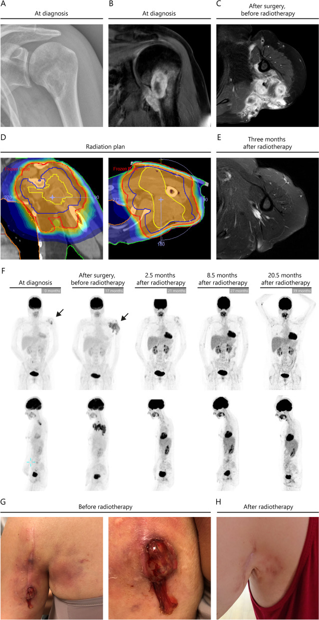
Case presentation: In this case report, we describe the initial presentation of a single BRAFV600E mutated osteolytic LCH lesion in the left proximal humerus of a 46-year-old previously healthy woman. Despite multiple surgical interventions, she unexpectedly experienced progressive disease manifestation with significant soft tissue extension to the surrounding musculature, subcutis and epidermis. Because the disease manifestation remained loco-regional, radiotherapy (RT) (total dose of 20 Gy in 10 fractions) was initiated.
Conclusion: The patient achieved a complete remission without any side effects. This case highlights that RT is a rational and relative mild local treatment option for patients with aggressive LCH affecting the bone and surrounding soft tissue.
Keywords: BRAF; Bone; LCH; Langerhans cell histiocytosis; Radiotherapy.
© 2022. The Author(s).
Conflict of interest statement
The authors declare no competing interests.
Figures

Similar articles
-
BRAF-mutated histiocytosis of the skull lacking the expression of Langerhans cell markers.Clin Neuropathol. 2020 Mar/Apr;39(2):64-69. doi: 10.5414/NP301225. Clin Neuropathol. 2020. PMID: 31661070
-
Gastrointestinal Langerhans cell histiocytosis with unifocal, single-system involvement in adults: Cases report and literature review.J Clin Lab Anal. 2022 Dec;36(12):e24765. doi: 10.1002/jcla.24765. Epub 2022 Nov 17. J Clin Lab Anal. 2022. PMID: 36397297 Free PMC article.
-
Langerhans cell histiocytosis of an intra-mammary lymph node in an 18-year-old woman.Pathologica. 2020 Mar;112(1):50-55. doi: 10.32074/1591-951X-27-19. Pathologica. 2020. PMID: 32202540 Free PMC article.
-
[Case of unifocal orbital langerhans cell histiocytosis in an adult].No Shinkei Geka. 2011 Dec;39(12):1183-8. No Shinkei Geka. 2011. PMID: 22128274 Review. Japanese.
-
Stereotactic Radiosurgery for Localized Cranial Langerhans Cell Histiocytosis: A Single Institution Experience and Review of Literature.World Neurosurg. 2023 Apr;172:e476-e482. doi: 10.1016/j.wneu.2023.01.053. Epub 2023 Jan 19. World Neurosurg. 2023. PMID: 36681322 Review.
References
Publication types
MeSH terms
LinkOut - more resources
Full Text Sources
Research Materials
