Review
. 2022 Aug 1;14(1):98.
doi: 10.1186/s13148-022-01305-8.

Therapeutic potential of inhibiting histone 3 lysine 27 demethylases: a review of the literature

Affiliations
Review

Therapeutic potential of inhibiting histone 3 lysine 27 demethylases: a review of the literature

Jeries Abu-Hanna et al. Clin Epigenetics. .

Abstract

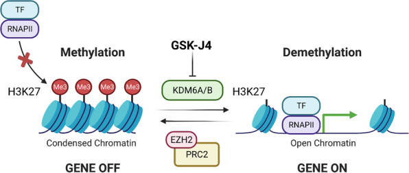
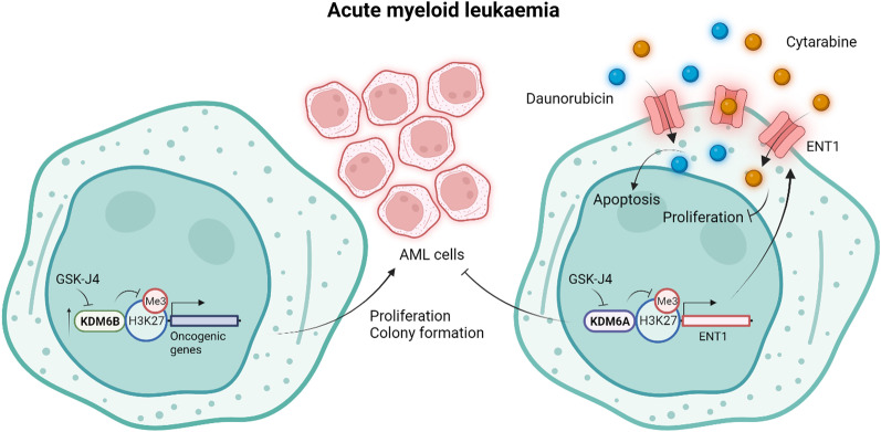
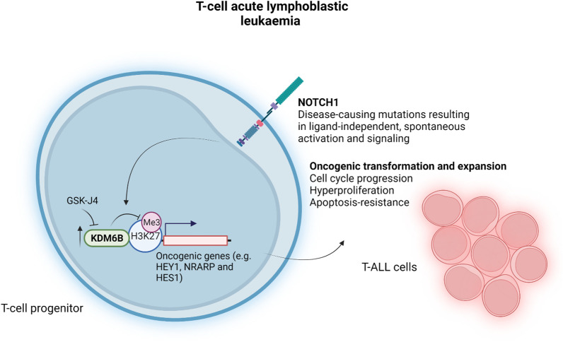
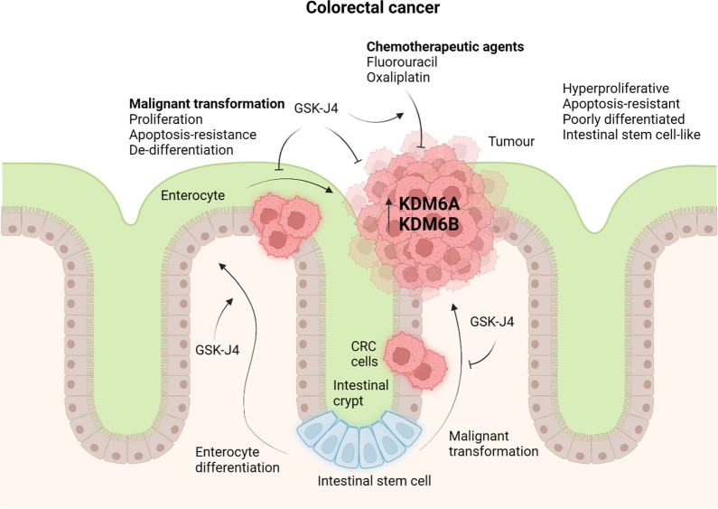
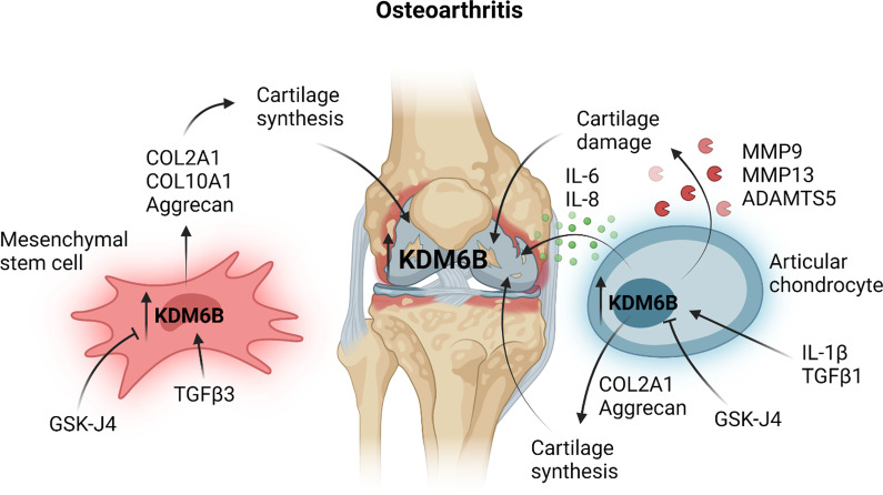
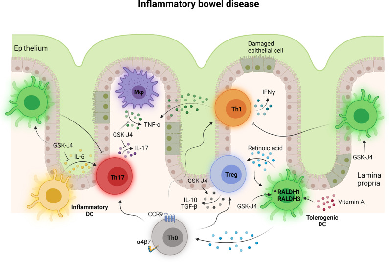
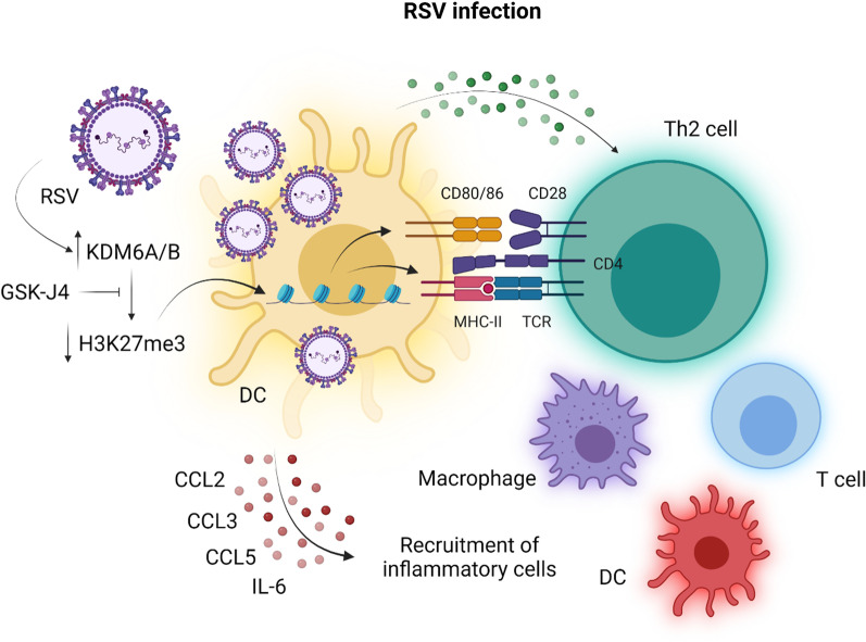
Histone 3 lysine 27 (H3K27) demethylation constitutes an important epigenetic mechanism of gene activation. It is mediated by the Jumonji C domain-containing lysine demethylases KDM6A and KDM6B, both of which have been implicated in a wide myriad of diseases, including blood and solid tumours, autoimmune and inflammatory disorders, and infectious diseases. Here, we review and summarise the pre-clinical evidence, both in vitro and in vivo, in support of the therapeutic potential of inhibiting H3K27-targeting demethylases, with a focus on the small-molecule inhibitor GSK-J4. In malignancies, KDM6A/B inhibition possesses the ability to inhibit proliferation, induce apoptosis, promote differentiation, and heighten sensitivity to currently employed chemotherapeutics. KDM6A/B inhibition also comprises a potent anti-inflammatory approach in inflammatory and autoimmune disorders associated with inappropriately exuberant inflammatory and autoimmune responses, restoring immunological homeostasis to inflamed tissues. With respect to infectious diseases, KDM6A/B inhibition can suppress the growth of infectious pathogens and attenuate the immunopathology precipitated by these pathogens. The pre-clinical in vitro and in vivo data, summarised in this review, suggest that inhibiting H3K27 demethylases holds immense therapeutic potential in many diseases.

Keywords: Autoimmune diseases; Cancer; Epigenetics; GSK-J4; H3K27; Histone lysine demethylase; Infectious diseases; Inflammation; JMJD3; KDM6A; KDM6B; UTX.


Conflict of interest statement

The authors declare that they have no competing interests.

Figures

Fig. 1
Fig. 1
Trimethyl H3K27 constitutes a repressive epigenetic mark of gene expression. When trimethylated at lysine 27 (K27) within its N terminus, histone 3 (H3) is intimately associated with inactive gene promoter regions, where the binding of RNA polymerase II (RNAPII) and transcription factors (TFs) is hampered. Trimethylation of H3K27 is unique in that it is exclusively catalysed by the H3K27-specific methyltransferase, enhancer of zeste homologue 2 (EZH2), a catalytic component of the polycomb repressive complex 2 (PRC2). Demethylation of H3K27, namely by the H3K27-specific lysine demethylases 6A (KDM6A) and 6B (KDM6B), serves to activate gene transcription by permitting the binding of RNAPII and TFs to promoter regions. This figure was created in BioRender.com
Fig. 2
Fig. 2
KDM6A/B inhibition in acute myeloid leukaemia. KDM6A and KDM6B have contrasting roles in the pathogenesis of acute myeloid leukaemia (AML). Increased KDM6B expression and occupancy at gene promoter regions in AML drives the transcription of genes involved in regulating the cell cycle, DNA replication, apoptosis and cellular differentiation, leading to the proliferation and colony formation of AML cells. Inhibition of KDM6B therefore attenuates AML cell proliferation and colony formation by promoting repressive H3K27 trimethylation, which downregulates the expression of these oncogenic genes. Contrastingly, through H3K27 demethylation, KDM6A upregulates the expression of the drug influx transporter ENT1, allowing the cellular entry of the chemotherapeutic agents cytarabine and daunorubicin and the consequent inhibition of AML cell proliferation and colony formation. Inhibition of KDM6A with GSK-J4 renders AML cells resistant to these chemotherapeutic agents by suppressing their cellular uptake through the downregulation of ENT1. This figure was created in BioRender.com
Fig. 3
Fig. 3
KDM6B inhibition in T cell acute lymphoblastic leukaemia. T cell acute lymphoblastic leukaemia (T-ALL) involves the oncogenic transformation and expansion of T cell progenitors. T-ALL cells overexpress KDM6B, which predominantly occupies the promoter regions of oncogenic genes that are targets of NOTCH1 signalling, including HEY1, NRARP and HES1. Indeed, in T-ALL, mutations in the NOTCH1 receptor result in ligand-independent, spontaneous receptor activation and signalling. GSK-J4 suppresses the growth of T-ALL cells by increasing the levels of the repressive mark H3K27me3 without influencing KDM6B occupancy. This figure was created in BioRender.com
Fig. 4
Fig. 4
KDM6A/KDM6B inhibition in colorectal cancer. KDM6A/B expression negatively correlates with survival in patients with colorectal cancer (CRC), suggesting a pathogenic role for these H3K27 demethylases. Inhibition of KDM6A/B with GSK-J4 attenuates the hyperproliferative, apoptosis-resistant and stem cell-like phenotype of CRC cells and promotes their enterocyte differentiation. GSK-J4 also inhibits the malignant transformation of intestinal stem cells, which reside in the intestinal crypts and are thought to be the source of most CRC cells, promoting instead their differentiation into enterocytes. In addition to its direct tumoricidal activity, GSK-J4 heightens the sensitivity of colorectal cancer cells to the chemotherapeutic agents fluorouracil and oxaliplatin. This figure was created in BioRender.com
Fig. 5
Fig. 5
KDM6B inhibition in rheumatoid arthritis. Rheumatoid arthritis is a chronic inflammatory disease of the joints characterised by synovial hyperplasia, inflammation and bone erosion. KDM6B is implicated in platelet-derived growth factor BB (PDGFBB)-induced proliferation and migration of fibroblast-like synoviocytes, a process that underlies hyperplasia of the synovial membrane. Through inhibition of KDM6B, GSK-J4 inhibits the expansion and migratory capacity of fibroblast-like synoviocytes to attenuate synovial hyperplasia. Synovial inflammation in rheumatoid arthritis is driven by the release of pro-inflammatory cytokines, including tumour necrosis factor α (TNFα) and interferon γ (IFNγ), from macrophages and natural killer (NK) cells. GSK-J4 inhibits the production of these cytokines to dampen the inflammatory response in rheumatoid arthritis. Bone erosion in rheumatoid arthritis is mediated by bone-resorbing osteoclasts, the differentiation of which is supported by NK cells. By inhibiting KDM6B in NK cells, GSK-J4 impairs the ability of NK cells to promote the differentiation of monocytes to osteoclasts, protecting arthritic joints from bone erosion. This figure was created in BioRender.com
Fig. 6
Fig. 6
KDM6B inhibition in osteoarthritis. KDM6B is overexpressed in damaged cartilage in the knees of patients with osteoarthritis, a likely consequence of chondrocyte stimulation with IL-1β or TGFβ1. In osteoarthritis, chondrocytes upregulate the expression of the proteases MMP9, MMP13 and ADMATS5 in response to stimulation with IL-1β to promote cartilage damage and erosion. IL-1β also induces the expression of IL-6 and IL-8 in chondrocytes to create a pro-inflammatory milieu in osteoarthritis. Inhibition of KDM6B with GSK-J4 attenuates cartilage loss and inflammation in OA by suppressing these chondrocyte responses to IL-1β and TGFβ1. KDM6B also drives the expression of the cartilage components COL2A1 and aggrecan and the bone constituent COL10A1 during TGFβ3-mediated chondrogenic differentiation of mesenchymal stem cells, contributing to cartilage and bone synthesis, respectively. Inhibiting KDM6B would not promote the replacement of damaged cartilage and bone in osteoarthritis. This figure was created in BioRender.com
Fig. 7
Fig. 7
KDM6A/B inhibition in inflammatory bowel disease. Chronic gut inflammation in inflammatory bowel disease (IBD) is driven by innate and adaptive immune cells with increased reactivity to self or foreign innocuous antigens. IBD also involves the loss of regulatory T (Treg) cell-mediated immunosuppression and tolerance further disrupting immunological homeostasis in the gut. KDM6A/B inhibition with GSK-J4 in IBD restores the balance between the pro-inflammatory and anti-inflammatory arms of the adaptive immune response. GSK-J4 promotes the generation of tolerogenic dendritic cells through the upregulation of retinaldehyde dehydrogenases 1 and 3 (RALDH1 and RALDH3) and the consequent production of retinoic acid from vitamin A. Retinoic acid then promotes the recruitment of naïve CD4+ T (Th0) cells by inducing the expression of the gut-homing receptors CCR9 and α4β7 and their differentiation into IL-10- and TGFβ-producing Tregs with enhanced lineage stability and immunosuppressive function while restricting the generation of pro-inflammatory TNFα- and IFNγ-producing Th1 cells and IL17-producing Th17 cells. GSK-J4 also dampens gut inflammation by inhibiting the production of IL-6 by inflammatory dendritic cells and the resultant generation of Th17 cells. This figure was created in BioRender.com
Fig. 8
Fig. 8
KDM6A/B inhibition in respiratory syncytial virus infection. Respiratory syncytial virus (RSV) infection is associated with the activation of dendritic cells (DCs), which promote a T helper 2 (Th2)-type response and a microenvironment within the lungs permissive for the recruitment of inflammatory cells, including macrophages, T cells and dendritic cells. DC activation by RSVs is accompanied by upregulation of the H3K27-specific demethylases KDM6A/B and a consequent decrease in H3K27 trimethylation (H3K27me3). Inhibition of KDM6A/B with GSK-J4 impairs the ability of DCs to present antigens to and activate Th2 cells through the downregulation of antigen-presenting MHC class II (MHC-II) and the costimulatory CD80/C86 molecules. GSK-J4 also inhibits the production of the pro-inflammatory cytokines and chemokines CCL2, CCL3, CCL5 and IL-6 by DCs to reduce the ingress of inflammatory cells into the lungs and attenuate the pulmonary immunopathology associated with the RSV infection. This figure was adapted from Malinczak et al. (2020) [77] and created in BioRender.com

Similar articles

Cited by

References

    1. Allis CD, Jenuwein T. The molecular hallmarks of epigenetic control. Nat Rev Genet. 2016;17(8):487–500. doi: 10.1038/nrg.2016.59. - DOI - PubMed
    1. Cheng Y, He C, Wang M, Ma X, Mo F, Yang S, Han J, Wei X. Targeting epigenetic regulators for cancer therapy: mechanisms and advances in clinical trials. Signal Transduct Target Ther. 2019;4(1):1–39. doi: 10.1038/s41392-019-0095-0. - DOI - PMC - PubMed
    1. Morera L, Lübbert M, Jung M. Targeting histone methyltransferases and demethylases in clinical trials for cancer therapy. Clin Epigenet. 2016;8(1):1–6. doi: 10.1186/s13148-016-0223-4. - DOI - PMC - PubMed
    1. Hyun K, Jeon J, Park K, Kim J. Writing, erasing and reading histone lysine methylations. Exp Mol Med. 2017;49(4):e324. doi: 10.1038/emm.2017.11. - DOI - PMC - PubMed
    1. Cheung P, Lau P. Epigenetic regulation by histone methylation and histone variants. Mol Endocrinol. 2005;19(3):563–573. doi: 10.1210/me.2004-0496. - DOI - PubMed

Publication types