. 2022 Sep;46(9):1665-1676.
doi: 10.1111/acer.14918. Epub 2022 Aug 11.

The ethanol inhibition of basolateral amygdala neuron spiking is mediated by a γ-aminobutyric acid type A-mediated tonic current

Affiliations

The ethanol inhibition of basolateral amygdala neuron spiking is mediated by a γ-aminobutyric acid type A-mediated tonic current

Sudarat Nimitvilai-Roberts et al. Alcohol Clin Exp Res. 2022 Sep.

Abstract

Background: The basolateral nucleus of the amygdala (BLA) plays an important role in the development of fear and anxiety-related behaviors. The BLA receives inputs from all sensory stimuli. After processing those stimuli, BLA neurons signal neurons within the central amygdala and other brain regions, including the ventral and dorsal striatum and frontal cortex. Studies suggest that the BLA is involved in drug dependence and in the reinforcing actions of ethanol. For example, acute exposure to ethanol reduces anxiety, while withdrawal from chronic ethanol exposure alters BLA synaptic transmission, which increases anxiety, a common underlying cause of relapse. Exposure to and withdrawal from chronic alcohol also disrupts many brain areas that connect with the BLA. Despite these important findings, the acute actions of alcohol on the intrinsic excitability of BLA neurons have not been fully characterized.

Methods: Brain slices containing the BLA were prepared from adult C57BL/6J male mice. Whole-cell and sharp electrode electrophysiological recordings were performed to characterize the effects of acute ethanol on BLA neuronal and astrocyte function, respectively.

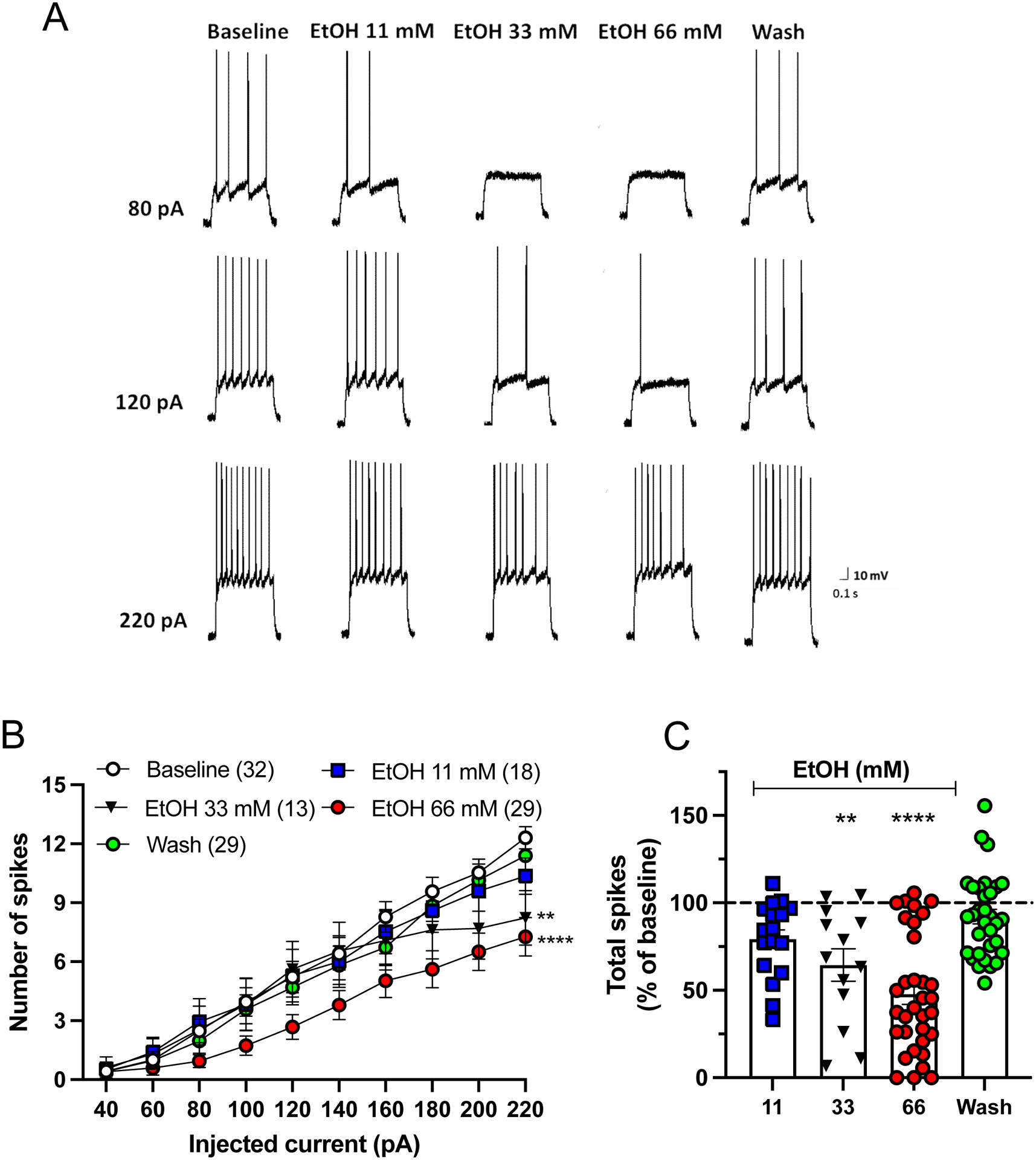
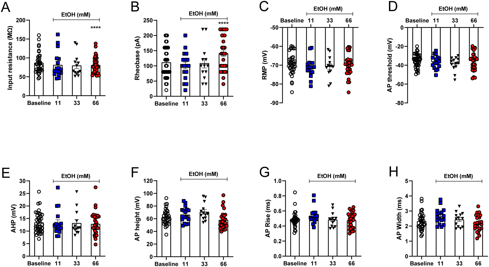
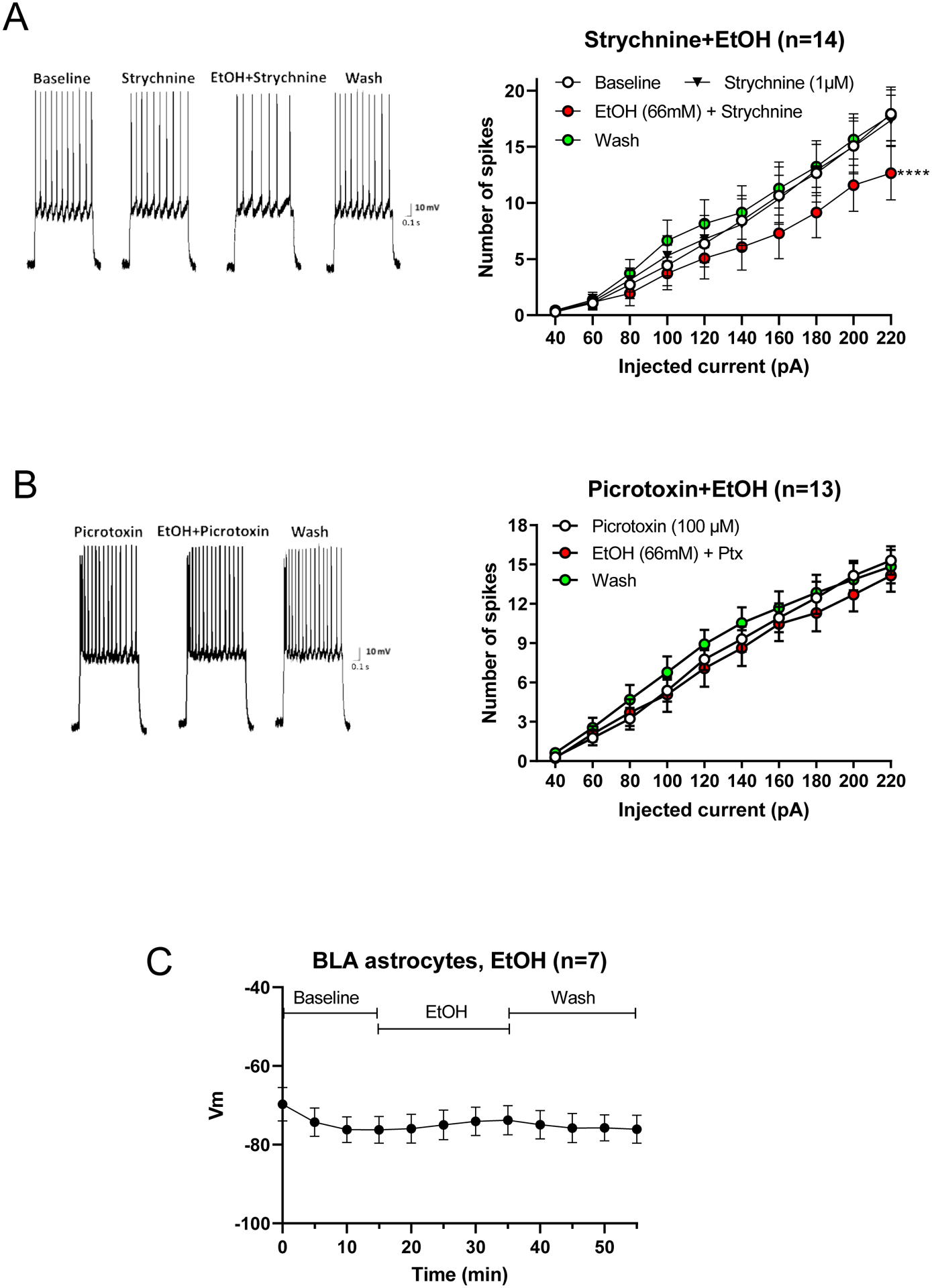
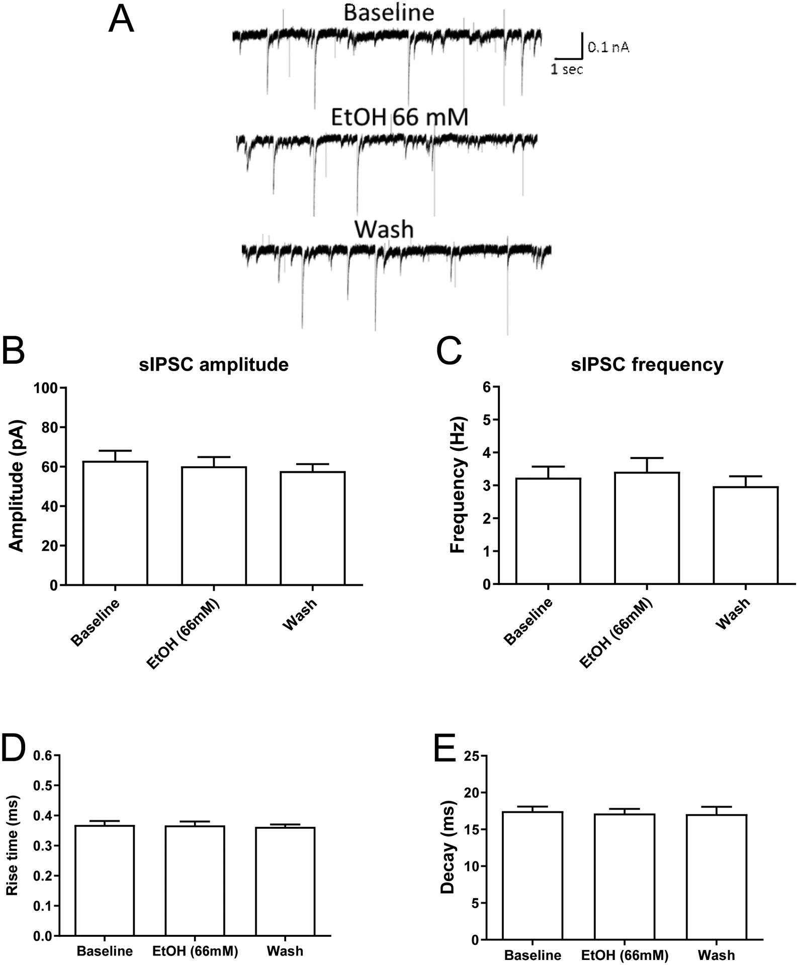
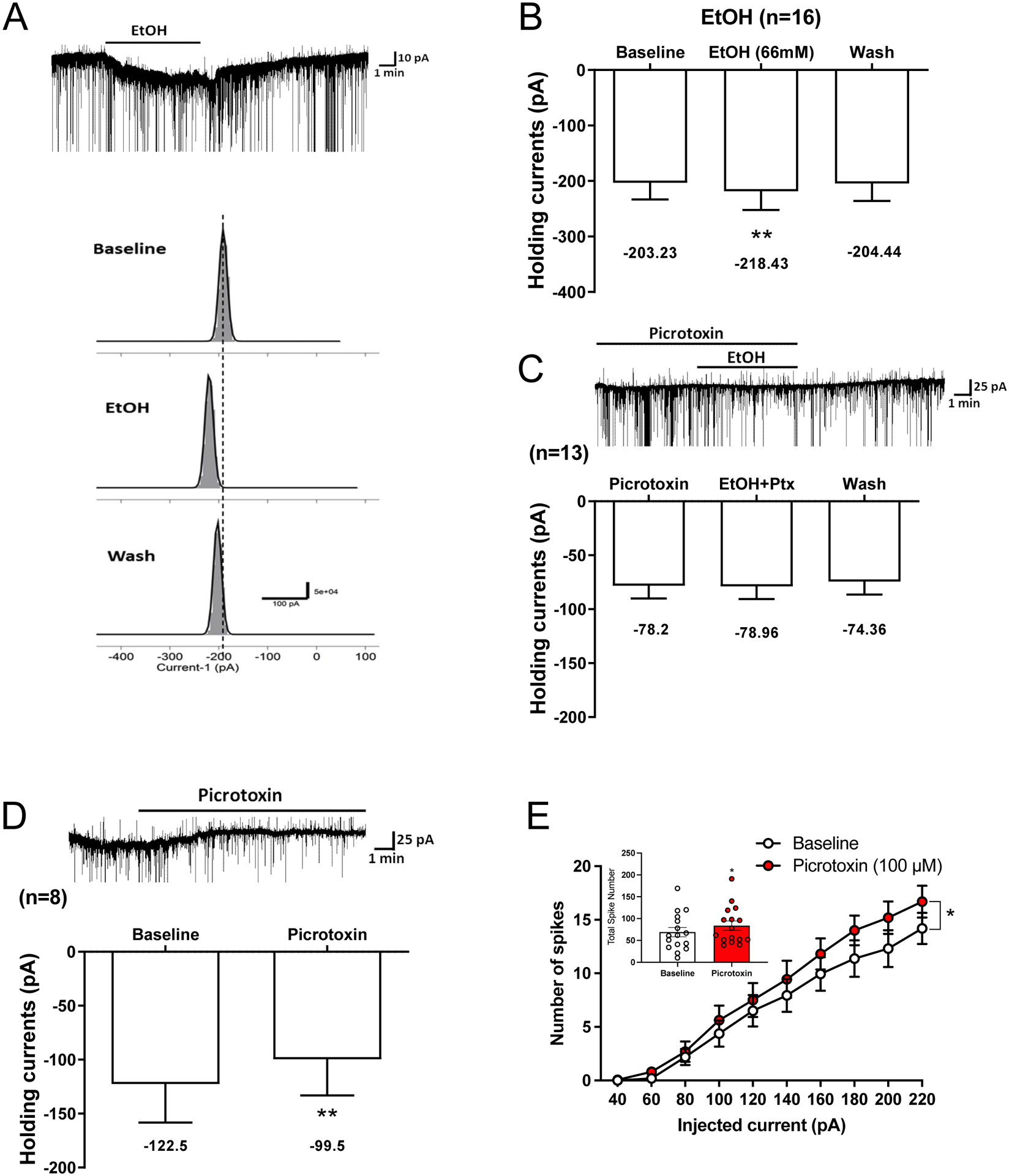
Results: Ethanol inhibited action potential (AP) firing of BLA neurons but had no effect on BLA astrocyte resting membrane potential. The ethanol-induced inhibition of firing was concentration-dependent (11 to 66 mM) and accompanied by a reduction in the input resistance and an increase in the rheobase of BLA neurons. The inhibitory effect of ethanol was suppressed by picrotoxin, which blocks both γ-aminobutyric acid type A (GABAA ) and glycine receptors, but not by the selective glycine receptor antagonist strychnine, which suggests an involvement of GABAA receptors. Ethanol did not affect spontaneous inhibitory postsynaptic currents suggesting that the inhibition of BLA neuronal excitability by ethanol was not due to an increase in GABAA -mediated synaptic transmission. However, acute ethanol enhanced the amplitude of the holding current of BLA neurons, an effect that was prevented by picrotoxin, which by itself reduced the holding current.

Conclusions: These results suggest that BLA neurons express a GABA-mediated tonic current that is enhanced by acute ethanol, which leads to reduced excitability of BLA neurons.

Keywords: GABAA-mediated tonic currents; basolateral amygdala; ethanol; intrinsic excitability.


Figures

Figure 1.
Figure 1.
Acute ethanol reduces current-evoked spiking in basolateral amygdala (BLA) neurons in a concentration-dependent manner. (A) Representative traces showing display action potential (AP) spiking in the absence and presence of ethanol (scale bars x= 0.1s, y=10 mV). (B) Effect of ethanol on AP spiking (mean ± SEM) induced by a series of current injections (40–220 pA). In comparison with control baseline, ethanol produced a significant decrease in basal firing rates of BLA neurons in a concentration-dependent manner (Two-way repeated measures ANOVA mixed model: main effect of ethanol, F(4,156) = 17.34, p<0.0001). The post hoc comparisons revealed significant differences in spike firing in response to 33 mM (q=3.48, **p=0.0025, n=13) or 66 mM (q=7.75, ****p<0.0001, n=29), but not 11 mM (q=1.74, p=0.27, n=18) of ethanol. Number in parentheses indicate number of recorded neurons. (C) Total number of APs in the absence and presence of ethanol. As compared to baseline, ethanol significantly decreased total APs recorded from BLA neurons (One-way repeated measures ANOVA mixed model: F(4,91) = 20.09, ****p<0.0001). Dunnett’s multiple comparison showed significant differences in the total number of spikes in response to 33 mM (q=3.77, **p=0.0012) and 66 mM (q=8.43, ****p<0.0001), but not 11 mM (q=1.95, p=0.18) of ethanol.
Figure 2.
Figure 2.
Electrophysiological characteristics of BLA neurons in response to acute ethanol. (A) Acute ethanol significantly decreased the input resistance of BLA neurons (One-way repeated measures ANOVA mixed model, main effect of ethanol F(3,56) = 10.50, ****p<0.0001). The post hoc comparisons revealed a significant difference in input resistance by the 66 mM ethanol (q=5.44, ****p<0.0001) but not by the 11 mM (q=0.61, p=0.89) or 33 mM concentration (q=2.20, p=0.088). (B) Ethanol (66 mM) increased the rheobase as compared to values obtained during baseline recording (One-way repeated measures ANOVA mixed model, main effect of ethanol F(3,56) = 9.54, ****p<0.0001). Post hoc comparisons reveal a significant difference in rheobase by the 66 mM ethanol (q=5.21, ****p<0.0001), but not by the 11 mM (q=0.26, p=0.99) or 33 mM concentration (q=2.07, p=0.12). In BLA neurons, ethanol does not affect resting membrane potential (RMP; C), AP threshold (D), AHP (E), AP height (F), AP rise time (G) or AP width (H; all One-way repeated measures ANOVA mixed model, p>0.05).
Figure 3.
Figure 3.
Effect of strychnine or picrotoxin on effect of ethanol (66 mM) on AP spiking (mean ± SEM). (A) Representative traces show AP spiking under control conditions or in the presence of strychnine or strychnine plus ethanol (scale bars x= 0.1s, y=10 mV). In the presence of strychnine (1 μM), ethanol significantly decreased AP number of BLA neurons as compared to strychnine baseline (Two-way repeated measures ANOVA: main effect of ethanol, F(3,39) = 10.11, q=5.58, **p=0.0017; n=14). Strychnine by itself did affect spiking as compared to aCSF baseline (q=0.24, p=0.99). (B) Representative traces showing lack of effect of ethanol on AP spiking in the presence of picrotoxin. When applied in the presence of picrotoxin (100 μM), ethanol did not alter firing of BLA neurons as compared to picrotoxin baseline (Two-way repeated measures ANOVA: main effect of ethanol, F(2,24) = 2.52, q=0.98, p=0.59; n=13). (C) Acute ethanol has no effect on the resting membrane potential (RMP) of BLA astrocytes. The RMP of BLA astrocytes after 15 min of recording in regular aCSF was −76.21 ± 3.4 mV and was −73.8 ± 3.74 mV following a 35 min exposure to ethanol. These two values were not significantly different from one another (one-way repeated measures ANOVA, F(11,66) = 5.56; Dunnett’s multiple comparisons 15 min value versus 35 min, q=2.22, p=0.20, n=7).
Figure 4.
Figure 4.
Acute ethanol does not alter GABAergic-mediated postsynaptic currents of BLA neurons. Recordings were performed in the presence of glutamate receptor blockers DL-AP5 (10 μM) and DNQX (10 μM) to isolate GABAA postsynaptic currents. (A) Representative traces showing sIPSCs before, during and after ethanol exposure. Summary graphs showing amplitude (B), frequency (E) and the kinetics (D, E) of sIPSCs before, during and after ethanol application. Ethanol (66 mM) did not alter sIPSC amplitude (B; One-way repeated measures ANOVA: main effect of ethanol, F(2,34) = 2.49, q=1.70, p=0.10; n=18, 9 mice) or frequency (C; One-way repeated measures ANOVA: main effect of ethanol, F(2,34) = 1.19, q=1.24, p=0.32) of BLA neurons. Likewise, ethanol had no effect on sIPSC rise time (D; One-way repeated measures ANOVA: main effect of ethanol, F(2,42) = 0.30, q=0.25, p=0.74) or decay time (E; One-way repeated measures ANOVA: main effect of ethanol, F(2,42) = 0.22, q=0.67, p=0.81).
Figure 5.
Figure 5.
Acute ethanol enhances a GABAA-mediated tonic current on BLA neurons. (A) Gaussian fit and representative trace of tonic current amplitudes measured before, during and after ethanol exposure are shown. Recordings were performed in the presence of glutamate receptor blockers DL-AP5 and DNQX to isolate GABAA postsynaptic currents. (B) Ethanol (66 mM) produced a small but significant increase (15.2 ± 5.4 pA) in the holding currents of BLA neurons as compared to pre-ethanol baseline (One-way repeated measures ANOVA, F(2,30) = 6.0, **p=0.0064, n=16). The average holding currents for baseline, ethanol and washout were −203.2 ± 29.93 pA, −218.4 ± 33.8 pA and −204.4 ± 31.24 pA, respectively. Dunnett’s multiple comparisons reveal a significant difference between ethanol versus baseline (q=3.12, **p<0.01). (C) Blocking GABAA receptors with the GABAA receptor blocker picrotoxin (100 μM) suppressed the ethanol-induced enhancement of the holding current (One-way repeated measures ANOVA, F(2,24) = 1.65, q=0.40, p=0.21, n=13). (D) Picrotoxin by itself produced a decrease in the holding currents of BLA neurons (Two-tailed paired t-test, t(7) = 5.04, **p=0.0015, n=8). (E) Picrotoxin (100 μM) significantly increased current-evoked spiking of BLA neurons (Two-way repeated measures ANOVA, main effect of picrotoxin, F(1,10) = 5.94; p=0.035). Inset shows total number of spikes evoked by the current steps was enhanced by picrotoxin (Two-tailed paired t-test, t15 = 2,87; p=0.012).

Similar articles

Cited by

References

    1. Ade KK, Janssen MJ, Ortinski PI, Vicini S (2008) Differential tonic GABA conductances in striatal medium spiny neurons. J Neurosci 28:1185–1197. - PMC - PubMed
    1. Angulo MC, Le Meur K, Kozlov AS, Charpak S, Audinat E (2008) GABA, a forgotten gliotransmitter. Prog Neurobiol 86:297–303. - PubMed
    1. Araque A, Parpura V, Sanzgiri RP, Haydon PG (1999) Tripartite synapses: glia, the unacknowledged partner. Trends Neurosci 22:208–215. - PubMed
    1. Ariwodola OJ, Weiner JL (2004) Ethanol potentiation of GABAergic synaptic transmission may be self-limiting: role of presynaptic GABA(B) receptors. J Neurosci 24:10679–10686. - PMC - PubMed
    1. Badanich KA, Mulholland PJ, Beckley JT, Trantham-Davidson H, Woodward JJ (2013) Ethanol reduces neuronal excitability of lateral orbitofrontal cortex neurons via a glycine receptor dependent mechanism. Neuropsychopharmacology 38:1176–1188. - PMC - PubMed

Publication types