. 2022 Jul 19:2022:7288729.
doi: 10.1155/2022/7288729. eCollection 2022.

ZNF354C Mediated by DNMT1 Ameliorates Lung Ischemia-Reperfusion Oxidative Stress Injury by Reducing TFPI Promoter Methylation to Upregulate TFPI

Affiliations

ZNF354C Mediated by DNMT1 Ameliorates Lung Ischemia-Reperfusion Oxidative Stress Injury by Reducing TFPI Promoter Methylation to Upregulate TFPI

Qi Shi et al. Oxid Med Cell Longev. .

Retraction in

Abstract

Background: Pulmonary ischemia reperfusion- (I/R-) induced dysfunction is a significant clinical problem after lung transplantation. In this study, we aim to explore the molecular mechanism of lung I/R injury (LIRI).

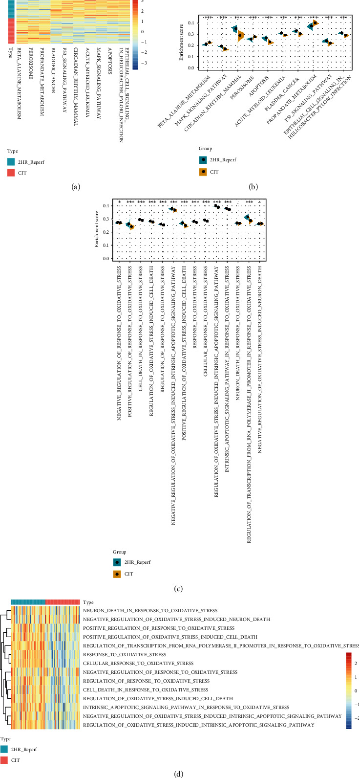
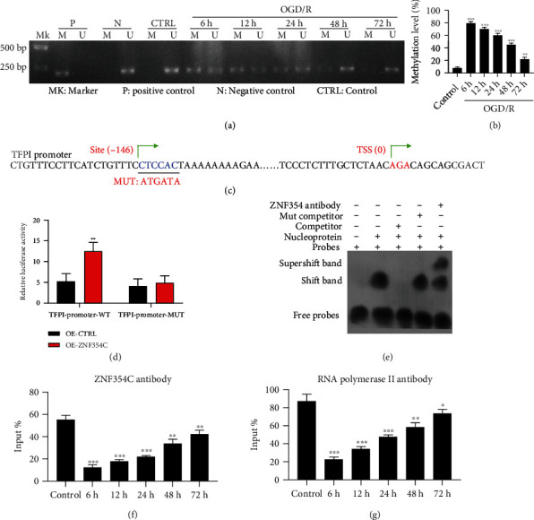
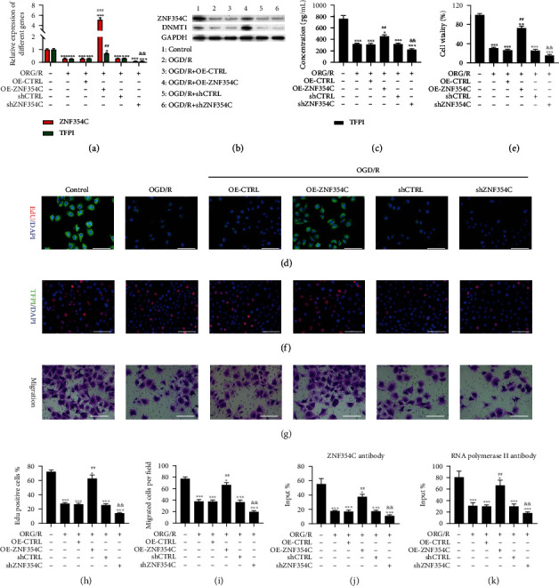
Methods: Bioinformatic analysis of gene involved in oxidative stress. A HUVEC oxygen glucose deprivation/reoxygenation (OGD/R) model and I/R mouse model were first established via I/R. The cellular proliferation, migration, reactive oxygen species (ROS), and parameters of lung injury were assessed via CCK-8, EdU staining, Transwell, cellular ROS kit, and H&E staining. We also confirmed related gene expressions and protein levels and the interaction between the tissue factor pathway inhibitor (TFPI) promotor and ZNF354C.

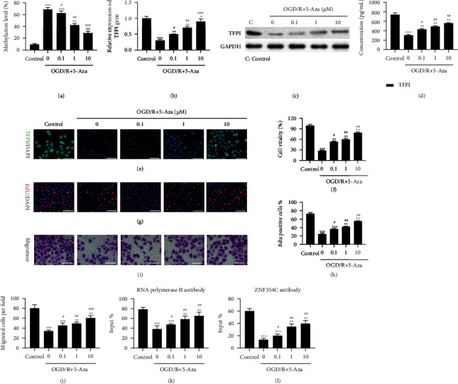
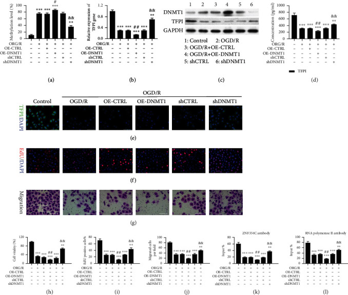
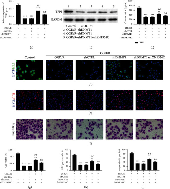
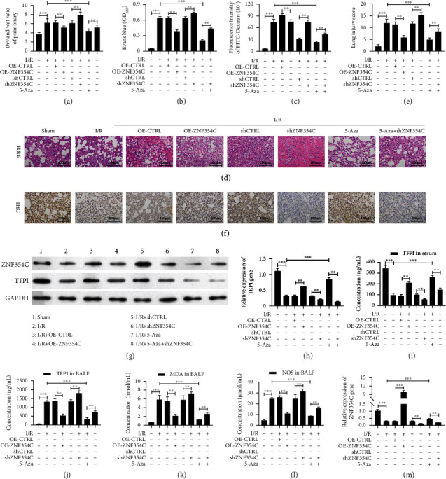
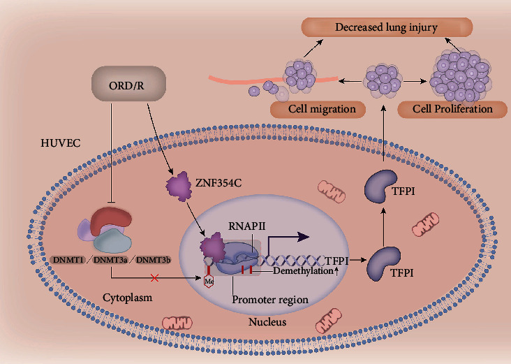
Results: Bioinformatic analysis results showed TFPI contributed to oxidative stress. OGD/R caused a reduction in cell viability and migration, hypermethylation of TFPI, increased ROS, and downregulation of ZNF354C, TFPI, and DNA methyltransferases (DNMTs) in HUVECs. Besides, ZNF354C could directly bind to the TFPI promoter, enhance proliferation and migration, and inhibit ROS in OGD/R-induced HUVECs by upregulating TFPI. More importantly, we discovered that 5-Aza could reduce TFPI methylation, upregulate TFPI, and enhance the binding of ZNF354C to the TFPI promoter in LIRI. Furthermore, DNMT1 silencing could induce proliferation and migration and prevent ROS in OGD/R-induced HUVECs by upregulating ZNF354C. Additionally, we verified that ZNF354C could alleviate LIRI by preventing DNA methylation in vivo.

Conclusions: ZNF354C overexpression induced proliferation and migration, as well as suppressed ROS in OGD/R-induced HUVECs, and alleviated LIRI in mice by inhibiting TFPI promoter methylation to upregulate TFPI. Therefore, ZNF354C and TFPI methylation might be promising molecular markers for LIRI therapy.


Conflict of interest statement

The authors declare no conflicts of interest.

Figures

Figure 1
Figure 1
Bioinformatic analysis of gene expression in peripheral human lung biopsies at the end of cold ischemia and after two hours of reperfusion following transplantation. (a) The top 10 KEGG pathways with the most significant differences were observed. (b) Peroxisome, beta alanine metabolism, and propanoate metabolism were significantly activated in the end of cold ischemia samples, whereas p53, MAPK signaling pathway, and pathways were significantly blocked. (c) Lower activity of oxidative stress-related biological processes was observed in the end of cold ischemia samples. (d) Twelve of the 14 oxidative stress-related biological processes were significantly lower in the end of cold ischemia samples.
Figure 2
Figure 2
The impact of OGD/R on cell viability, migration, DNA methyltransferase, and TFPI in HUVECs. HUVECs were exposed to OGD for 4 h and reoxygenated for 6, 12, 24, 48, and 72 h. (a) Cell viability was tested by CCK-8. (b, c) HUVECs were subjected to EdU staining, and EdU-positive cells were calculated in each group. (d, e) Migrated cells were confirmed via Transwell, and five fields were summarized and counted. TFPI level was monitored with (f) RT-qPCR, (g) ELISA, and (h) IF in HUVECs after OGD/R. (i) Western blotting analysis of TFPI, ZNF345C, DNMT1, DMNT3a, and DMNT3b levels. (j) ROS level was tested using DCFDA/H2DCFDA-Cellular ROS Assay in each group. (k) Quantitative analysis of ROS relative fluorescence intensity. The experiments were repeated three times independently.
Figure 3
Figure 3
OGD/R induced hypermethylation of TFPI in HUVECs, and TFPI could interact with ZNF354C. (a) Methylation level of TFPI promoter was tested with MSP in OGD/R-treated HUVECs. (b) The methylation level was quantified based on MSP results. (c) The predicated binding sites between TFPI and ZNF354C. (d) Luciferase activity of TFPI promoter in ZNF354C-overexpressed HUVECs was determined by applying dual luciferase reporter gene. (e) EMSA was utilized to test the binding of TFPI promoter and ZNF354C. (f) The interaction between ZNF354C antibody and TFPI promoter was confirmed with ChIP in HUVECs under OGD/R. (g) HUVECs were incubated with Poly II antibody and beads, and the binding of RNA polymerase II on TFPI 5′UTR was examined using ChIP in HUVECs under OGD/R. The experiments were repeated three times independently.
Figure 4
Figure 4
ZNF354C overexpression induced proliferation and migration through upregulating TFPI in OGD/R-induced HUVECs. OGD/R-treated HUVECs were transfected with ZNF354C overexpression plasmids or shZNF354C, respectively. ZNF354C and TFPI expressions were assessed via (a) RT-qPCR and (b) western blot. (c) TFPI level was analyzed with ELISA kit. (d) IF staining of TFPI in the processed HUVECs. (e) Cell viability was evaluated by CCK-8. (f) HUVECs in each group were subjected to EdU staining. (g) The images of cell migration were obtained using Transwell. (h) EdU-positive cells were quantitated in line with EdU staining results. (i) Quantitative analysis of migratory cells. (j) The binding of ZNF354C and TFPI promoter was confirmed through ChIP in each group. (k) The binding of RNA polymerase II on TFPI 5′UTR was examined via ChIP. The experiments were repeated three times independently.
Figure 5
Figure 5
5-Aza decreased TFPI methylation, increased TFPI expression, accelerated proliferation and migration, and enhanced the combination of TFPI and ZNF354C in OGD/R-treated HUVECs. OGD/R-mediated HUVECs were treated with 5-Aza (low: 0.1 μM, mid: 1 μM, high: 10 μM) for 24 h. (a) MSP was utilized to identify the methylation level of TFPI promoter. TFPI level was certified by applying (b) RT-qPCR, (c) western blot, (d) ELISA, and (e) IF staining in the processed HUVECs. Cell proliferation was determined with (f) CCK-8 and (g) EdU staining. (i) Migration in HUVECs was tested with Transwell. (h) EdU-positive cells and (j) migrated cells were counted. (k) The impact of 5-Aza on the binding of ZNF354C and TFPI promoter was confirmed by ChIP. (l) ChIP was adopted to analyze the effect of 5-Aza on the binding of RNA polymerase II on TFPI 5′UTR. The experiments were repeated three times independently.
Figure 6
Figure 6
DNMT1 suppressed proliferation and migration and reduced the interaction of TFPI and ZNF354C in OGD/R-induced HUVECs. HUVECs under OGD/R were transfected with DNMT1 overexpression plasmids or shDNMT1, respectively. (a) MSP displayed the change in the methylation level of TFPI promoter. The level of TFPI was tested through (b) RT-qPCR and (c) western blot, (d) ELISA, and (e) IF staining. (f) EdU staining was utilized to confirm the change of proliferation. (g) Transwell exhibited the change of cell migration. (h) CCK-8 presented the change in cell viability. (i) Based on the EdU staining data, EdU-positive cells were quantitated. (j) Migratory cells were quantitated in line with Transwell data. (k) ChIP demonstrated the impact of DNMT1 on the binding of ZNF354C and TFPI promoter. (l) ChIP showed the influence of DNMT1 on the binding of RNA polymerase II on TFPI 5′UTR. The experiments were repeated three times independently.
Figure 7
Figure 7
Silencing of DNMT1 accelerated proliferation and migration by regulating ZNF354C to target TFPI in OGD/R-induced HUVECs. OGD/R-induced HUVECs were transfected with shDNMT1 or/and shZNF354C. TFPI expression was identified through (a) RT-qPCR and (b) western blot, (c) ELISA, and (d) IF staining. (e, h) The change of cell proliferation was confirmed with EdU staining, and EdU-positive cells were quantitated. (f, i) The change of cell migration was monitored with Transwell, and migratory cells were quantitated. (g) CCK-8 presented the change of cell viability. The experiments were repeated three times independently.
Figure 8
Figure 8
ZNF354C alleviated LIRI through suppressing DNA methylation in vivo. The I/R model was established and then was disposed of ZNF354C overexpression plasmids, shZNF354C lentivirus, and/or 5-Aza (each group contained 5 mice). (a) After sacrifice, wet and dry lung weights of mice were measured and compared with wet-to-dry ratios. (b) Evans Blue was injected via the jugular vein 1 h before sacrifice, and the absorbance of the supernatant was tested at 620 nm. (c) The fluorescence intensity of FITC-Dextran was confirmed with FITC dextran assay. (d) The pathologic structure of lung tissues was assessed by H&E staining. (e) The lung injury score was also calculated by ImageJ based on the H&E staining results. (f) The expression of TFPI was measured by IHC. (g) The expressions of ZNF354C and TFPI were identified by Western blot. (h, i) And ZNF354C and TFPI expressions were quantified. EILISA kits were adopted to evaluate the levels of (j) TFPI in serum, (l) TFPI in BALF, (l) MDA in BALF, and (m) NOS in BALF.
Figure 9
Figure 9
Schematic overview of the mechanism between ZNF354C, TFPI, and DNMT1 based on the findings of the study. ZNF354C interacts with TFPI and upregulates TFPI by preventing TFPI promoter methylation in OGD/R-induced HUVECs. DNMT1 reduces the interaction of TFPI and ZNF354C. Functionally, ZNF354C ameliorates LIRI by preventing TFPI methylation to upregulate TFPI.

Similar articles

Cited by

References

    1. Laubach V. E., Sharma A. K. Mechanisms of lung ischemia-reperfusion injury. Current Opinion in Organ Transplantation . 2016;21(3):246–252. doi: 10.1097/MOT.0000000000000304. - DOI - PMC - PubMed
    1. Ovechkin A. V., Lominadze D., Sedoris K. C., Robinson T. W., Tyagi S. C., Roberts A. M. Lung ischemia-reperfusion injury: implications of oxidative stress and platelet-arteriolar wall interactions. Archives of Physiology and Biochemistry . 2007;113(1):1–12. doi: 10.1080/13813450601118976. - DOI - PMC - PubMed
    1. Tang J., Xu L., Zeng Y., Gong F. Effect of gut microbiota on LPS-induced acute lung injury by regulating the TLR4/NF-kB signaling pathway. International Immunopharmacology . 2021;91, article 107272 doi: 10.1016/j.intimp.2020.107272. - DOI - PubMed
    1. Shah R. J., Diamond J. M. Primary graft dysfunction (PGD) following lung transplantation. Seminars in Respiratory and Critical Care Medicine . 2018;39(2):148–154. doi: 10.1055/s-0037-1615797. - DOI - PubMed
    1. den Hengst W. A., Gielis J. F., Lin J. Y., Van Schil P. E., De Windt L. J., Moens A. L. Lung ischemia-reperfusion injury: a molecular and clinical view on a complex pathophysiological process. American journal of physiology Heart and circulatory physiology . 2010;299(5):H1283–H1299. doi: 10.1152/ajpheart.00251.2010. - DOI - PubMed

Publication types

LinkOut - more resources