

Temperature-Responsive Liposome-Linked Immunosorbent Assay for the Rapid Detection of SARS-CoV-2 Using Immunoliposomes
- PMID: 35915635
- PMCID: PMC9328125
- DOI: 10.1021/acsomega.2c03597
Temperature-Responsive Liposome-Linked Immunosorbent Assay for the Rapid Detection of SARS-CoV-2 Using Immunoliposomes
Abstract
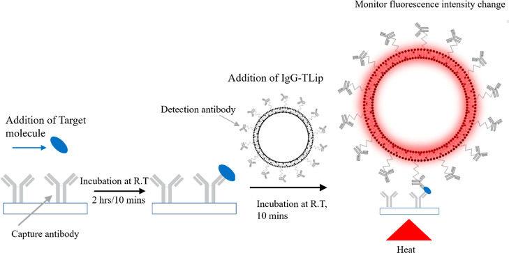
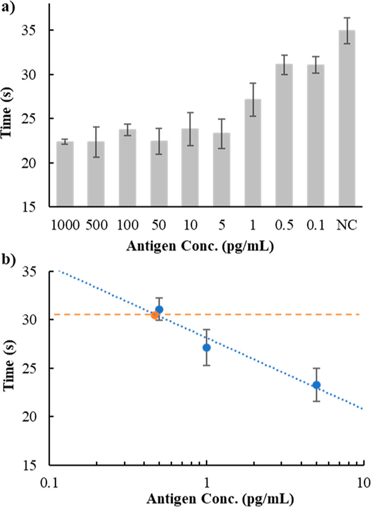
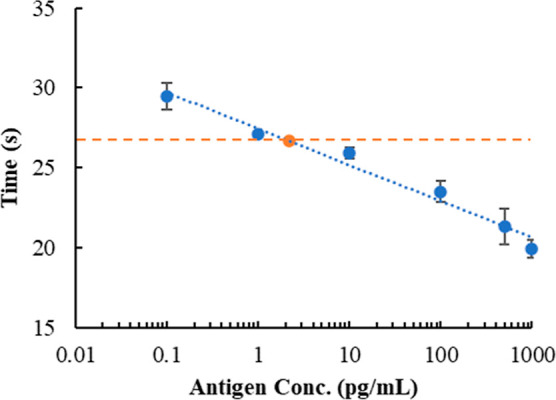
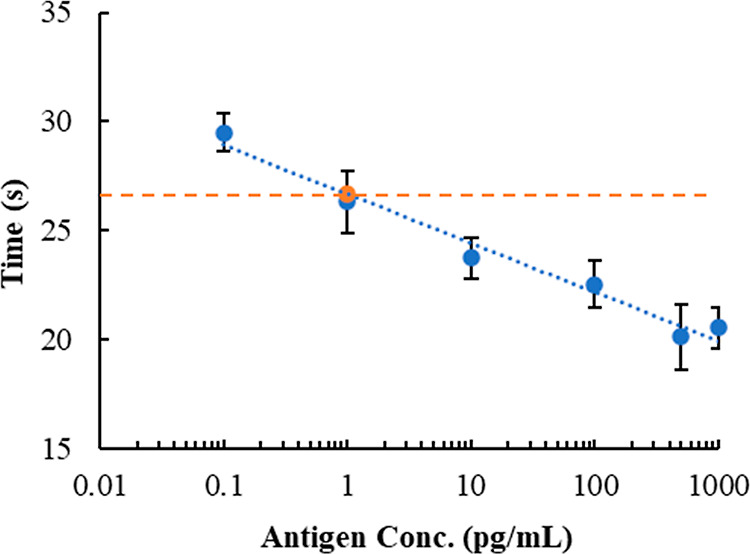
Severe acute respiratory syndrome coronavirus 2 (SARS-CoV-2), which is the etiological agent of coronavirus disease 2019 (COVID-19), has infected more than 340 million people since the outbreak of the pandemic in 2019, resulting in approximately 55 million deaths. The rapid and effective diagnosis of COVID-19 patients is vital to prevent the spread of the disease. In a previous study, we reported a novel temperature-responsive liposome-linked immunosorbent assay (TLip-LISA) using biotinylated-TLip that exhibited high detection sensitivity for the prostate-specific antigen. Herein, we used immunoglobulin-TLip (IgG-TLip), in which the antibodies were directly conjugated to the liposomal surface to simplify pretreatment procedures and reduce the detection time for SARS-CoV-2. The results indicated that TLip-LISA could detect the recombinant nucleocapsid protein and the nucleocapsid protein in inactivated virus with 20 min incubation time in total, and the limit of detection was calculated to be 2.2 and 1.0 pg/mL, respectively. Therefore, TLip-LISA has high potential to be used in clinic for rapid diagnosis and disease control.
© 2022 The Authors. Published by American Chemical Society.
Conflict of interest statement
The authors declare the following competing financial interest(s): R.H., K.S., and S.T. are inventors of the method for detecting antigens using the temperature-responsive liposomes applied in this study and named as inventors on the PCT patent application (PCT/JP2020/021358). The other authors have no conflict of interest to declare in this study.
Figures
Similar articles
-
A rapid and highly sensitive biomarker detection platform based on a temperature-responsive liposome-linked immunosorbent assay.Sci Rep. 2020 Oct 22;10(1):18086. doi: 10.1038/s41598-020-75011-x. Sci Rep. 2020. PMID: 33093468 Free PMC article.
-
IgG antibody titers against SARS-CoV-2 nucleocapsid protein correlate with the severity of COVID-19 patients.BMC Microbiol. 2021 Dec 18;21(1):351. doi: 10.1186/s12866-021-02401-0. BMC Microbiol. 2021. PMID: 34922455 Free PMC article.
-
Sensitive and specific serological ELISA for the detection of SARS-CoV-2 infections.Virol J. 2022 Mar 19;19(1):50. doi: 10.1186/s12985-022-01768-4. Virol J. 2022. PMID: 35305688 Free PMC article.
-
Fcγ-Receptor-Based Enzyme-Linked Immunosorbent Assays for Sensitive, Specific, and Persistent Detection of Anti-SARS-CoV-2 Nucleocapsid Protein IgG Antibodies in Human Sera.J Clin Microbiol. 2022 Jun 15;60(6):e0007522. doi: 10.1128/jcm.00075-22. Epub 2022 May 16. J Clin Microbiol. 2022. PMID: 35574677 Free PMC article.
-
The utility of SARS-CoV-2 nucleocapsid protein in laboratory diagnosis.J Clin Lab Anal. 2022 Jul;36(7):e24534. doi: 10.1002/jcla.24534. Epub 2022 Jun 3. J Clin Lab Anal. 2022. PMID: 35657146 Free PMC article. Review.
Cited by
-
Recent updates on liposomal formulations for detection, prevention and treatment of coronavirus disease (COVID-19).Int J Pharm. 2023 Jan 5;630:122421. doi: 10.1016/j.ijpharm.2022.122421. Epub 2022 Nov 19. Int J Pharm. 2023. PMID: 36410670 Free PMC article. Review.
-
Red-Emitting Latex Nanoparticles by Stepwise Entrapment of β-Diketonate Europium Complexes.Int J Mol Sci. 2022 Dec 15;23(24):15954. doi: 10.3390/ijms232415954. Int J Mol Sci. 2022. PMID: 36555596 Free PMC article.
-
Neuroinflammation in the Evolution of Motor Function in Stroke and Trauma Patients: Treatment and Potential Biomarkers.Curr Issues Mol Biol. 2023 Oct 25;45(11):8552-8585. doi: 10.3390/cimb45110539. Curr Issues Mol Biol. 2023. PMID: 37998716 Free PMC article. Review.
References
-
- Marra M. A.; Jones S. J.; Astell C. R.; Holt R. A.; Brooks-Wilson A.; Butterfield Y. S.; Khattra J.; Asano J. K.; Barber S. A.; Chan S. Y.; Cloutier A.; Coughlin S. M.; Freeman D.; Girn N.; Griffith O. L.; Leach S. R.; Mayo M.; McDonald H.; Montgomery S. B.; Pandoh P. K.; Petrescu A. S.; Robertson A. G.; Schein J. E.; Siddiqui A.; Smailus D. E.; Stott J. M.; Yang G. S.; Plummer F.; Andonov A.; Artsob H.; Bastien N.; Bernard K.; Booth T. F.; Bowness D.; Czub M.; Drebot M.; Fernando L.; Flick R.; Garbutt M.; Gray M.; Grolla A.; Jones S.; Feldmann H.; Meyers A.; Kabani A.; Li Y.; Normand S.; Stroher U.; Tipples G. A.; Tyler S.; Vogrig R.; Ward D.; Watson B.; Brunham R. C.; Krajden M.; Petric M.; Skowronski D. M.; Upton C.; Roper R. L. The Genome Sequence of the SARS-associated Coronavirus. Science 2003, 300, 1399–1404. 10.1126/science.1085953. - DOI - PubMed
LinkOut - more resources
Full Text Sources
Miscellaneous
