. 2022 May 8;4(4):408-412.
doi: 10.1093/jbi/wbac019. eCollection 2022 Jul-Aug.

Nipple Adenoma: Correlation of Imaging Findings and Histopathology

Affiliations

Nipple Adenoma: Correlation of Imaging Findings and Histopathology

Madeline E Leo et al. J Breast Imaging. .

Abstract

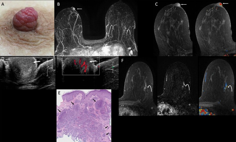
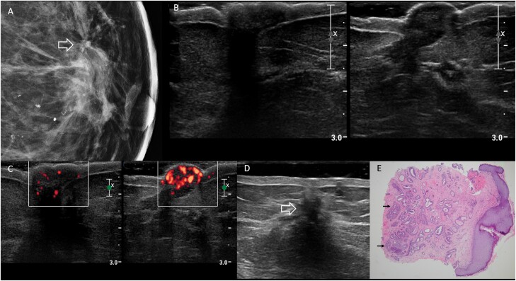
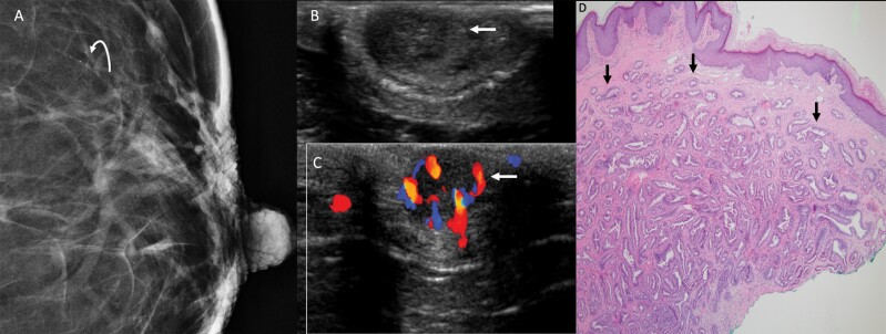
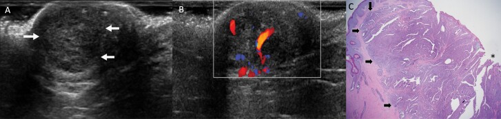
Nipple adenomas (NAs) are benign neoplasms composed of papillary hyperplasia of the epithelium of the major lactiferous ducts. Patients with NA may report bloody nipple discharge and clinically may resemble Paget disease, raising concern for malignancy. Mammographically, NAs are often occult. US can show a hypervascular circumscribed mass centered within the nipple with varying echogenicity. Diagnosis is usually made on punch biopsy or excision, but breast radiologists should be aware of this entity. Malignancy can be found elsewhere in the ipsilateral or contralateral breast, or very rarely may directly extend to involve an NA, but published experience with concurrent malignancies is small. We describe the radiologic-pathologic correlation of NAs.

Keywords: histopathology; nipple adenoma; nipple discharge.


Figures

Figure 1.
Figure 1.
Images of a 54-year-old female with a six-month history of spontaneous bloody right nipple discharge following poor healing of a nipple laceration. After initial mammographic and sonographic evaluation failed to show an etiology for the bloody discharge, she was referred for MRI. A: Clinical photograph demonstrates a prominent right nipple with hyperemic “raspberry-like” distortion of the nipple. B: Axial maximum intensity projection (MIP) image of first post-contrast MRI subtraction shows asymmetric strong enhancement of the right nipple (arrow) and non-mass enhancement in the outer left breast (curved arrow). C: Axial T1-weighted, fat-suppressed first post-contrast breast MRI shows a rim-enhancing 10-mm oval mass in the right nipple (left, arrow) with washout kinetics (right, arrow). D: MRI-directed transverse US of the right nipple (left) demonstrates a subtle, slightly hypoechoic, heterogeneous, 1-cm, ovoid mass within the right nipple (arrow), with internal vascularity on power Doppler (right, arrow). E: Histopathology (hematoxylin and eosin, 4x) from punch biopsy shows proliferating ductal structures with usual ductal hyperplasia consistent with nipple adenoma (arrows). Myoepithelial cell nuclei (which can be highlighted with immunohistochemical stain for p63) surround each individual ductal structure. F: Axial T1-weighted, fat-suppressed contrast-enhanced breast MRI with kinetic overlay better demonstrates a 3.3-cm linear non-mass enhancement in the outer left breast (curved arrow). The patient underwent MRI-guided biopsy, followed by breast-conserving surgery, with final pathology demonstrating two foci of invasive lobular carcinoma, measuring 1.6 cm and 0.8 cm, on a background of lobular carcinoma in situ. Two left axillary lymph nodes were negative for carcinoma. Surgical pathology from the right nipple confirmed nipple adenoma.
Figure 2.
Figure 2.
Images of a 69-year-old woman with bloody left nipple discharge, retraction, and discoloration due to nipple adenoma. Physical examination reported firmness and bluish discoloration of the left nipple. A: Spot magnification mediolateral mammographic image of the left breast demonstrates an area of architectural distortion at the 12-o’clock position in the retroareolar left breast (arrow) with a few associated amorphous calcifications. The left nipple was mammographically unremarkable. B: On US, no discrete mass was seen in either nipple (right on left and left on right). C: Transverse power Doppler image shows hypervascularity of the left nipple (right-hand image) compared to the right. D: Additional transverse sonographic evaluation of the left breast (using a standoff pad) revealed a 15-mm hypoechoic, irregular mass at the 12-o’clock position, 1 cm from the nipple (open arrow), with posterior shadowing, corresponding to the mammographic distortion. US-guided core-needle biopsy showed radial scar with microcalcifications, ductal epithelial hyperplasia, and sclerosing adenosis; associated grade 1 ductal carcinoma in situ was found at excision. E: Histopathology (hematoxylin and eosin, 4x) from punch biopsy of the left nipple shows proliferation of irregular ductal structures (arrows), some of which demonstrate usual ductal hyperplasia, extending to multiple margins of the biopsy, consistent with nipple adenoma.
Figure 3.
Figure 3.
This 66-year-old woman presented with non-spontaneous left bloody nipple discharge, with a nodular mass in the left nipple on exam. A: Spot magnification craniocaudal view of the left breast demonstrates fine linear calcifications spanning 5 mm approximately 3 cm from the nipple in the 1-o’clock position (curved arrow). No obvious nipple abnormality was evident mammographically. Transverse US of the left nipple (B) demonstrated a subtle, circumscribed, hypoechoic, 9-mm, oval mass (arrow) centered within the nipple, with minimal posterior enhancement, that was hypervascular on color Doppler (C) (arrow). D: Histopathology (hematoxylin and eosin, 4x) from nipple punch biopsy shows proliferation of irregular ductal structures (arrows) with usual ductal hyperplasia, consistent with nipple adenoma. This patient also underwent stereotactic left breast biopsy of the calcifications, which demonstrated ductal carcinoma in situ (DCIS), nuclear grade 2. Left segmental mastectomy including left nipple resection confirmed nipple adenoma. Atypical ductal hyperplasia, lobular carcinoma in situ, and atypical lobular hyperplasia were also seen in the 1-o’clock position, as well as multiple intraductal papillomas without atypia, ductal epithelial hyperplasia, and fibrocystic changes. No additional DCIS was seen in the surgical specimen.
Figure 4.
Figure 4.
Images of a 68-year-old postmenopausal female with itching and bleeding of the left nipple for several months due to nipple adenoma. On exam, the left nipple was swollen and erythematous with excoriation (not shown). Mammogram was unrevealing (not shown). Transverse US of the left breast (A) shows a subtle round circumscribed isoechoic mass (arrows) within the nipple, with posterior enhancement, and color Doppler (B) demonstrates increased internal vascularity within the mass. The patient was referred to surgery and underwent an excisional biopsy of the left nipple, which demonstrated nipple adenoma with focal skin ulceration, negative for malignancy. C: Histopathology (hematoxylin and eosin, 2x) demonstrates a predominantly papillary growth pattern (arrows) with epidermal ulceration (asterisk), consistent with nipple adenoma.

Similar articles

Cited by

References

    1. Jones DB. Florid papillomatosis of the nipple ducts. Cancer 1955;8(2):315–319. - PubMed
    1. Lester SC, Lee AHS.. Nipple adenoma. In: World Health Organization Classification of Tumours, Breast Tumours. 5th ed. Vol 2. Lyons, France: International Agency for Research on Cancer, 2019:182–183.
    1. Di Bonito M, Cantile M, Collina F, et al. . Adenoma of the nipple: a clinicopathological report of 13 cases. Oncol Lett 2014;7(6):1839–1842. - PMC - PubMed
    1. Perzin KH, Lattes R.. Papillary adenoma of the nipple (florid papillomatosis, adenoma, adenomatosis). A clinicopathologic study. Cancer 1972;29(4):996–1009. - PubMed
    1. Wang C, Wang X, Ma R.. Diagnosis and surgical treatment of nipple adenoma. ANZ J Surg 2015;85(6):444–447. - PubMed