Review
. 2022 Oct;11(20):e2201583.
doi: 10.1002/adhm.202201583. Epub 2022 Aug 15.

Genetically Engineered Viral Vectors and Organic-Based Non-Viral Nanocarriers for Drug Delivery Applications

Affiliations
Review

Genetically Engineered Viral Vectors and Organic-Based Non-Viral Nanocarriers for Drug Delivery Applications

Sakineh Hajebi et al. Adv Healthc Mater. 2022 Oct.

Abstract

Conventional drug delivery systems are challenged by concerns related to systemic toxicity, repetitive doses, drug concentrations fluctuation, and adverse effects. Various drug delivery systems are developed to overcome these limitations. Nanomaterials are employed in a variety of biomedical applications such as therapeutics delivery, cancer therapy, and tissue engineering. Physiochemical nanoparticle assembly techniques involve the application of solvents and potentially harmful chemicals, commonly at high temperatures. Genetically engineered organisms have the potential to be used as promising candidates for greener, efficient, and more adaptable platforms for the synthesis and assembly of nanomaterials. Genetically engineered carriers are precisely designed and constructed in shape and size, enabling precise control over drug attachment sites. The high accuracy of these novel advanced materials, biocompatibility, and stimuli-responsiveness, elucidate their emerging application in controlled drug delivery. The current article represents the research progress in developing various genetically engineered carriers. Organic-based nanoparticles including cellulose, collagen, silk-like polymers, elastin-like protein, silk-elastin-like protein, and inorganic-based nanoparticles are discussed in detail. Afterward, viral-based carriers are classified, and their potential for targeted therapeutics delivery is highlighted. Finally, the challenges and prospects of these delivery systems are concluded.

Keywords: abiotic nanomaterials; genetically manipulation; nanoparticles; non-viral vectors; surface modifications.


Conflict of interest statement

The authors declare no conflict of interest.

Figures

Figure 1
Figure 1
Schematic illustration of the synthesis of the genetically engineered organic, inorganic, and virus‐like nanostructures for drug delivery applications.
Figure 2
Figure 2
A) Schematic and B) representing SEM images of the self‐assembly of pure collagen (CMP), (GCD/CMP), (GLH/CMP). Reproduced with permission.[ 43 ] Copyright 2021, Springer Nature. Abbreviation: CMP: Mimetic peptide; GCD/CMP: Glycidol‐modified CMP. GLH/CMP: Y‐glycidyl ether oxypropyl trimethoxysilane‐modified CMP.
Figure 3
Figure 3
A) Synthesis of ELP‐modified liposomes. B) Confocal images of HeLa cells incubated in the culture media with DOX: a) Control, b) PEG‐liposomes, and c) ELP‐modified liposomes. C) Mean fluorescence intensity of liposomes identified through the flow cytometry assay: a) Control, b) PEG‐liposomes, and c) ELP‐modified liposomes. Mean and S.D. are shown (n = 3). Reproduced with permission.[ 49 ] Copyright 2012, Elsevier. Abbreviation: ELP: Elastin‐like protein; DOX: Doxorubicin; PEG: Polyethylene glycol.
Figure 4
Figure 4
Self‐assembly of micellar‐like nanoparticles in T‐responsive. Reproduced with permission.[ 58 ] Copyright 2011, American Chemical Society.
Figure 5
Figure 5
A) Bioengineering of the spider silk. B) Cellular uptake of the silk spheres on the SKOV3 and SKBR3 cells, which were treated with spheres prepared of fluorescently labeled control MS1 and silk proteins H2.1MS1 and H2.2MS1. Scale bar = 10 µm. C) Behavior of release of DOX. Reproduced with permission.[ 59 ] Copyright 2014, American Chemical Society.
Figure 6
Figure 6
The primary structures of customized xanthan in Xanthomonas campestris and their biosynthetic pathways.[ 80 ]
Figure 7
Figure 7
Thermoresponsive plasmonic gold/silk‐elastin protein core–shell nanoparticles. A) Schematic of functionalized gold nanoparticles with SELPs. B) (a) Representative TEM images of functionalized gold nanoparticles in the individual form at 25 °C and (b) in aggregated form when heated to 60 °C. C,D) Variable‐temperature UV−vis extinction spectra of the functionalized gold nanoparticles at room temperature and when heated to 60 °C. E) Thermal cycles of functionalized gold nanoparticles between 25 and 60 °C were monitored by UV−vis spectroscopy indicating good reversibility. Reproduced with permission.[ 17 ] Copyright 2014, American Chemical Society.
Figure 8
Figure 8
A) Schematic illustration on the preparation of stimuli‐responsive graphene‐based materials functionalized with elastin‐like polypeptides. B) Images of ELP(V50)‐treated graphene oxide solutions before and after the inverse temperature transition (a); image of significant and reversible aggregation of ELP(V50GB)‐treated graphene oxide after the inverse temperature transition (b). c) The nIR‐induced aggregation of ELP(V50GB)‐treated graphene oxide is shown in a series of photos taken over the course of 4 min. The arrow indicates the nIR beam's position. C) Fluorescent images of cells remaining on a) RGD‐ELP(V40GB)‐treated reduced graphene oxide and b) ELP(V40GB)‐treated reduced graphene oxide. c) Average cell number and area of cells on treated reduced graphene oxides. Reproduced with permission.[ 95 ] Copyright 2014, American Chemical Society. Abbreviation: ELP: Elastin‐like protein.
Figure 9
Figure 9
Virus‐like particles (VLPs) act as highly effective drug delivery vehicles. The surface of VLPs modified and functionalized with biomolecules. These ligands can be used to target specific cells, which can overcome the numerous challenges with delivering therapeutic cargo. After extravasation, VLPs target specific cells and trigger internalization while escaping the immune system. The VLPs escape the endosome after endocytosis and subsequently disassemble to release their cargo.
Figure 10
Figure 10
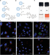
Bacteriophage MS2 virus‐Like particle‐based cell‐specific delivery induces selective cytotoxicity in tumor cell. A) The procedure of developing HCC‐specific MS2 VLPs that encapsulate different therapeutic and imaging components is depicted in this diagram. Using a suitable cross‐linker, nanoparticles (e.g., quantum dots), protein toxins, and DOX are attached to the pac site. Targeting peptides can be added to cargo‐loaded VLPs to increase selective internalization by cancer cells, fusogenic peptides can be added to promote endosomal escape of internalized VLPs, and PEG can be added to minimize nonspecific interactions and the humoral immune response to coat protein. Using a heterobifunctional cross‐linker with a PEG spacer arm, peptides having a C‐terminal cysteine residue are attached to lysine residues (red) on the outer capsid surface (yellow). B) Internalization of Alexa Fluor 555‐labeled SP94‐targeted VLPs (red) occurs via an endocytotic pathway, as evidenced by their punctate presence within HCC. The positive Pearson's correlation coefficient (r) between SP94‐targeted VLPs and Alexa Fluor 488‐labeled LAMP‐1 (green) as well as the near‐zero R‐value between SP94‐targeted VLPs and Alexa Fluor 647‐labeled Rab11a (white), a marker for recycling endosomes, show that SP94‐targeted VLPs are directed to lysosomes upon endocytosis by HCC. C) Hep3B cells were treated to SP94‐targeted VLPs (red) for 15 min or 1 h. In (i) and (iii), VLPs were co‐modified with SP94 peptide and H5WYG peptide, while in (ii) and (iv), VLPs were co‐modified with SP94 peptide alone. VLPs co‐modified with the SP94 targeting peptide and the H5WYG fusogenic peptide get disseminated in the cytoplasm of Hep3B cells after endocytosis, but VLPs changed with just SP94 remain in endosomes. Alexa Fluor 555 was used to label VLPs. Hoechst 33342 and CellTracker Green CMDFA were used to label the cells. Scale bars = 10 µm. Reproduced with permission.[ 101 ] Copyright 2011, Elsevier. Abbreviation: HCC: Human hepatocellular carcinoma; LAMP‐1: Lysosomal‐associated membrane protein 1; Rab11a: Ras‐related protein Rab‐11A.
Figure 11
Figure 11
Schematic illustration of axitinib‐loaded VLN synthesis. A) Axitinib and the CRISPR/Cas9 system are loaded into the mesoporous silica nanoparticle (MSN)‐based core, which is further enclosed with a lipid shell in VLN's core–shell structure. B) In response to the reductive microenvironment, VLN releases the CRISPR/Cas9 system and drug. Reproduced with permission.[ 102 ] Copyright 2020, Elsevier. Abbreviation: GSH: Glutathione; Axi: Axitinib; VLN: Virus‐like nanoparticle.
Figure 12
Figure 12
The VLN‐sgPD‐L1 anti‐tumor activity in melanoma mice model. A) Administration timelines and procedures for various formulations. B) Tumor growth curves in PBS, VLN‐sg (carrying a sgRNA without PD‐L1 targeting sequence), free RNP‐sgPD‐L1, RMSN‐sgPD‐L1, and VLN‐sgPD‐L1 mice. C) Survival curves for mice received several formulations. D) Body weight changes in mice after treatment with various formulations. E) Analysis of PD‐L1 immunofluorescence in each treatment group. Alex Fluor 594 (red) and DAPI (blue) were used to counterstain PD‐L1 and nuclei, respectively. Scale bars = 100 m. F) Levels of IFN‐ʏ and TNF‐α in mice treated with various formulations. Representative plots (G) and quantitative analysis (H) of CD8+ tumor infiltrating lymphocytes infiltration in treated tumors assessed by flow cytometry (gated on CD45+ CD3+ cells). I) Immunofluorescence image of the tumors revealed infiltrating CD4+ T cells (red) and infiltrated CD8+ T cells (green). The nuclei of the cells were counterstained with DAPI (blue). Scale bars = 100 µm. Data are presented as mean ± s.d. The significant levels are shown as **p < 0.01, and *** p < 0.001. Reproduced with permission.[ 102 ] Copyright 2020, Elsevier. Abbreviation: VLN; Virus‐like nanoparticle; sgRNA: Single guide RNA; PBS: Phosphate buffered saline; PDL‐1: Programmed death‐ ligand‐1; INF: Interferons; TNF: Tumor necrosis factor; RNP: Ribonucleoprotein; MSN: Mrsoporous silica nanoparticle.
Figure 13
Figure 13
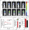
TGN‐HBc nanocages efficiently target the brain. A) In vivo imaging of Cy5.5‐labeled TGN‐HBc nanocages in the brain and HBc nanocages as a control at various time points. B) The fluorescence intensity of the brain region was used to quantify TGN‐HBc nanocages and HBc nanocages. C) The ex vivo brain images and fluorescence data from two mentioned groups (i) and the rate of fluorescence retention in brains of each group (ii). *p ˂ 0.05, **p ˂ 0.01). (n = 3). Reproduced with permission.[ 105 ] Copyright 2020, Elsevier. Abbrevition: HBc: Hepatitis B core.
Figure 14
Figure 14
The conjugation process for preparing A) CPMV‐DOX and B) CPMV‐SS‐DOX. C) Aqueous suspensions of wild‐type CPMV (a) and CPMV‐DOX (b) are depicted schematically. D) Time course study using confocal scanning microscopy showing cellular uptake of CPMV (Green: CPMV, red: Endolysosomes, blue: nuclei). Reproduced with permission.[ 113 ] Copyright 2013, American Chemical Society. Abbreviation: CPMV: Cowpea mosaic virus; DOX: Doxorubicin.
Figure 15
Figure 15
RR‐TMV DOXPEG conjugates are characterized. A) RR‐TMV disks were loaded with EMCH DOX at their S123C positions and aminophenolPEG5k at their N‐terminal prolines. B) Significant cell death observed after 72 h of incubation of U87MG glioblastoma cells with varying concentrations of RR‐TMVDOX‑PEG conjugates. C) U87MG cell uptake of RR‐TMVDOX‑PEG conjugates at 48 h. Reproduced with permission.[ 117 ] Copyright 2016, American Chemical Society. Abbreviation: DOX: Doxorubicin; TMV: Tobacco mosaic virus; PEG: Polyethylene glycol.
Figure 16
Figure 16
A) The development of the chimeric drug delivery mechanism is depicted schematically. The CCMV virus's customized capsid proteins were folic acid, dye, and DOX separately. The right combination of these ligand bioconjugated capsids was constructed with DOX conjugated AuNPs at its center to create nanoparticulate targeted delivery vehicles. B) Cytotoxicity of various drug delivery system formulations toward MCF7 cells by XTT Assay. Reproduced with permission.[ 118 ] Copyright 2016, Nature. Abbreviation: DOX: Doxorubicin; CCMV: Cowpea chlorotic stripe mosaic virus.
Figure 17
Figure 17
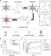
A) Focusing on the VLPs of Macrobrachium rosenbergii (M. rosenbergii) nodavirus, a schematic image of a drug delivery mechanism is seen (MrNVLP). Applying EDC and N‐hydroxysulfo‐succinimide, carboxylic acid groups of folic acid were conjugated with the amines of lysine residues found on the surface of MrNVLP (sulfo‐NHS). Via associations with the RNA molecules encapsidated within the nanoparticles, DOX biomolecules were injected into the cavity of FA‐conjugated MrNVLP. B) UV–vis spectra of the virus‐like particle of M. rosenbergii nodavirus (MrNVLP), free doxorubicin (DOX), DOX‐loaded MrNVLP (MrNv‐DOX), and DOX‐loaded‐and‐folic acid (FA)‐conjugated MrNVLP (FA‐MrNVLP‐DOX). C) Drug release profiles of DOX, MrNVLP‐DOX, and FA‐MrNVLP‐DOX for the first 18 h at 37 and 43 °C. Reproduced with permission.[ 125 ] Copyright 2019, Nature. Abbreviation: FA: Folic acid; EDC: 1‐ethyl‐3‐(3‐dimethyl aminopropyl) carbodiimide hydrochloride; Sulfo‐NHS: N‐hydroxysulfosuccinimide; DOX: Doxorubicin.
Figure 18
Figure 18
A) Biodistribution study of antibody‐MS2 capsid conjugates in cancer models. B) Formation of MS2‐antibody conjugates. C) In vivo biodistribution of Cu‐labeled MS2 in nude mice with HCC1954 orthotopic breast cancer tumors. PET scans were attained at 24 h after injection. Biodistribution studies were done 24 h after injection through gathering the main organs. The error bars display the standard deviation from samples. Reproduced with permission.[ 146 ] Copyright 2016 American Chemical Society.

Similar articles

Cited by

References

    1. Chen M., Hu J., Wang L., Li Y., Zhu C., Chen C., Shi M., Ju Z., Cao X., Zhang Z., Sci. Rep. 2020, 10, 14447. - PMC - PubMed
    1. Dey A. D., Bigham A., Esmaeili Y., Ashrafizadeh M., Moghaddam F. D., Tan S. C., Yousefiasl S., Sharma S., Maleki A., Rabiee N., Semin. Cancer Biol. 2022, 10.1016/j.semcancer.2022.06.003. - DOI - PubMed
    1. Yetisgin A. A., Cetinel S., Zuvin M., Kosar A., Kutlu O., Molecules 2020, 25, 2193. - PMC - PubMed
    1. Chamundeeswari M., Jeslin J. G., Verma M. L., Environ. Chem. Lett. 2018, 17, 849.
    1. Tabasi H., Babaei M., Abnous K., Taghdisi S. M., Saljooghi A. S., Ramezani M., Alibolandi M., J. Nanostruct. Chem. 2021, 11, 501.

LinkOut - more resources