

Experimental platform for the functional investigation of membrane proteins in giant unilamellar vesicles
- PMID: 35916307
- PMCID: PMC9364335
- DOI: 10.1039/d2sm00551d
Experimental platform for the functional investigation of membrane proteins in giant unilamellar vesicles
Abstract
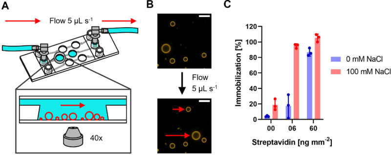
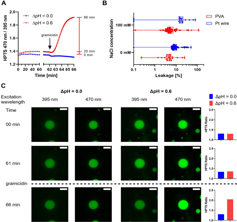
Giant unilamellar vesicles (GUVs) are micrometer-sized model membrane systems that can be viewed directly under the microscope. They serve as scaffolds for the bottom-up creation of synthetic cells, targeted drug delivery and have been widely used to study membrane related phenomena in vitro. GUVs are also of interest for the functional investigation of membrane proteins that carry out many key cellular functions. A major hurdle to a wider application of GUVs in this field is the diversity of existing protocols that are optimized for individual proteins. Here, we compare PVA assisted and electroformation techniques for GUV formation under physiologically relevant conditions, and analyze the effect of immobilization on vesicle structure and membrane tightness towards small substrates and protons. There, differences in terms of yield, size, and leakage of GUVs produced by PVA assisted swelling and electroformation were found, dependent on salt and buffer composition. Using fusion of oppositely charged membranes to reconstitute a model membrane protein, we find that empty vesicles and proteoliposomes show similar fusion behavior, which allows for a rapid estimation of protein incorporation using fluorescent lipids.
Conflict of interest statement
There are no conflicts to declare.
Figures


Similar articles
-
Electroformation of giant unilamellar vesicles from erythrocyte membranes under low-salt conditions.Anal Biochem. 2013 Apr 15;435(2):174-80. doi: 10.1016/j.ab.2013.01.001. Epub 2013 Jan 17. Anal Biochem. 2013. PMID: 23333270
-
Electroformation of giant unilamellar vesicles in saline solution.Colloids Surf B Biointerfaces. 2016 Nov 1;147:368-375. doi: 10.1016/j.colsurfb.2016.08.018. Epub 2016 Aug 16. Colloids Surf B Biointerfaces. 2016. PMID: 27566225
-
Secondary Ion Mass Spectrometry of Single Giant Unilamellar Vesicles Reveals Compositional Variability.J Am Chem Soc. 2023 Dec 20;145(50):27521-27530. doi: 10.1021/jacs.3c09039. Epub 2023 Dec 6. J Am Chem Soc. 2023. PMID: 38056605 Free PMC article.
-
Protein Reconstitution Inside Giant Unilamellar Vesicles.Annu Rev Biophys. 2021 May 6;50:525-548. doi: 10.1146/annurev-biophys-100620-114132. Epub 2021 Mar 5. Annu Rev Biophys. 2021. PMID: 33667121 Review.
-
Advances in giant unilamellar vesicle preparation techniques and applications.Adv Colloid Interface Sci. 2023 Aug;318:102935. doi: 10.1016/j.cis.2023.102935. Epub 2023 Jun 7. Adv Colloid Interface Sci. 2023. PMID: 37320960 Review.
Cited by
-
Molecular Mechanisms of Action of Dendrimers with Antibacterial Activities on Model Lipid Membranes.Polymers (Basel). 2025 Mar 29;17(7):929. doi: 10.3390/polym17070929. Polymers (Basel). 2025. PMID: 40219317 Free PMC article.
-
Genetic encoding and expression of RNA origami cytoskeletons in synthetic cells.Nat Nanotechnol. 2025 May;20(5):664-671. doi: 10.1038/s41565-025-01879-3. Epub 2025 Mar 17. Nat Nanotechnol. 2025. PMID: 40097648 Free PMC article.
-
Photoreceptor-Like Signal Transduction Between Polymer-Based Protocells.Adv Mater. 2025 Jan;37(3):e2413981. doi: 10.1002/adma.202413981. Epub 2024 Nov 3. Adv Mater. 2025. PMID: 39491508 Free PMC article.
-
Assessing the mechanism of facilitated proton transport across GUVs trapped in a microfluidic device.Biophys J. 2024 Sep 17;123(18):3267-3274. doi: 10.1016/j.bpj.2024.07.030. Epub 2024 Jul 26. Biophys J. 2024. PMID: 39066477 Free PMC article.
-
Function Investigations and Applications of Membrane Proteins on Artificial Lipid Membranes.Int J Mol Sci. 2023 Apr 13;24(8):7231. doi: 10.3390/ijms24087231. Int J Mol Sci. 2023. PMID: 37108393 Free PMC article. Review.
References
-
- Dimova R. and Marques C., in The Giant Vesicle Book. ed. R. Dimova and C. Marques, CRC Press, Boca Raton, FL, USA, 1st edn, 2019, vol. 53, p. 676
-
- Wang X. Du H. Wang Z. Mu W. Han X. Versatile Phospholipid Assemblies for Functional Synthetic Cells and Artificial Tissues. Adv. Mater. 2021;33(6):1–23. - PubMed
-
- Staufer O. Antona S. Zhang D. Csatári J. Schröter M. Janiesch J. W. et al., Microfluidic production and characterization of biofunctionalized giant unilamellar vesicles for targeted intracellular cargo delivery. Biomaterials. 2021;264(October 2019):120203. doi: 10.1016/j.biomaterials.2020.120203. doi: 10.1016/j.biomaterials.2020.120203. - DOI - DOI - PubMed
MeSH terms
Substances
LinkOut - more resources
Full Text Sources
Miscellaneous