. 2022 Sep;32(9):660-678.
doi: 10.1002/hipo.23458. Epub 2022 Aug 2.

Assessing mild cognitive impairment using object-location memory in immersive virtual environments

Affiliations

Assessing mild cognitive impairment using object-location memory in immersive virtual environments

Andrea Castegnaro et al. Hippocampus. 2022 Sep.

Abstract

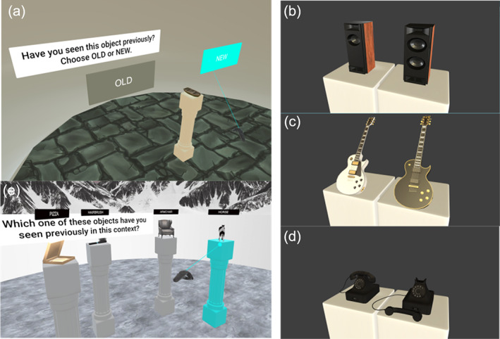
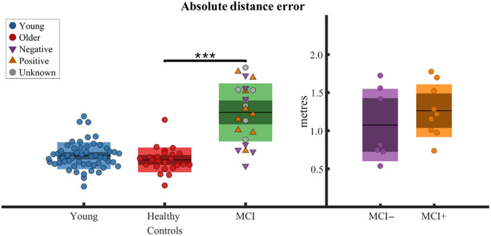
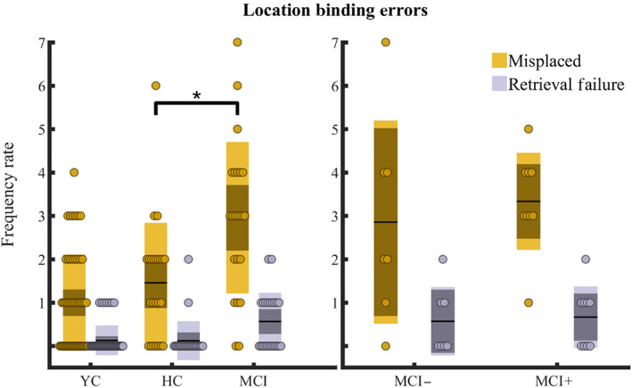
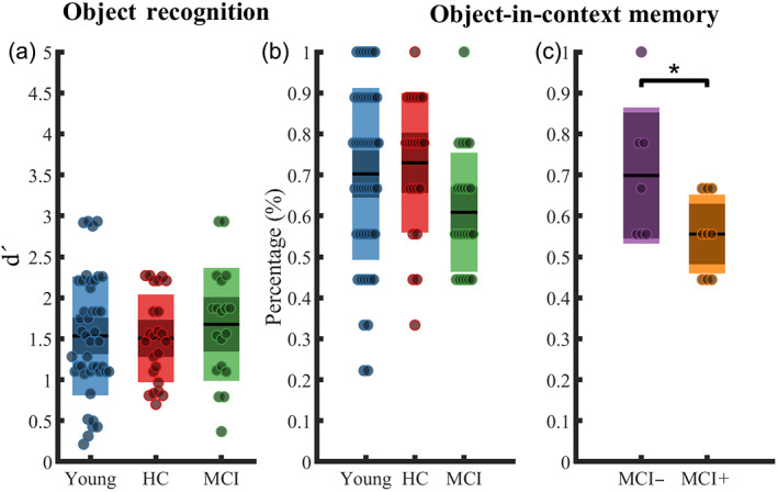
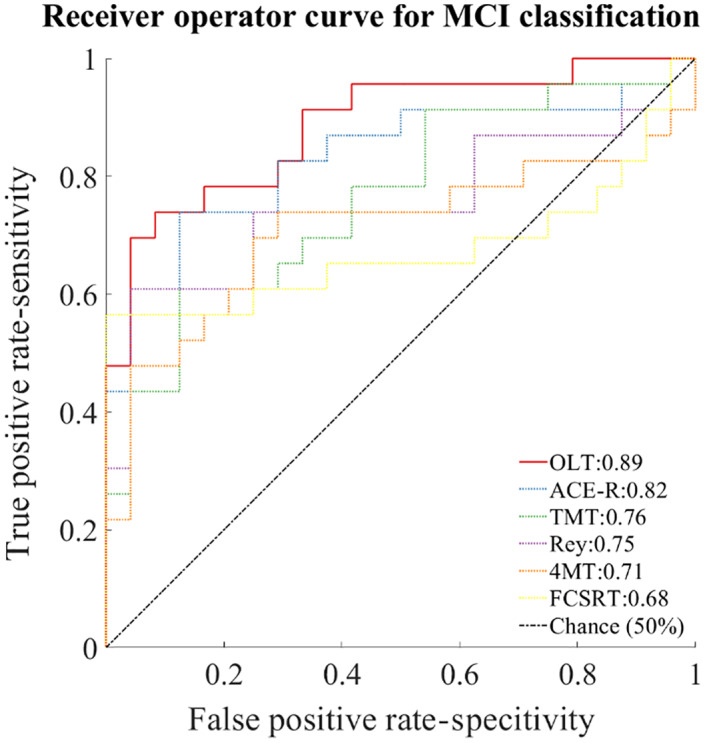
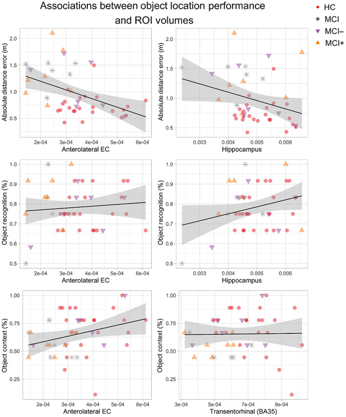
Pathological changes in the medial temporal lobe (MTL) are found in the early stages of Alzheimer's disease (AD) and aging. The earliest pathological accumulation of tau colocalizes with the areas of the MTL involved in object processing as part of a wider anterolateral network. Here, we sought to assess the diagnostic potential of memory for object locations in iVR environments in individuals at high risk of AD dementia (amnestic mild cognitive impairment [aMCI] n = 23) as compared to age-related cognitive decline. Consistent with our primary hypothesis that early AD would be associated with impaired object location, aMCI patients exhibited impaired spatial feature binding. Compared to both older (n = 24) and younger (n = 53) controls, aMCI patients, recalled object locations with significantly less accuracy (p < .001), with a trend toward an impaired identification of the object's correct context (p = .05). Importantly, these findings were not explained by deficits in object recognition (p = .6). These deficits differentiated aMCI from controls with greater accuracy (AUC = 0.89) than the standard neuropsychological tests. Within the aMCI group, 16 had CSF biomarkers indicative of their likely AD status (MCI+ n = 9 vs. MCI- n = 7). MCI+ showed lower accuracy in the object-context association than MCI- (p = .03) suggesting a selective deficit in object-context binding postulated to be associated with anterior-temporal areas. MRI volumetric analysis across healthy older participants and aMCI revealed that test performance positively correlates with lateral entorhinal cortex volumes (p < .05) and hippocampus volumes (p < .01), consistent with their hypothesized role in binding contextual and spatial information with object identity. Our results indicate that tests relying on the anterolateral object processing stream, and in particular requiring successful binding of an object with spatial information, may aid detection of pre-dementia AD due to the underlying early spread of tau pathology.

Keywords: Alzheimer's disease; entorhinal cortex; spatial cognition; virtual reality.


Conflict of interest statement

The authors declare no conflict of interests.

Figures

FIGURE 1
FIGURE 1
Object location memory subtask design. Design structure for the object location memory subtask. For each environment, participants underwent an encode and recall phase. During the encode phase participants walked to four different objects, one at a time (example in the view below the first encode box from the left). During the recall phase participants were asked to replace the previously seen objects by using the handheld controller which acted as a laser pointer (example in the view below the recall box). During the task participants visited three visually distinct environments featuring a 10 virtual meters circular colorless wall placed at the center of the virtual reality tracked area (one virtual meter corresponds to one real meter). Each environment had distinct distant landmarks (mountains with different textures and shape), near landmarks, light setting (daylight, night, and sunset), and sky texture
FIGURE 2
FIGURE 2
Object recognition and object‐in‐context subtasks. During the object recognition subtask, participants were kept in the “waiting” room devoid of any contextual cue (a). An object was placed on a pedestal in front of the participants along with two virtual cards labeled “Old”/“New.” Participants selected one of the cards using the handheld controller acting as a laser pointer depending on whether the object was previously seen in any of the environment (“Old”) or not (“New”). After the selection, participants were asked to give a four‐scale confidence rating to their choice with the following choices: “Certain”, “High Confidence,” “Low Confidence,” and “Guess”. “New” objects were created by pairing with “Old” objects and allowing one of the following changes: change in the number of a featured part (b), a texture change (c), and pose change (d). During the object‐in‐context subtask participants were placed back in one the previously visited environment and were shown four different objects at four different pedestals (e). Only one of the objects shown belonged to that environment. Participants made the selection using the handheld controller acting as a laser pointer
FIGURE 3
FIGURE 3
Object replacement accuracy. Absolute distance error per participant between veridical object locations and participant's responses averaged across trials reported for different groups. On the left panel groups presented are young, healthy older age‐matched controls, and pooled MCI (containing positive, negative, and unknown CSF status); on the right panel groups presented are MCI with negative/positive CSF status. Boxplots report the group mean as the thick black horizontal line, standard error of the mean as the darker area, and confidence interval as the external limits of the boxes. Significance bar indicates the main effect of MCI status on the absolute distance error found in the linear mixed effect model run across older age‐matched healthy controls and MCI and where each trial was inserted as a separate observation (***p < .001)
FIGURE 4
FIGURE 4
Binding errors frequency rate. Frequencies of the detected location binding errors were summed for each participant and reported for different groups. On the left panel groups presented are young, older age‐matched healthy controls (HC), and pooled MCI (positive, negative, and unknown CSF status). On the right panel groups presented are MCI with negative/positive CSF status. A binding error occurs when a participant replaced an object A in proximity of the veridical location of an object B in the same configuration. A heuristic in‐house algorithm (see Supplementary information S1) was developed to detect binding errors. When the algorithm detected at least three misplaced objects in a configuration, a retrieval error was registered. Yellow and gray circles indicate the sum of detected binding errors and retrieval errors, respectively, per participant. Boxplots report the group mean as the thick black horizontal line, standard error of the mean as the darker area, and confidence interval as the external limits of the boxes. Significance bar indicates the main effect of MCI status on the binding error count found in a generalized linear model run across older age‐matched healthy controls and MCI and where each configuration was inserted as a separate observation (*p < .05)
FIGURE 5
FIGURE 5
Object recognition and object‐in‐context memory performances. (a) Object recognition subtask performance. (b–c) Percentage of correct answers in the object‐in‐context memory subtask. Groups presented are young, older healthy age‐matched older controls (HC) and pooled MCI (positive, negative, and unknown) in (a, b), and MCI with negative/positive CSF status in (c). Mean is reported as a thick black horizontal line, standard error of the mean as the darker area, and confidence interval as the external limits of the boxes. Significance bar indicates the main effect of CSF status on the percentage of correct responses in the object‐in‐context subtask in a general linear model run across MCI+ and MCI−, and where each percentage score was inserted as a separate observation (*p < .05)
FIGURE 6
FIGURE 6
Receiver operating characteristics (ROC) curve. A logistic classifier has been trained to assess the ability of the neuropsychological tests in determining the MCI status against the healthy age‐matched controls. Pooled MCI (including patients with positive, negative, and unknown CSF biomarkers status) have been used for training the classifier. Only neuropsychological tests with performances that have been found statistically different between the two groups have been used for training the classifier. The classifier for the OLT has been trained with the performance from the object replacement subtask. The classifier for free and cued selective reminding test has been trained with the performance from the free immediate recall and the delayed free recall. The classifier for the Rey–Osterrieth figure recall test has been trained with the performance from the immediate recall only. ACE‐R, Addenbrooke cognitive examination‐revised; TMT, trail making test part B; 4MT, four mountain test. Chance level is represented by black dash‐dot line
FIGURE 7
FIGURE 7
Associations between object location subtasks performance and regions of interest volumetry. Included graphs reflect the a‐priori selected ROIs, except for the association between object recognition and the hippocampus which was a far stronger predicted of object recognition than the hypothesized perirhinal (BA35) volume. In each graph the main outcome of the subtask is reported alongside the y axis while the x axis represents the extracted volumes corrected by estimated intracranial volumes. The black line reported is the result of a simple regression between the two reported quantities without controlling for age, sex, years in education, and diagnostic status, thus the slope might not capture the results of the multilevel modeling adopted for the volumetric analysis reported in Table 2. In line with our hypothesis, anterolateral EC volumes is associated with object‐in‐context performance and approach significance in the location memory subtask. The hippocampus, but not the entorhinal or perirhinal volumes, were predictive of object recognition performance

Similar articles

Cited by

References

    1. Adams, J. N. , Maass, A. , Harrison, T. M. , Baker, S. L. , & Jagust, W. J. (2019). Cortical tau deposition follows patterns of entorhinal functional connectivity in aging. eLife, 8, e49132. 10.7554/ELIFE.49132 - DOI - PMC - PubMed
    1. Auger, S. D. , Mullally, S. L. , & Maguire, E. A. (2012). Retrosplenial cortex codes for permanent landmarks. PLoS One, 7(8), e43620. 10.1371/journal.pone.0043620 - DOI - PMC - PubMed
    1. Backman, L. , Jones, S. , Berger, A. K. , Laukka, E. J. , & Small, B. J. (2004). Multiple cognitive deficits during the transition to Alzheimer's disease. Journal of Internal Medicine, 256(3), 195–204. 10.1111/j.1365-2796.2004.01386.x - DOI - PubMed
    1. Barense, M. D. , Gaffan, D. , & Graham, K. S. (2007). The human medial temporal lobe processes online representations of complex objects. Neuropsychologia, 45(13), 2963–2974. 10.1016/J.NEUROPSYCHOLOGIA.2007.05.023 - DOI - PubMed
    1. Barense, M. D. , Henson, R. N. A. , & Graham, K. S. (2011). Perception and conception: Temporal lobe activity during complex discriminations of familiar and novel faces and objects. Journal of Cognitive Neuroscience, 23(10), 3052–3067. 10.1162/JOCN_A_00010 - DOI - PubMed

Publication types

MeSH terms