. 2022 Sep 16;479(17):1785-1806.
doi: 10.1042/BCJ20220244.

Structural and functional characterisation of a stable, broad-specificity multimeric sialidase from the oral pathogen Tannerella forsythia

Affiliations

Structural and functional characterisation of a stable, broad-specificity multimeric sialidase from the oral pathogen Tannerella forsythia

Marianne J Satur et al. Biochem J. .

Abstract

Sialidases are glycosyl hydrolase enzymes targeting the glycosidic bond between terminal sialic acids and underlying sugars. The NanH sialidase of Tannerella forsythia, one of the bacteria associated with severe periodontal disease plays a role in virulence. Here, we show that this broad-specificity enzyme (but higher affinity for α2,3 over α2,6 linked sialic acids) digests complex glycans but not those containing Neu5,9Ac. Furthermore, we show it to be a highly stable dimeric enzyme and present a thorough structural analysis of the native enzyme in its apo-form and in complex with a sialic acid analogue/ inhibitor (Oseltamivir). We also use non-catalytic (D237A) variant to characterise molecular interactions while in complex with the natural substrates 3- and 6-siallylactose. This dataset also reveals the NanH carbohydrate-binding module (CBM, CAZy CBM 93) has a novel fold made of antiparallel beta-strands. The catalytic domain structure contains novel features that include a non-prolyl cis-peptide and an uncommon arginine sidechain rotamer (R306) proximal to the active site. Via a mutagenesis programme, we identified key active site residues (D237, R212 and Y518) and probed the effects of mutation of residues in proximity to the glycosidic linkage within 2,3 and 2,6-linked substrates. These data revealed that mutagenesis of R306 and residues S235 and V236 adjacent to the acid-base catalyst D237 influence the linkage specificity preference of this bacterial sialidase, opening up possibilities for enzyme engineering for glycotechology applications and providing key structural information that for in silico design of specific inhibitors of this enzyme for the treatment of periodontitis.

Keywords: enzyme; periodontitis; sialic acid; sialidase.


Conflict of interest statement

The authors declare that there are no competing interests associated with the manuscript.

Figures

Figure 1.
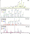
Figure 1.. Ability of NanH to digest complex glycans.
(A). LC-FLR traces of procainamide labelled sialidase digestions of FA2G2S2. Undigested (sialylated) FA2G2S2 (black trace). NanH 1h and O/N digests (green traces) are shown with known glycans. All symbols follow the Consortium for Functional Glycomics conventions and are shown in the Key: Gal-Galactose, Glc-Glucose, GalNAc-N-acetyl galactosamine, GlcNAc-N-acetyl glucosamine, Man-Mannose NeuAc-N-acetly-neuraminic/sialic acid. (B) LC-FLR traces of procainamide labelled human plasma N-glycans (black, control) and after overnight (O/N) incubation with NanH (0.1 mg ml−1).
Figure 2.
Figure 2.. Effect of Neu5,9Ac glycans on digestion with NanH.
N-glycan profile of erythropoietin N-glycans before (top panel, see Supplementary Table S1 for peak assignments) and after (lower panels) incubation with NanH (0.1 mg ml−1 o/n) overlaid with M/S traces for Neu5Ac (green), Neu5Ac-Mono-OAc (red) and Di-OAC (purple). Peak numbers in the upper panel are assigned according to Supplementary Table S1 and all symbols as shown in Figure 1.
Figure 3.
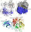
Figure 3.. Crystal model of NanH-apo.
(A) Each subunit (A in grey and B in blue) comprises a Beta-propellor (β-propellor) containing the active site and FRIP motif (red), Asp-boxes (orange) and an N-terminal Carbohydrate-binding module (CBM). Shown in cartoon and surface visualisation from Pymol with open cleft between subunits labelled. (B) Subunit A of NanH-apo, highlighting individual blades of the β-propeller domain in rainbow representation and with active site labelled.
Figure 4.
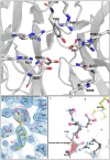
Figure 4.. Detailed view of the NanH active site.
(A) Shows the catalytic arginine triad of R212, R487 and R423 alongside the catalytic nucleophile Y518, D237 and E407 as well as other key residues. (B) Displays the bound HEPES molecule in yellow with the –COOH group circled. (C) Location of the non-proline cis-peptide at Q239 in relation to the bound HEPES.
Figure 5.
Figure 5.. The CBM of T.forsythia forms a novel CBM family.
CBM (A) and entire enzyme (B) with conserved putative binding site residues (as shown in Supplementary Figure S5) highlighted in red. (C) T. forsythia NanH alongside equivalent predicted CBM from the closely related P. distasonis (PDB: 4FJ6) and BTSA from B. thetaiotaomicron (PDB: 4BBW) and the CBM40 sialic acid binding CBM from S. pneumoniae (PDB: 4ZXK), RgNanH from R. gnavus (PDB: 6ER2) and NanB from S. pneumoniae (PDB: 2VW0).
Figure 6.
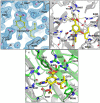
Figure 6.. NanH active site bound in complex with the inhibitor oseltamivir.
(A) Oseltamivir (yellow) was modelled into the electron density (blue) of the F0 − FC double difference map at an rmsd of 1.04. (B) NanH sialidase in cartoon representation (grey), with the catalytic residues (grey sticks) and oseltamivir (yellow sticks). A single water molecule is highlighted as a red sphere and the yellow dashed lines represent the hydrogen bonding network; (C) Overlay of NanH-apo (green carbons) with NanH-oseltamivir (grey carbons) illustrating the movement of W308 (highlighted by bracket) upon the binding of oseltamivir (yellow) to the NanH active site.
Figure 7.
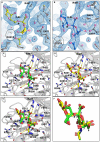
Figure 7.. NanH-D237A bound in complex with 3- and 6-SL.
(A) 3-SL (yellow) was modelled into the electron density (blue) of the 2F0 − FC double difference map at an RMSD of 1.02. (B) 6-SL (dark blue) was modelled into the electron density (blue) of the 2F0 − FC double difference map at an RMSD of 0.99. (C) NanH-D237A (grey cartoon) in complex with 3-SL (green sticks). (D) NanH-D237A in complex with 6-SL (yellow sticks). (E) NanH-D237A in complex with 3-SL (green sticks) overlaid with NanH-D237A in complex with 6-SL (yellow sticks). (F) Overlay of 3-SL (green sticks) and 6-SL (yellow sticks) isolated from the protein backbone (carbon atoms numbered). In all panels, the NanH-D237A protein is shown in grey in cartoon representation with all the key residues around the catalytic centre added in stick representation, and hydrogen bonding (yellow dashes) shown. D237A is circled in all panels while the glycosidic linkage is highlighted with an asterisk.
Figure 8.
Figure 8.. Structure–function mutagenesis of NanH.
(A) Illustration of the catalytic domain in isolation showing the location of residues targeted by mutagenesis in relation to bound 3-SL or 6-SL. (B) Kinetic parameters of NanH and variants with 3-SL or 6-SL as assayed by TBA assay. The kcat/KM (efficiency) values are presented in the bar-chart on the left and in tabular form on the right, alongside the ratio of 3-SL : 6-SL. N/A indicates non-active.

References

    1. Cross, B.W. and Ruhl, S. (2018) Glycan recognition at the saliva - oral microbiome interface. Cell. Immunol. 333, 19–33 10.1016/j.cellimm.2018.08.008 - DOI - PMC - PubMed
    1. Lewis, A.L. and Lewis, W.G. (2012) Host sialoglycans and bacterial sialidases: a mucosal perspective. Cell. Microbiol. 14, 1174–1182 10.1111/j.1462-5822.2012.01807.x - DOI - PubMed
    1. Li, C., Yang, X., Pan, Y., Yu, N., Xu, X., Tong, T.et al. (2017) A sialidase-deficient Porphyromonas gingivalis mutant strain induces less interleukin-1beta and tumor necrosis factor-alpha in Epi4 cells than W83 strain through regulation of c-Jun N-terminal kinase pathway. J. Periodontol. 88, e129–e139 10.1902/jop.2017.160815 - DOI - PubMed
    1. Roy, S., Honma, K., Douglas, C.W.I., Sharma, A. and Stafford, G.P. (2011) Role of sialidase in glycoprotein utilization by Tannerella forsythia. Microbiology 157, 3195–3202 10.1099/mic.0.052498-0 - DOI - PMC - PubMed
    1. Moustafa, I., Connaris, H., Taylor, M., Zaitsev, V., Wilson, J.C., Kiefel, M.J.et al. (2004) Sialic acid recognition by Vibrio cholerae neuraminidase. J. Biol. Chem. 279, 40819–40826 10.1074/jbc.M404965200 - DOI - PubMed

Publication types