. 2022 Aug 24;96(16):e0055922.
doi: 10.1128/jvi.00559-22. Epub 2022 Aug 2.

Induction of Interferon-Stimulated Genes Correlates with Reduced Growth of Influenza A Virus in Lungs after RIG-I Agonist Treatment of Ferrets

Affiliations

Induction of Interferon-Stimulated Genes Correlates with Reduced Growth of Influenza A Virus in Lungs after RIG-I Agonist Treatment of Ferrets

Lara S U Schwab et al. J Virol. .

Abstract

Intracellular RIG-I receptors represent key innate sensors of RNA virus infection, and RIG-I activation results in the induction of hundreds of host effector genes, including interferon-stimulated genes (ISGs). Synthetic RNA agonists targeting RIG-I have shown promise as antivirals against a broad spectrum of viruses, including influenza A virus (IAV), in both in vitro and mouse models of infection. Herein, we demonstrate that treatment of a ferret airway epithelial (FRL) cell line with a RIG-I agonist rapidly and potently induced expression of a broad range of ISGs and resulted in potent inhibition of growth of different IAV strains. In ferrets, a single intravenous injection of RIG-I agonist was associated with upregulated ISG expression in peripheral blood mononuclear cells and lung tissue, but not in nasal tissues. In a ferret model of viral contact transmission, a single treatment of recipient animals 24 h prior to cohousing with IAV-infected donors did not reduce virus transmission and shedding but did result in reduced lung virus titers 6 days after treatment. A single treatment of the IAV-infected donor animals also resulted in reduced virus titers in the lungs 2 days later. Thus, a single intravenous treatment with RIG-I agonist prior to infection or to ferrets with an established IAV infection can reduce virus growth in the lungs. These findings support further development of RIG-I agonists as effective antiviral treatments to limit the impact of IAV infections, particularly in reducing virus replication in the lower airways. IMPORTANCE RIG-I agonists have shown potential as broad-spectrum antivirals in vitro and in mouse models of infection. However, their antiviral potential has not been reported in outbred animals such as ferrets, which are widely regarded as the gold standard small animal model for human IAV infections. Herein, we demonstrate that RIG-I agonist treatment of a ferret airway cell line resulted in ISG induction and inhibition of a broad range of human influenza viruses. A single intravenous treatment of ferrets also resulted in systemic induction of ISGs, including in lung tissue, and when delivered to animals prior to IAV exposure or to animals with established IAV infection treatment resulted in reduced virus replication in the lungs. These data demonstrate the effectiveness of single RIG-I treatment against IAV in the ferret model and highlight the importance of future studies to optimize treatment regimens and delivery routes to maximize their ability to ameliorate IAV infections.

Keywords: RIG-I; antiviral agents; ferret; influenza; innate immunity; mouse model.


Conflict of interest statement

The authors declare a conflict of interest. G.H. holds patents on RIG-I and is one of the founders of the Rigontec GmbH.

Figures
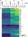
FIG 1
FIG 1
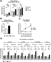
Treatment of FRL cells with RIG-I agonist or ferret IFN-α induces upregulation of multiple ISGs. FRL cells were transfected with RIG-I agonist (3pRNA, 200 ng/mL) or with control RNA (ctrl RNA, 200 ng/mL). Untreated cells were included for comparison. (A) Total RNA was isolated at various times after treatment, and expression of ISGs was assessed by qRT-PCR. Expression was normalized to GAPDH and expressed as fold induction relative to untreated cells. (B) In an independent experiment, FRL cells were treated with 3pRNA or ctrl RNA, as for panel A, or with recombinant ferret IFN-α (50 ng/mL). Untreated cells were included for comparison. After 6 h, total RNA was isolated from triplicate samples in each group and sequenced with a 3′ mRNA-Seq kit on a NovaSeq 6000 instrument. The heatmap shows the normalized gene expression for most significant differentially expressed genes in control, IFN-α, 3pRNA, and untreated groups for all pairwise comparisons. Colors indicate the prevalence of transcript expression from low (dark blue) to high (bright yellow).
FIG 2
FIG 2
RIG-I agonist treatment inhibits IAV infection and growth in FRL cells. FRL cells were transfected with RIG-I agonist (3pRNA, 200 ng/mL) or control RNA (ctrl RNA, 200 ng/mL), or treated with ferret IFN-α (50 ng/mL). Untreated cells were included for comparison. (A) After 24 h, cells were infected for 1 h at 37°C with IAV (strain HKx31, H3N2) at an MOI of 1, 5 or 20. Cells were incubated an additional 7 h, then fixed and stained for intracellular expression of the viral NP and analyzed by flow cytometry. (B) At 24 h after transfection, cells were washed and cultured in complete medium with 5% FCS. After 4 days (e.g., 5 days after transfection), cells were infected with HKx31 (MOI = 10) for 1 h, washed, inoculated for an additional 7 h, fixed, and stained for intracellular expression of the viral NP and analyzed by flow cytometry. (C) Cells were inoculated for 1 h with HKx31 (MOI = 0.01), washed, and incubated in the presence of exogenous trypsin (0.5 μg/mL). Titers of infectious virus were determined in clarified supernatants at various times postinfection using a Virospot assay on MDCK cells. (D) Cells were inoculated with the indicated viruses (MOI = 0.1): A/Udorn/307/1972, A/Beijing/353/89, A/Brazil/11/1978, A/New Caledonia/20/1999, A/Perth/65/09, A/Fiji/2/2016, or B/Brisbane/20/2008 in the presence of 0.5 μg/mL TPCK trypsin, and cell-free supernatant was harvested after 2 h or 48 h. Titers of infectious virus were determined in clarified supernatants using a Virospot assay on MDCK cells. The dotted line represents the level of detection. Fold increase was determined by the change of virus titer from 48 hpi compared to 2 hpi. All data show means (± SD) from triplicate samples and are representative of 2 or more independent experiments. Statitsical evaluation used a two-way analysis of variance (ANOVA) with Bonferroni’s multiple-comparison test (A and D) or a Student’s unpaired t test (B and C). *, P < 0.05; **, P < 0.01; ***, P < 0.001; ****, P < 0.0001; ns, not significant.
FIG 3
FIG 3
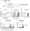
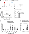
A single intravenous treatment of ferrets with RIG-I agonist 24 h prior to cohousing with IAV-infected donor animals resulted in reduced virus replication in the lung but not the nasal tissues. (A) Recipient animals (n = 4/group) received a single intravenous injection of 3pRNA or ctrl RNA (0.3 mg/kg) 24 h prior to cohousing with IAV-infected donor animals that had received 500 μL containing 105 TCID50 of A/Perth/265/2009 (H1N1pdm09) 24 h earlier. After cohousing for 48 h, donor animals were removed and recipients were monitored for an additional 3 days. Nasal wash samples were collected daily, and animals were killed 5 days after cohousing commenced. (B) Body temperature was recorded over time (mean ± SEM). The dotted line represents average baseline temperature. (C to F) Titers of infectious virus in nasal washes (days 1 to 5 post-cohousing) (C) and nasal tissues (day 5 post cohousing) (D) from 3pRNA- or ctrl RNA-treated animals. Individual animals are shown. Data points represent viral titers isolated from individual lung lobes from each individual 3pRNA- or ctrl RNA-treated animal (5 lobes per animal) (E) or viral titers obtained from the five individual lung lobes from all four 3pRNA- or ctrl RNA-treated animals pooled (F). Bars represent the mean (±SD) viral titers per group. Dashed lines represent the limit of detection (<0.2 log10 TCID50/mL), and samples below the limit of detection were assigned values of 0.1 log10TCID50/mL for statistical analysis. Statistical evaluation used a two-tailed Mann-Whitney U test to compare viral loads in nasal wash over time (C) or an unpaired Student's t test to compare the viral load in respiratory tissue (D and F) after 3pRNA or ctrl RNA treatment. *, P < 0.05; ns, not significant.
FIG 4
FIG 4
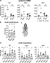
A single intravenous treatment of ferrets with RIG-I agonist resulted in systemic upregulation of ISGs, as well as upregulation of ISGs in the lung. Ferrets received a single i.v. injection with 3pRNA or ctrl RNA (0.3 mg/kg). After 24 h, blood (for PBMC isolation) and respiratory tissues were collected for RNA isolation and then examined for expression of Mx1, ISG15, and OAS1 by qRT-PCR. (A) ISG expression in PBMCs collected prior to i.v. injection (pre) compared to levels 24 h after 3pRNA or ctrl RNA injection (post). Levels of each ISG are expressed relative to GAPDH and symbols represent individual animals. Horizontal lines represent median values. (B) ISG expression in nasal tissue. Data show the fold change in ISG expression, defined as the expression level in 3pRNA-treated animals compared to ctrl RNA-treated animals. (C) Schematic of ferret lung lobes (adapted from Chan et al. [56]). (D) ISG expression in individual lung lobes, as defined for panel C. Bars in panels B and D represent means ± SD (n = 4 animals/group). Statistical evaluation used a paired Student's t test to compare pre- versus posttreatment (A) or an unpaired Student's t test to compare 3pRNA- versus ctrl RNA-treated animals (B and D). *, P < 0.05; **, P < 0.01; ***, P < 0.001; ns, not significant.
FIG 5
FIG 5
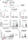
A single intravenous treatment of ferrets with RIG-I agonist 24 h prior to cohousing with IAV-infected donor animals resulted in reduced virus replication in the lung but not the nasal tissues. (A) Recipient animals (n = 4/group) received a single intravenous injection of 3pRNA or ctrl RNA (0.3 mg/kg) 24 h prior to cohousing with IAV-infected donor animals, which had received 500 μL containing 105 TCID50 of A/Perth/265/2009 (H1N1pdm09) intranasally 24 h earlier. After cohousing for 48 h, donor animals were removed and recipients monitored for an additional 3 days. Nasal wash samples were collected daily, and animals were killed 5 days after cohousing commenced. (B) Whole blood was collected and PBMCs were isolated prior to (pre) and 24 h after (post) i.v. injection with 3pRNA or ctrl RNA. qRT-PCR was used to determine ISG levels, which are expressed relative to GAPDH. Data from individual animals are shown, and horizontal lines indicate the median values. Red boxes are shown to highlight recipient animal 2, which showed the lowest induction of all ISGs tested. (C) Body temperature was recorded over time (mean ± SEM). The dotted line represents average baseline temperature. (D and E) Titers of infectious virus in nasal washes (days 1 to 5 post cohousing) (D) and nasal tissues (day 5 post cohousing) from 3pRNA- or ctrl RNA-treated animals (E). Individual animals are shown. (F and G) Data points in represent individual lung lobes from individual 3pRNA- or ctrl RNA-treated animals (F) and individual lung lobes from all 3pRNA- or ctrl RNA-treated animals pooled (G). Bars in panels D to G represent mean (± SD) viral titers per group. Dashed lines represent the limit of detection (<0.2 log10 TCID50/mL) and samples below that limit were assigned values of 0.1 log10 TCID50/mL for statistical analysis. Statistical evaluation used a paired Student's t test (B), two-tailed Mann-Whitney U test to compare viral loads in nasal wash over time (D), or an unpaired Student's t test to compare the viral load in respiratory tissue after 3pRNA or ctrl RNA treatment (E and G). *, P < 0.05; ns, not significant.
FIG 6
FIG 6
A single intravenous injection of RIG-I agonist to donor ferrets with established IAV infection results in reduced virus replication in the lung but not in the nasal tissue. (A) Donor animals were infected via the intranasal route with 105 TCID50 of A/Perth/265/2009 (H1N1pdm09) in 500 μL. At 3 dpi, animals received an i.v. injection of 3pRNA or ctrl RNA (0.3 mg/kg). (B) Body temperature was recorded over time (mean ± SEM). The dotted line represents average baseline temperature. (C to E) At 5 dpi (i.e., 2 days after treatment), ferrets were euthanized, and nasal tissue (C) and lungs (D and E) were collected and homogenized, and virus titers in clarified homogenates were determined by TCID50 assay. The data in panel D represent individual lung lobes from 3pRNA- or ctrl RNA-treated animals, and the data in panel E represent pooled data from individual lung lobes from all 3pRNA- or ctrl RNA-treated animals (viral titers ± SD). Dashed lines represent the limit of detection. Samples below detection (<0.2 log10 TCID50/mL) were assigned values of 0.1 log10 TCID50/mL for statistical analysis (unpaired Student's t test to compare the viral loads after 3pRNA versus ctrl RNA treatment). ****, P < 0.0001; ns, not significant.
FIG 7
FIG 7
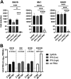
Treatment of primary ferret nasal epithelial cells (NECs) with RIG-I agonist or ferret IFN-α induced upregulation of multiple ISGs. NECs were transfected with RIG-I agonist (3pRNA, 200 ng/mL), control RNA (ctrl RNA, 200 ng/mL), or recombinant ferret IFN-α at a high (50 ng/mL) or low (10 ng/mL) concentration. Untreated cells were included for comparison. (A) After 24 h, total RNA was isolated at various times after treatment and expression of ISGs was assessed by qRT-PCR. Expression is normalized to GAPDH and expressed as fold induction relative to untreated cells. (B) Cells were inoculated with Perth/09 (H1N1, MOI = 0.1) in the presence of 0.5 μg/mL TPCK trypsin, and cell-free supernatant was harvested after 2 h or 48 h. Titers of infectious virus were determined in clarified supernatants using a Virospot assay on MDCK cells. The dotted line represents the level of detection. Fold increase was determined by the change of virus titer from 48 hpi compared to 2 hpi. All data show means (± SD) from triplicate samples and are representative of 2 or more independent experiments. Statistical evaluation used a two-way ANOVA with Bonferroni’s multiple-comparison test (A) or a two-way ANOVA with Bonferroni’s multiple-comparison (B). *, P < 0.05; **, P < 0.01; ***, P < 0.001; ****, P < 0.0001.

Similar articles

Cited by

References

    1. Kell AM, Gale M, Jr.. 2015. RIG-I in RNA virus recognition. Virology 479–480:110–121. 10.1016/j.virol.2015.02.017. - DOI - PMC - PubMed
    1. Loo YM, Gale M, Jr.. 2011. Immune signaling by RIG-I-like receptors. Immunity 34:680–692. 10.1016/j.immuni.2011.05.003. - DOI - PMC - PubMed
    1. Schoggins JW, Rice CM. 2011. Interferon-stimulated genes and their antiviral effector functions. Curr Opin Virol 1:519–525. 10.1016/j.coviro.2011.10.008. - DOI - PMC - PubMed
    1. Hornung V, Ellegast J, Kim S, Brzozka K, Jung A, Kato H, Poeck H, Akira S, Conzelmann KK, Schlee M, Endres S, Hartmann G. 2006. 5'-Triphosphate RNA is the ligand for RIG-I. Science 314:994–997. 10.1126/science.1132505. - DOI - PubMed
    1. Schlee M, Roth A, Hornung V, Hagmann CA, Wimmenauer V, Barchet W, Coch C, Janke M, Mihailovic A, Wardle G, Juranek S, Kato H, Kawai T, Poeck H, Fitzgerald KA, Takeuchi O, Akira S, Tuschl T, Latz E, Ludwig J, Hartmann G. 2009. Recognition of 5' triphosphate by RIG-I helicase requires short blunt double-stranded RNA as contained in panhandle of negative-strand virus. Immunity 31:25–34. 10.1016/j.immuni.2009.05.008. - DOI - PMC - PubMed

Publication types