. 2022 Oct 4;82(19):3486-3498.
doi: 10.1158/0008-5472.CAN-22-0408.

Selenium Modulates Cancer Cell Response to Pharmacologic Ascorbate

Affiliations

Selenium Modulates Cancer Cell Response to Pharmacologic Ascorbate

Connor S R Jankowski et al. Cancer Res. .

Abstract

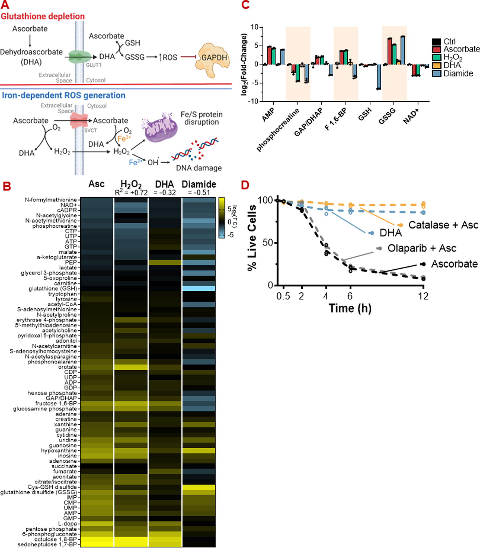
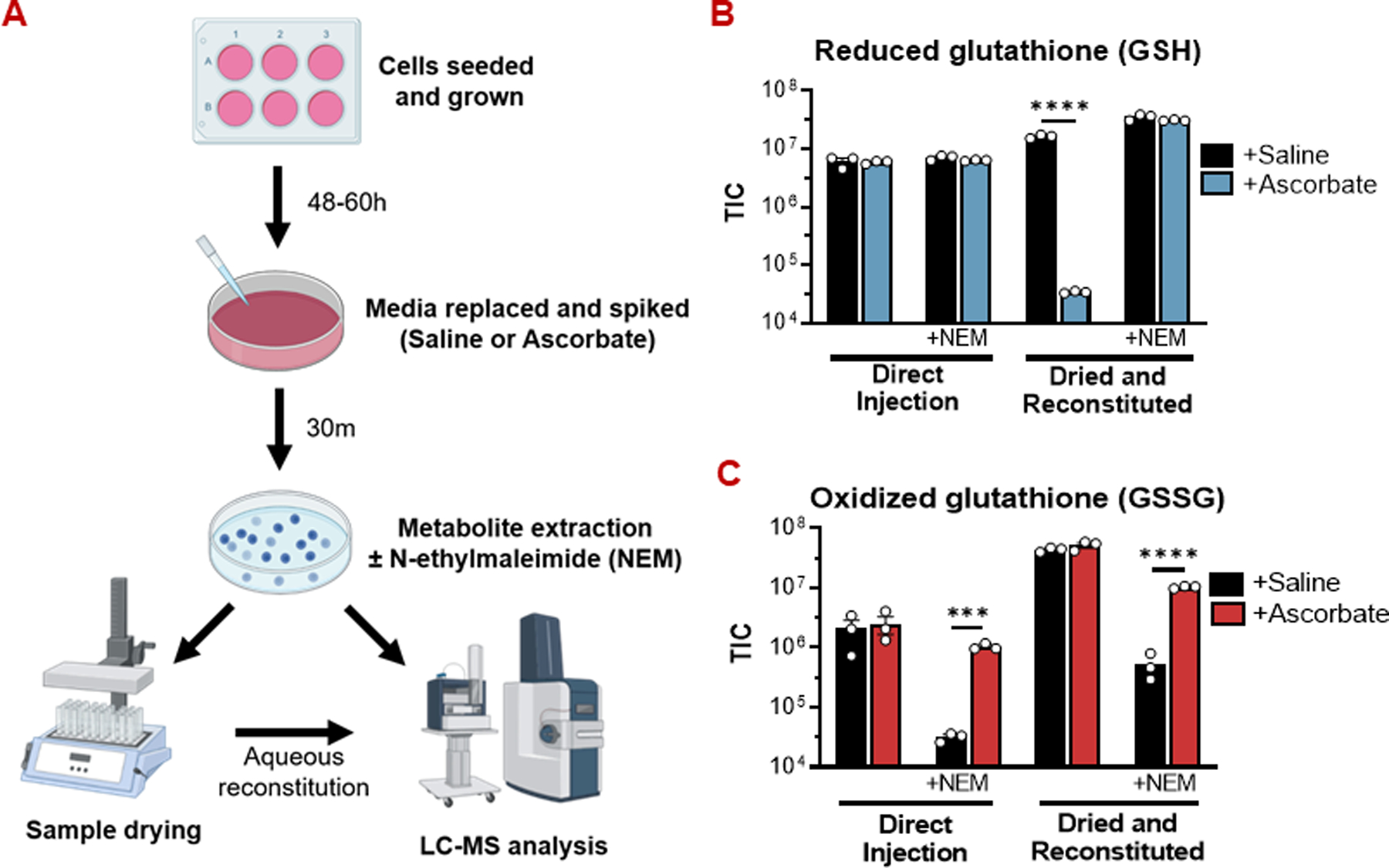
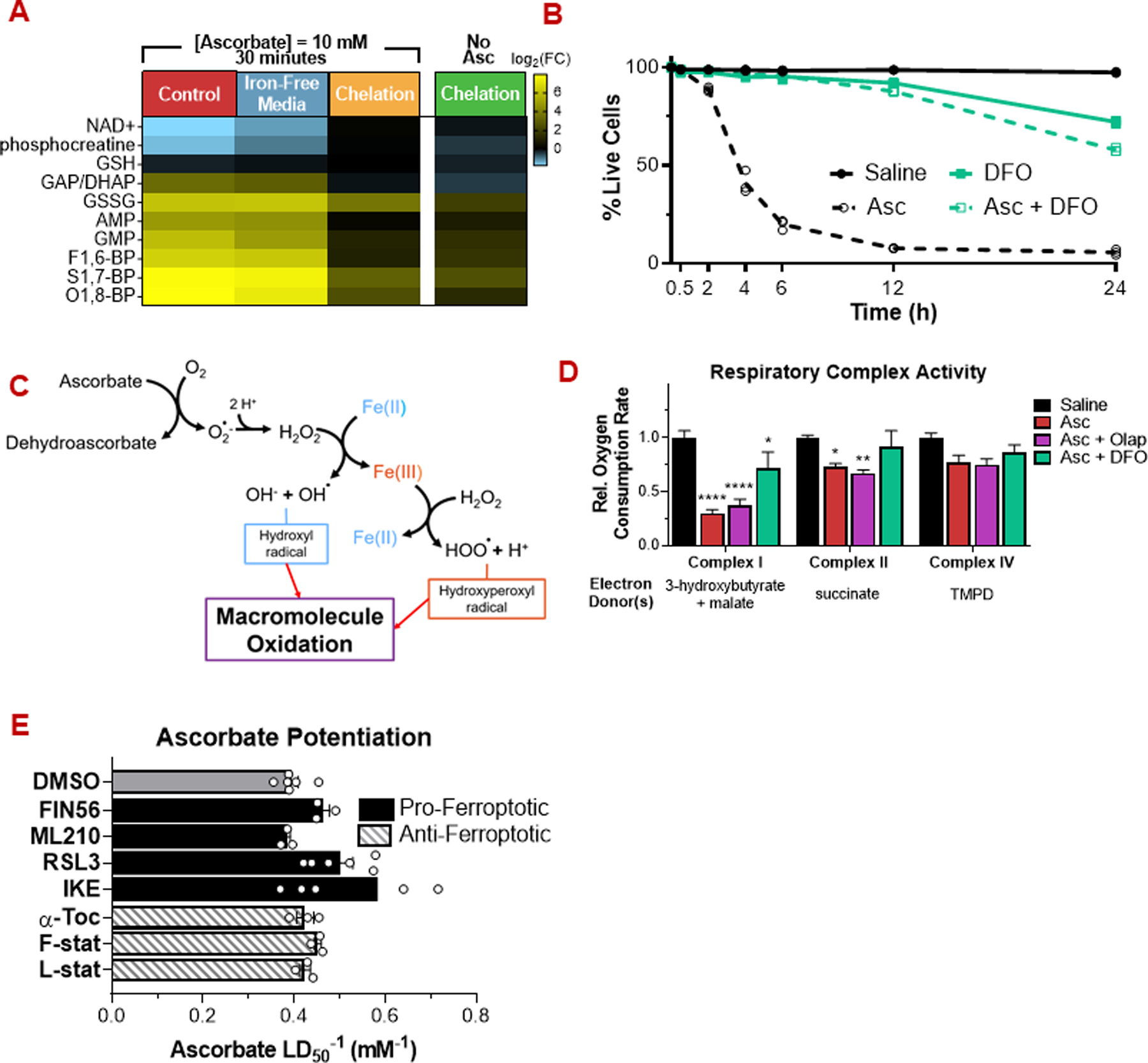
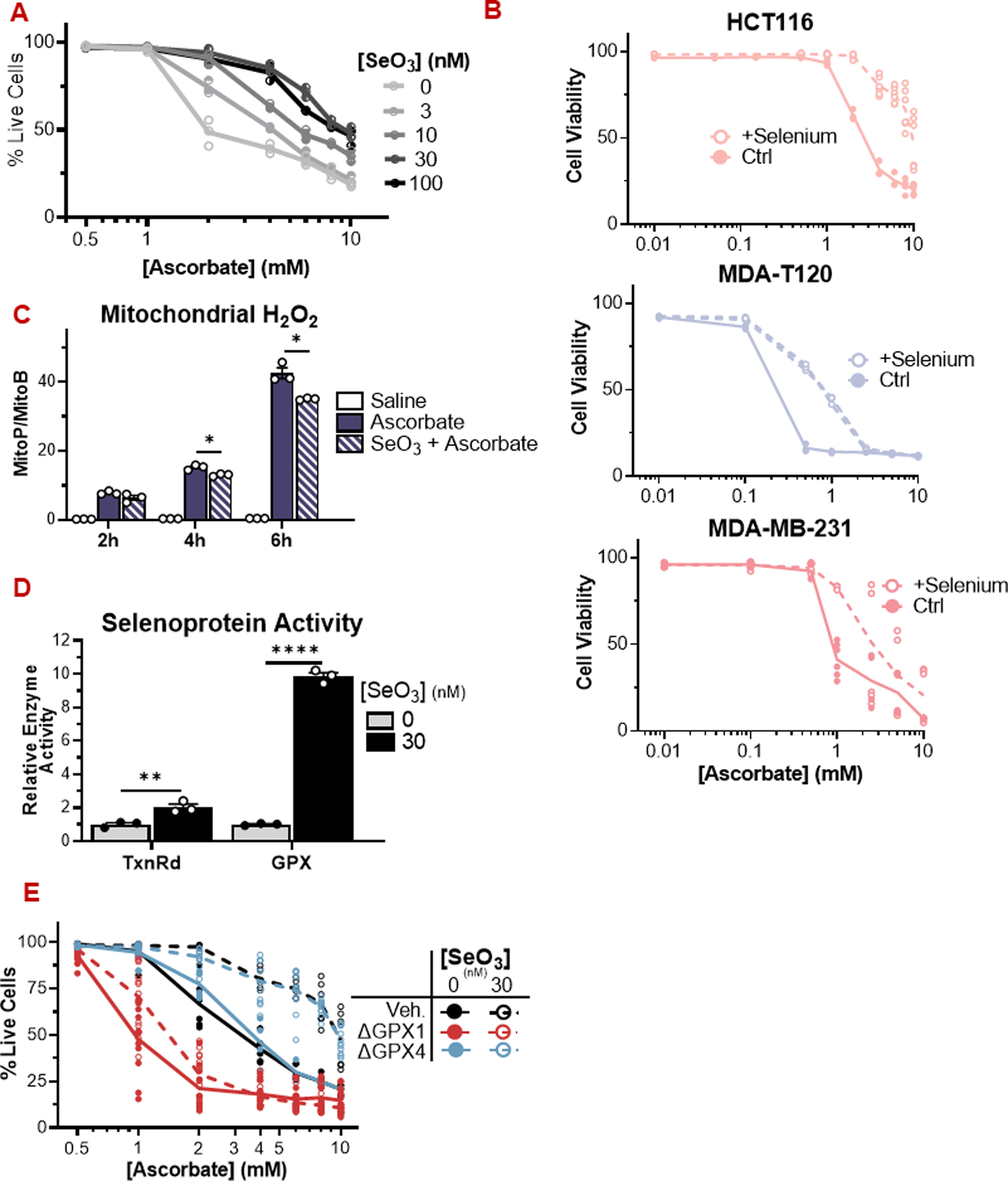
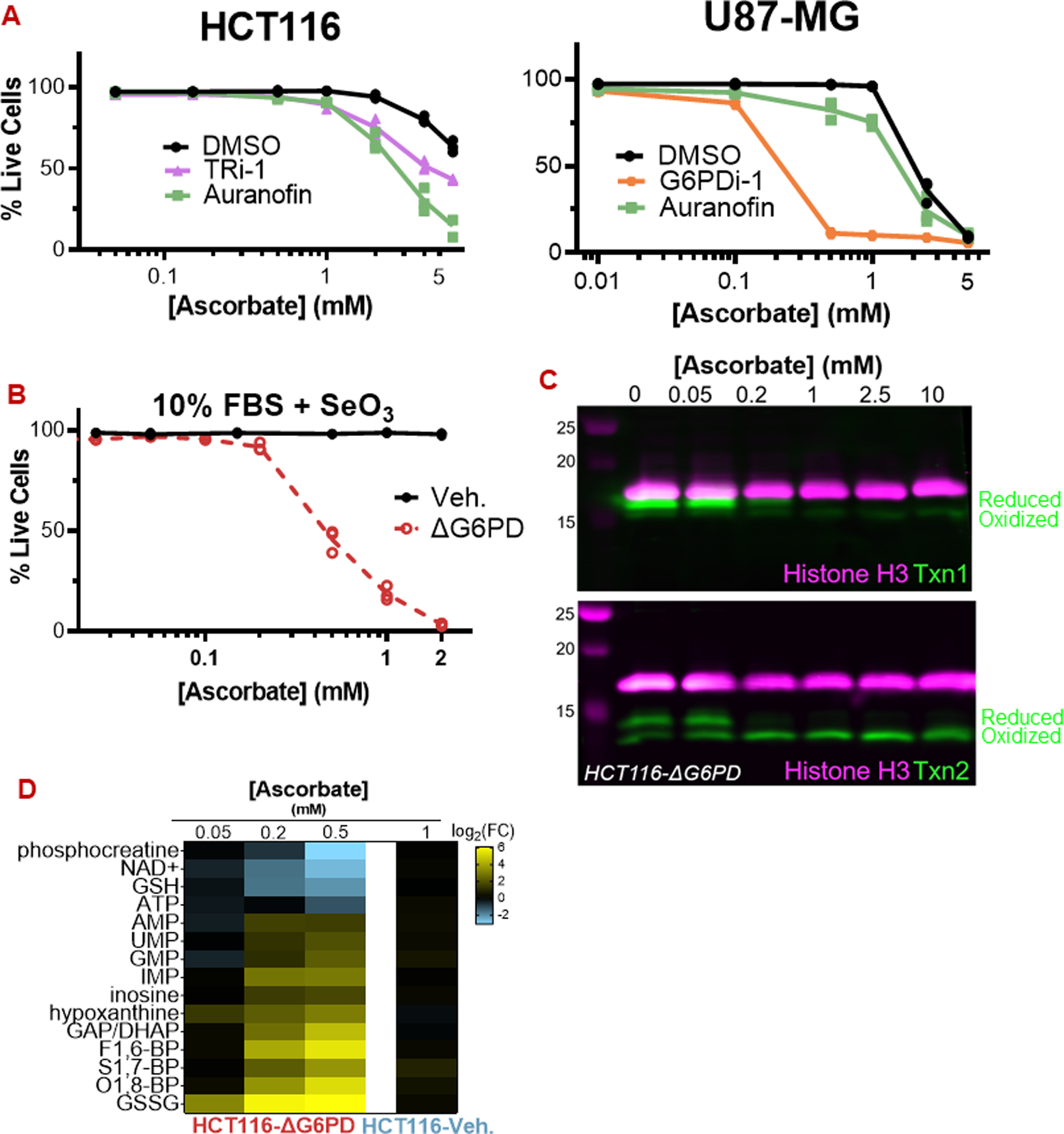
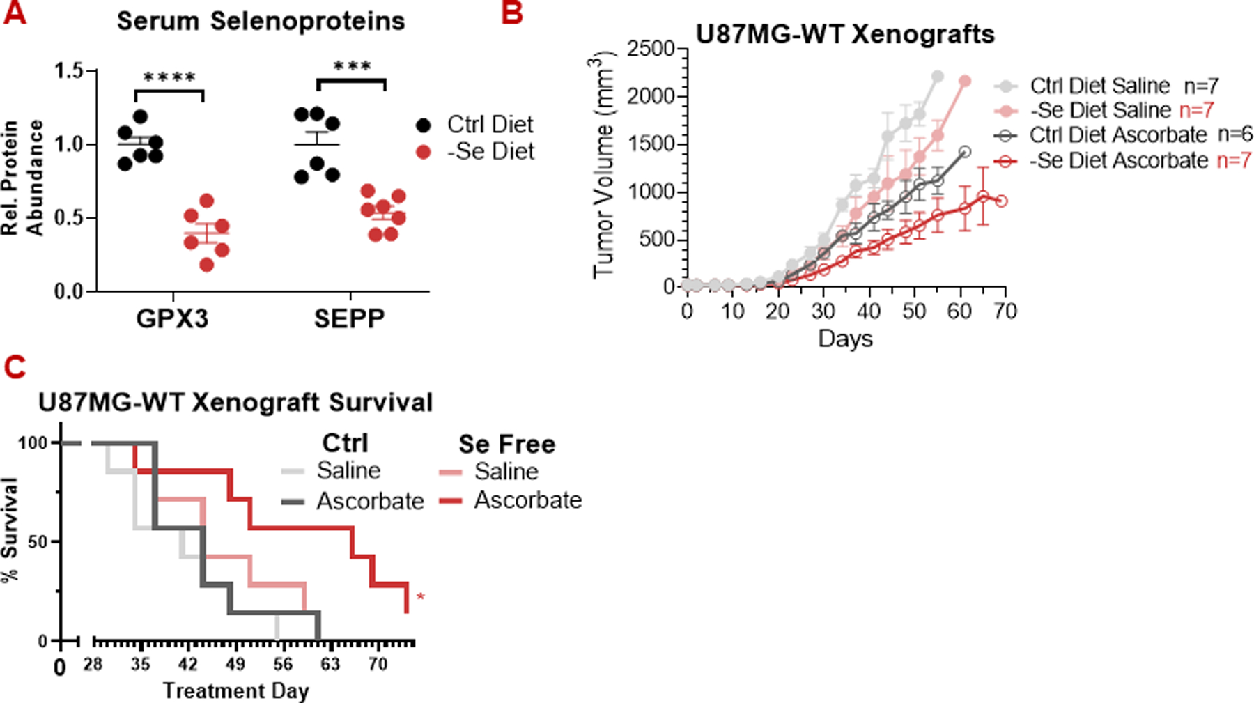
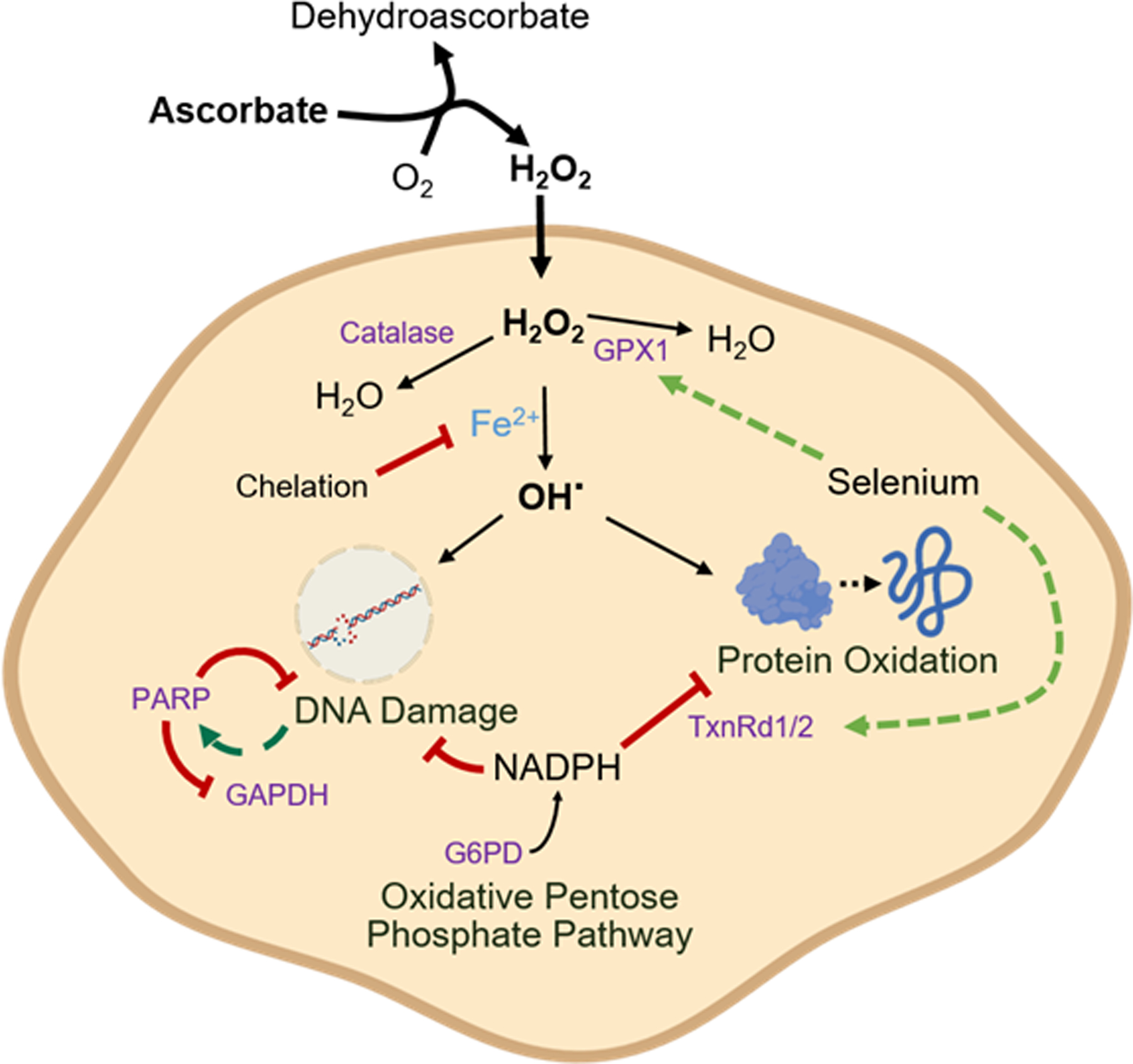
High-dose ascorbate (vitamin C) has shown promising anticancer activity. Two redox mechanisms have been proposed: hydrogen peroxide generation by ascorbate itself or glutathione depletion by dehydroascorbate (formed by ascorbate oxidation). Here we show that the metabolic effects and cytotoxicity of high-dose ascorbate in vitro result from hydrogen peroxide independently of dehydroascorbate. These effects were suppressed by selenium through antioxidant selenoenzymes including glutathione peroxidase 1 (GPX1) but not the classic ferroptosis-inhibiting selenoenzyme GPX4. Selenium-mediated protection from ascorbate was powered by NADPH from the pentose phosphate pathway. In vivo, dietary selenium deficiency resulted in significant enhancement of ascorbate activity against glioblastoma xenografts. These data establish selenoproteins as key mediators of cancer redox homeostasis. Cancer sensitivity to free radical-inducing therapies, including ascorbate, may depend on selenium, providing a dietary approach for improving their anticancer efficacy.

Significance: Selenium restriction augments ascorbate efficacy and extends lifespan in a mouse xenograft model of glioblastoma, suggesting that targeting selenium-mediated antioxidant defenses merits clinical evaluation in combination with ascorbate and other pro-oxidant therapies.


Conflict of interest statement

Competing Interests

J.D.R. is a paid adviser and/or stockholder in Colorado Research Partners, Kadmon Pharmaceuticals, L.E.A.F. Pharmaceuticals, Rafael Pharmaceuticals and its subsidiaries, Empress, and Agios Pharmaceuticals; a paid consultant of Pfizer; a founder, director, and stockholder of Farber Partners and Serien Therpeutics. C.S.R.J. declares no conflicts of interest.

Figures

Figure 1:
Figure 1:. The metabolic phenotype of pharmacologic ascorbate mirrors H2O2 but differs from dehydroascorbate (DHA) or diamide
(A) Proposed mechanisms of high-dose ascorbate toxicity. Top: DHA-dependent glutathione depletion. Bottom: Iron-dependent ROS generation. (B) Metabolic profiles of HCT116 cells challenged with ascorbate (10 mM), H2O2 (200 µM), DHA (10 mM), or diamide (1 mM) for 30 minutes, quantitated by LC-MS. (C) Bar graphs of selected metabolites from (B) highlighting the overlap in metabolic profile between ascorbate and H2O2 treatment. (D) Extracellular H2O2 scavenging by catalase (10 U/mL), but not PARP inhibition (10 µM olaparib), protects HCT116 cells from ascorbate (10 mM). DHA (10 mM) itself is markedly less toxic than ascorbate. ROS: Reactive oxygen species; DHA: Dehydroascorbate; PARP: poly (ADP-ribose) polymerase For viability assays and metabolomics, n = 3 biological replicates.
Figure 2:
Figure 2:. Glutathione oxidation by pharmacologic ascorbate is an abiotic phenomenon.
(A) Experimental workflow for preparing metabolite extracts for LC-MS. (B) Drying metabolite extracts from ascorbate-treated cells in the absence of N-ethylmaleimide (NEM) causes a dramatic reduction in reduced glutathione (GSH) abundance. In experiments where NEM was included in the extraction solvent, the GSH-NEM adduct was measured. Otherwise, free GSH was measured. (C) Drying metabolite extracts without NEM protection masks the true extent of oxidized glutathione (GSSG) accumulation in ascorbate-treated cells. For all GSH/GSSG measurements, n = 3 biological replicates. All data are mean ± SEM. **** p < 0.0001 by unpaired t-test.
Figure 3:
Figure 3:. Ascorbate toxicity is iron-dependent but does not occur via ferroptosis
(A) Depletion of the intracellular labile iron pool by chelation (deferoxamine (DFO), 500 µM, 12 h) prevents induction of energetic stress by pharmacologic ascorbate (10 mM, 30 min). In contrast, depletion of the extracellular iron pool by exposure in iron-free media has a minimal effect. (B) Deferoxamine prevents ascorbate cytotoxicity. (C) Schematic of ROS generation by ascorbate and iron. (D) Ascorbate (10 mM, 2h) decreases respiratory chain complex activity, in a manner rescuable by deferoxamine but not PARP inhibition (10 µM olaparib). (E) Pro-ferroptotic (10 µM) and anti-ferroptotic (1 µM) small molecules have a minimal effect on ascorbate cytotoxicity. For metabolomics and viability assays, n ≥ 3 biological replicates. For respiratory complex activity assays, n = 5 biological replicates. All data are mean ± SEM. * p < 0.05; ** p < 0.005; *** p < 0.0005; **** p < 0.0001 by unpared t-test or two-way ANOVA.
Figure 4:
Figure 4:. Selenium availability suppresses ascorbate cytotoxicity by enhancing selenoprotein activity.
(A) HCT116 cells grown in media supplemented with selenite are resistant to ascorbate (24 h). (B) HCT116 (colorectal), MDA-T120 (thyroid), and MDA-MB-231 (breast) cancer cells are more resistant to ascorbate when grown in DMEM supplemented with 30 nM sodium selenite. (C) HCT116 cells grown in selenite-supplemented media display a reduced mitochondrial peroxide load over time compared to control cells exposed to pharmacologic ascorbate (1 mM). (D) Cells grown in selenite-supplemented media display increased activity of the antioxidant selenozyme sytems TxnRd and GPX. The activity assays measure gross TxnRd and GPX activity, reflecting the combined activities of TxnRd1/2 and multiple GPX isoforms in each assay. (E) HCT116 cells lacking GPX1, but not GPX4, are sensitized to pharmacologic ascorbate under low and high selenium conditions. GPX: Glutathione peroxidase; TxnRd: Thioredoxin reductase; SeO3: selenite. For enzymatic assays, n = 3 biological replicates. For viability assays, n ≥ 3 biological replicates. All data are mean ± SEM. ^ p < 0.1; * p < 0.05; ** p < 0.005; *** p < 0.0005; **** p < 0.0001 by unpaired t-test or two-way ANOVA.
Figure 5:
Figure 5:. The pentose phosphate pathway defends against ascorbate via selenoproteins
(A) Inhibition of TxnRd1 by TRi-1 (10 µM) and auranofin (5 µM) sensitize HCT116 and U87-MG cells to ascorbate. G6PDi-1 (50 µM) also sensitized U87-MG cells. (B) HCT116-ΔG6PD cells are highly sensitive to ascorbate even when grown with 30 nM selenite. (C) Ascorbate (30 min) causes degradation of cytosolic Txn1 and oxidation of mitochondrial Txn2 in HCT116-ΔG6PD. (D) Ascorbate (30 min) induces energetic stress and GSH depletion at sub-millimolar concentrations in HCT116-ΔG6PD cells. For all data, n = 3 biological replicates. Representative immunoblots are shown. All data are mean ± SEM.
Figure 6:
Figure 6:. Dietary selenium restriction enhances ascorbate activity against U87-MG glioblastoma tumors.
(A) Maintaining mice on a selenium-free diet for 3-weeks prior to xenograft implantation decreased serum glutathione peroxidase 3 (GPX3) and selenoprotein P (SEPP) levels. (B) Pharmacologic ascorbate (4 g/kg daily) slowed the growth of U87-MG xenografts in both control and selenium-free diets. Selenium restriction alone did not significantly decrease tumor size, but it did lead to stronger response to ascorbate. (C) In mice harboring U87-MG xenografts, the combination of dietary selenium restriction and pharmacologic ascorbate (4 g/kg daily) enhanced survival. All data, n ≥ 6 mice. All data are mean ± SEM. * p < 0.05; ** p < 0.005; *** p < 0.0005; **** p < 0.0001 by unpaired t-test or Kaplan-Meier survival analysis.
Figure 7:
Figure 7:. Iron-dependent, selenium-repressible ascorbate toxicity.
Ascorbate generates H2O2 in the extracellular space, which crosses the plasma membrane. Inside the cell, H2O2 reacts with free iron (Fe2+), generating the reactive hydroxyl radical (OH·). H2O2 is detoxified by catalase and GPX1, and selenium availability drives GPX1 expression. The hydroxyl radical causes both protein oxidation, which is repaired by the thioredoxin reductase (TxnRd) system, and DNA damage, which activates PARP, consumes NAD, and thereby inhibits GAPDH. The cellular defense against these oxidative injuries ultimately relies on NADPH generated in the pentose phosphate pathway by the activity of glucose 6-phosphate dehydrogenase (G6PD).

Similar articles

Cited by

References

    1. Espey MG, Chen P, Chalmers B, Drisko J, Sun AY, Levine M, et al. Pharmacologic ascorbate synergizes with gemcitabine in preclinical models of pancreatic cancer. Free Radic Biol Med 2011; 50: 1610–19 - PMC - PubMed
    1. Yun J, Mullarky E, Lu C, Bosch KN, Kavalier A, Rivera K, et al. Vitamin C selectively kills KRAS and BRAF mutant colorectal cancer cells by targeting GAPDH. Nature 2015; 350: 1391–6 - PMC - PubMed
    1. Schoenfeld JD, Sibenaller ZA, Mapuskar KR, Wagner BA, Cramer-Morales KL, Furqan M, et al. O2 and H2O2-mediated disruption of Fe metabolism causes the differential susceptibility of NSCLC and GBM cancer cells to pharmacological ascorbate. Cancer Cell 2017; 31: 487–500 - PMC - PubMed
    1. Polireddy K, Dong R, Reed G, Yu J, Chen P, Williamson S, et al. High dose parenteral ascorbate inhibited pancreatic cancer growth and metastasis: mechanisms and a phase I/IIa study. Sci Rep 2017; 7:17188. - PMC - PubMed
    1. Ching SYL, Prins AW, and Beilby JP. Stability of ascorbic acid in serum and plasma prior to analysis. Ann Clin Biochem 2002; 39: 518–20 - PubMed

Publication types