

The role of foreign body response in peri-implantitis: What is the evidence?
- PMID: 35916872
- PMCID: PMC9804527
- DOI: 10.1111/prd.12456
The role of foreign body response in peri-implantitis: What is the evidence?
Abstract
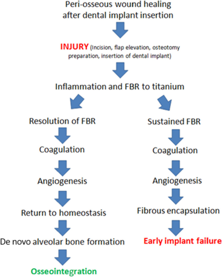
Historically, there has been broad consensus that osseointegration represents a homeostasis between a titanium dental implant and the surrounding bone, and that the crestal bone loss characteristic of peri-implantitis is a plaque-induced inflammatory process. However, this notion has been challenged over the past decade by proponents of a theory that considers osseointegration an inflammatory process characterized by a foreign body reaction and peri-implant bone loss as an exacerbation of this inflammatory response. A key difference in these two schools of thought is the perception of the relative importance of dental plaque in the pathogenesis of crestal bone loss around implants, with obvious implications for treatment. This review investigates the evidence for a persistent foreign body reaction at osseointegrated dental implants and its possible role in crestal bone loss characteristic of peri-implantitis. Further, the role of implant-related material release within the surrounding tissue, particularly titanium particles and corrosion by-products, in the establishment and progression in peri-implantitis is explored. While it is acknowledged that these issues require further investigation, the available evidence suggests that osseointegration is a state of homeostasis between the titanium implant and surrounding tissues, with little evidence that a persistent foreign body reaction is responsible for peri-implant bone loss after osseointegration is established. Further, there is a lack of evidence for a unidirectional causative role of corrosion by-products and titanium particles as possible non-plaque related factors in the etiology of peri-implantitis.
Keywords: foreign body reaction; macrophages; osseointegration; peri-implantitis; titanium particles.
© 2022 The Authors. Periodontology 2000 published by John Wiley & Sons Ltd.
Figures
References
-
- Berglundh T, Armitage G, Araujo MG, et al. Peri‐implant diseases and conditions: consensus report of Workgroup 4 of the 2017 World Workshop on the Classification of Periodontal and Peri‐Implant Diseases and Conditions. J Clin Periodontol. 2018;45(Suppl 20):S286‐S291. - PubMed
-
- Pontoriero R, Tonelli MP, Carnevale G, Mombelli A, Nyman SR, Lang NP. Experimentally induced peri‐implant mucositis. A clinical study in humans. Clin Oral Implants Res. 1994;5(4):254‐259. - PubMed
-
- Salvi GE, Aglietta M, Eick S, Sculean A, Lang NP, Ramseier CA. Reversibility of experimental peri‐implant mucositis compared with experimental gingivitis in humans. Clin Oral Implants Res. 2012;23(2):182‐190. - PubMed
-
- Zitzmann NU, Berglundh T, Marinello CP, Lindhe J. Experimental peri‐implant mucositis in man. J Clin Periodontol. 2001;28(6):517‐523. - PubMed
Publication types
MeSH terms
Substances
LinkOut - more resources
Full Text Sources
Medical
Miscellaneous
