Review
. 2022 Oct;24(10):1439-1453.
doi: 10.1007/s11886-022-01758-6. Epub 2022 Aug 2.

Multimodality Imaging of Constrictive Pericarditis: Pathophysiology and New Concepts

Affiliations
Review

Multimodality Imaging of Constrictive Pericarditis: Pathophysiology and New Concepts

Sumit Sohal et al. Curr Cardiol Rep. 2022 Oct.

Abstract

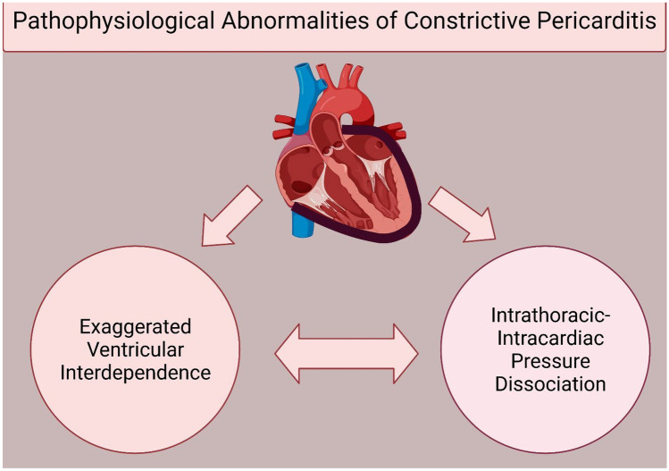
Purpose of review: The unique pathophysiological changes of constrictive pericarditis (CP) can now be identified with better imaging modalities, thereby helping in its early diagnosis. Through this review, we outline the pathophysiology of CP and its translation into symptomology and various imaging findings which then are used for both diagnosis and guiding treatment options for CP.

Recent findings: Multimodality imaging has provided us with the capability to recognize early stages of the disease and identify patients with a potential for reversibility and can be treated with medical management. Additionally, peri-procedural planning and prediction of post-operative complications has been made possible with the use of advanced imaging techniques. Advanced imaging has the potential to play a greater role in identification of patients with reversible disease process and provide peri-procedural risk stratification, thereby improving outcomes for patients with CP.

Keywords: Cardiac magnetic resonance imaging; Constrictive pericarditis; Multimodality imaging; Pathophysiology; Pre-operative imaging.


Conflict of interest statement

The authors report no disclosures pertaining to this article.

Figures

Fig. 1
Fig. 1
Schematic representation of pathophysiology of constrictive pericarditis. The phenomena of pericardial thickening (in black), exaggerated ventricular interdependence, and intracardiac-intrathoracic pressure dissociation play role in the clinical features and diagnosis of constrictive pericarditis. (Created with BioRender.com)
Fig. 2
Fig. 2
Schematic representation of intracardiac-intrathoracic pressure dissociation. In apnea, the pressure gradient between PV and LA/LV leads to ventricular filling. During inspiration in a normal heart, reduction in intrathoracic pressure is equally distributed to extra-pericardial structures (PV) and intrapericardial structures (LA/LV) but the gradient remains. In constrictive pericarditis, the reduction in ITP is not transferred to intrapericardial structures due to thick pericardium leading to reduction of gradient and thus decreased ventricular filling. (Abbreviations: LA, left atrium; LV, left ventricle; PV, pulmonary vein; ITP, intrathoracic pressure) (Created with BioRender.com)
Fig. 3
Fig. 3
Schematic representation of ventricular interdependence. During inspiration (green color), the blood return to the right side (white arrow) increases leading to higher tricuspid valve inflow velocities (green TV Doppler) but thickened pericardium prevents outward expansion of right ventricle leading to shifting of septum (yellow arrow) towards left side, thereby contributing to decreased blood flow on left side (blue arrow) and lower mitral inflow velocities (green MV Doppler). During expiration (purple color), the inflow to left ventricle increases (white arrow) depicted by increased mitral inflow velocity (purple MV Doppler), causing shifting of septum (yellow arrow) towards right side contributing to decreased blood flow on right side (blue arrow) and lower tricuspid inflow velocities (purple TV Doppler) (Created with BioRender.com)
Fig. 4
Fig. 4
Fluoroscopic (A) and chest X-ray (B) images of a patient with constrictive pericarditis showing calcification (arrows)
Fig. 5
Fig. 5
Respiration dependent and independent echocardiographic markers of constrictive pericarditis
Fig. 6
Fig. 6
Echocardiographic representation of the phenomenon of ventricular interdependence. Yellow arrow shows leftward shift of septum during inspiration (A) and blue arrow shows rightward shift of septum during expiration (B)
Fig. 7
Fig. 7
Phenomenon of septal bounce (red arrow) on M mode echocardiography
Fig. 8
Fig. 8
Tissue Doppler imaging on echocardiography demonstrating the phenomenon of annulus reversus. The Doppler velocity of medial annulus here is higher (13 cm/s) than lateral annulus (10 cm/s)
Fig. 9
Fig. 9
Calcification (arrows) of pericardium on computed tomographic imaging
Fig. 10
Fig. 10
Phenomenon of ventricular interdependence on free-breathing protocol of magnetic resonance imaging (MRI). Left panel shows short axis views whereas right panel shows long axis views during MRI red arrow shows leftward shift of septum during inspiration (A, B) and blue arrow shows rightward shift of septum during expiration (C, D)
Fig. 11
Fig. 11
Delayed enhancement of pericardium on cardiac magnetic resonance imaging in 2 patients. A Severe intensity of pericardial delayed enhancement before (left) and moderate intensity after (right) anti-inflammatory medication in a 55-year-old man with constrictive pericarditis who recovered clinically and echocardiographically after medical therapy. B Mild intensity of pericardial delayed enhancement before (left) and persistent mild intensity after (right) anti-inflammatory medication in a 61-year-old man who underwent pericardiectomy because of persistent symptoms and clinical and echocardiographic evidence of constrictive pericarditis after medical therapy. Subendocardial myocardial infarct was also noticed in the inferior and inferolateral walls. (From: DaLi Feng, et al. Circulation. 2011 Oct 25;124(17):1830–7, with permission of Wolters Kluwer Health, Inc.) [85]

References

    1. Welch TD, Oh JK. Constrictive pericarditis. Cardiol Clin. 2017;35:539–549. doi: 10.1016/j.ccl.2017.07.007. - DOI - PubMed
    1. Nishimura RA. Constrictive pericarditis in the modern era: a diagnostic dilemma. Heart. 2001;86:619–623. doi: 10.1136/heart.86.6.619. - DOI - PMC - PubMed
    1. Sengupta PP, Eleid MF, Khandheria BK. Constrictive pericarditis. Circ J. 2008;72:1555–1562. doi: 10.1253/circj.CJ-08-0654. - DOI - PubMed
    1. Alajaji W, Xu B, Sripariwuth A, Menon V, Kumar A, Schleicher M, Isma’eel H, Cremer PC, Bolen MA, Klein AL. Noninvasive multimodality imaging for the diagnosis of constrictive pericarditis. Circ Cardiovasc Imaging. 2018;11:e007878. doi: 10.1161/CIRCIMAGING.118.007878. - DOI - PubMed
    1. Brandt RR, Oh JK. Constrictive pericarditis: role of echocardiography and magnetic resonance imaging. E-Journal Cardiol Pract. 2017;15:22.

MeSH terms