. 2022 Sep;19(5):1546-1565.
doi: 10.1007/s13311-022-01275-y. Epub 2022 Aug 2.

Synthetic PPAR Agonist DTMB Alleviates Alzheimer's Disease Pathology by Inhibition of Chronic Microglial Inflammation in 5xFAD Mice

Affiliations

Synthetic PPAR Agonist DTMB Alleviates Alzheimer's Disease Pathology by Inhibition of Chronic Microglial Inflammation in 5xFAD Mice

Eunji Oh et al. Neurotherapeutics. 2022 Sep.

Abstract

Abnormal productions of amyloid beta (Aβ) plaque and chronic neuroinflammation are commonly observed in the brain of patients with Alzheimer's disease, and both of which induce neuronal cell death, loss of memory, and cognitive dysfunction. However, many of the drugs targeting the production of Aβ peptides have been unsuccessful in treating Alzheimer's disease. In this study, we identified synthetic novel peroxisome proliferator-activating receptor (PPAR) agonist, DTMB, which can ameliorate the chronic inflammation and Aβ pathological progression of Alzheimer's disease. We discovered that DTMB attenuated the proinflammatory cytokine production of microglia by reducing the protein level of NF-κB. DTMB also improved the learning and memory defects and reduced the amount of Aβ plaque in the brain of 5xFAD mice. This reduction in Aβ pathology was attributed to the changes in gliosis and chronic inflammation level. Additionally, bulk RNA-sequencing showed that genes related to inflammation and cognitive function were changed in the hippocampus and cortex of DTMB-treated mice. Our findings demonstrate that DTMB has the potential to be a novel therapeutic agent for Alzheimer's disease.

Keywords: Alzheimer's disease; Astrocyte; Microglia; Neuroinflammation; PPAR.


Figures

Fig. 1
Fig. 1
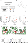
Identification of DTMB as a PPAR α/δ/γ agonist. a Schematic flowchart of the overall strategy for PPAR agonist screening. b Chemical structure of DTMB. c Gal4-transactivation assay. pcDNA5-GAL4-PPAR LBD, pG5-luc, and Rluc vector (as control) were transfected into HEK293A cells, which were then treated with various concentrations of DTMB for 24 h, followed by measurement of relative luciferase activity using dual-luciferase system. Cells were treated with each known PPAR ligands as positive control to compare with the response elicited by DTMB. Data are the mean ± standard error of mean (SEM, n = 6). d Molecular docking analysis. Docking positions and binding energy of DTMB to PPAR LBD (PPARα: 4BCR, 3VI8, 5HYK; PPARδ: 5U3Q, 5U46; PPARγ: 3U9Q, 5YCP, 5JI0) were predicted using PyRx and PyMol software. DTMB was predicted to have hydrogen bond to Ser280 of PPARα LBD, Arg248 and Ala306 of PPARδ, and Ser289 of PPARγ. e CNBr-bead pull down assay. Recombinant human PPAR LBD protein directly binds to the DTMB conjugated CNBr-bead
Fig. 2
Fig. 2
Pharmacokinetics and brain distribution of DTMB. a Correlations between brain DTMB concentrations and plasma DTMB concentrations after oral administration of DTMB in mice. Lines were generated from linear regression analysis and 90% confidence intervals around the geometric mean value. r2 represents the correlation coefficient and p represents the statistical significance for the regression analysis. b Plasma and brain concentration vs. time profiles of DTMB after single oral administration of DTMB (50 mg/kg) in mice. c Pharmacokinetic parameters of DTMB in mice after oral administrations of DTMB. Data represent mean ± standard deviation (n = 8 per each data point). *p < 0.05 by t test
Fig. 3
Fig. 3
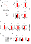
DTMB has an anti-inflammatory effect on microglia by reducing NF-κB protein level. a Griess assay data. RAW 264.7 macrophages were treated with LPS (1 µg/mL) to induce NO production. Cells were treated with the indicated drug for 24 h, and nitrite concentration was measured using a Griess reaction kit (n = 6). b, c Anti-inflammatory effect of DTMB on LPS-treated BV2 cells or primary microglia. LPS (0.5 µg/mL) was used to induce proinflammatory cytokines either with DMSO (0.1%) or DTMB for 24 h. d, e Anti-inflammatory effect of DTMB on pre-aggregated Aβ-treated BV2 cells or primary microglia. Human Aβ1-42 were pre-aggregated for 24 h in an incubation chamber. Aggregated Aβ (4 µg/mL) was then used to induce inflammation in cells treated with either DMSO (0.1%) or DTMB for 24 h, and the production of proinflammatory cytokines was detected. f, g Representative western blot data of NF-κB, NLRP3, and ASC from primary microglia treated with either DMSO (0.1%) or DTMB (25 µM) for 24 h. Quantification of blot intensity was determined using ImageJ. Data represent the mean ± standard error of the mean (SEM) of three independent experiments (*p < 0.05; **p < 0.01; ***p < 0.001, by one-way ANOVA)
Fig. 4
Fig. 4
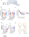
DTMB improves spatial memory and learning in 5xFAD mice. a Timeline of the in vivo experiment. Oral drug administration was started at the 8th week and continued for 3 months. Y-maze test and Morris water maze test were conducted to evaluate learning and memory of 5xFAD mouse. At the end of testing, all mouse brains were prepared for further biochemical and histological analysis. b Y-maze test. The percentage of Y-maze alternation decreased to almost 50% in transgenic mice. There were no changes of total path. ce Morris water maze test. c Training process for the Morris water maze. Each mouse went through three trials of training per day to find the platform in the target quadrant. The probe test was conducted on day 6. d, e Target quadrant occupancy (%) and mouse movement were measured automatically using a Smart v2.5 video tracking system. There was no change of total path. All data are the mean ± standard error of the mean (SEM) (n = 25 mice per group). **p < 0.01, ***p < 0.001 by two-way ANOVA
Fig. 5
Fig. 5
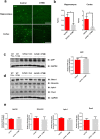
Aβ pathology in DTMB-treated 5xFAD mice. a, b Aβ plaques were detected using 6e10 antibody in both the hippocampal and the cortical tissue samples. Fluorescence (% area) was measured using ImageJ, scale bar = 200 µm. c Representative image of the Western blot is shown. Total APP level in the hippocampal tissue was measured through Western blotting to compare mice between the 5xFAD control group and DTMB-treated 5xFAD group. d, e Representative Western blot data to confirm the protein level of enzymes related to APP processing are shown (BACE1 for β-secretase, nicastrin1, aph-1, and pen2 for γ-secretase). Quantification was analyzed using ImageJ. All data were shown as the means ± standard error of the mean (SEM) **p < 0.01, ***p < 0.001 by t test
Fig. 6
Fig. 6
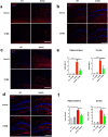
DTMB reduces chronic inflammation pathology in 5xFAD mouse brain. a, b Representative immunohistochemical images of Iba-1 staining in the cortex and hippocampus of 5xFAD mice treated with either vehicle or DTMB. Red fluorescence intensity represents the level of microglia gliosis. c, d Representative immunohistochemical images of GFAP staining in the cortex and hippocampus of 5xFAD mice treated with either vehicle or DTMB. Red fluorescent intensity shows the level of astrocyte gliosis. e, f Quantification graph of the immunohistochemical images showing the percentage area of fluorescence analyzed by Image. g Quantitative PCR data of proinflammatory cytokines and related enzyme. h, i Protein levels of inflammasome-related proteins (NLRP3, NF-κB, and ASC) in hippocampal brain lysate by western blot. All data are shown as mean ± standard error of the mean (SEM) **p < 0.01, ***p < 0.001 by one-way ANOVA and t test
Fig. 6
Fig. 6
DTMB reduces chronic inflammation pathology in 5xFAD mouse brain. a, b Representative immunohistochemical images of Iba-1 staining in the cortex and hippocampus of 5xFAD mice treated with either vehicle or DTMB. Red fluorescence intensity represents the level of microglia gliosis. c, d Representative immunohistochemical images of GFAP staining in the cortex and hippocampus of 5xFAD mice treated with either vehicle or DTMB. Red fluorescent intensity shows the level of astrocyte gliosis. e, f Quantification graph of the immunohistochemical images showing the percentage area of fluorescence analyzed by Image. g Quantitative PCR data of proinflammatory cytokines and related enzyme. h, i Protein levels of inflammasome-related proteins (NLRP3, NF-κB, and ASC) in hippocampal brain lysate by western blot. All data are shown as mean ± standard error of the mean (SEM) **p < 0.01, ***p < 0.001 by one-way ANOVA and t test
Fig. 7
Fig. 7
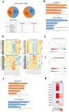
Bulk RNA-seq data of DTMB-treated 5xFAD mouse brain. a Overview of differentially expressed genes (DEGs). b Heat map of DEGs from hippocampus and cortex. c, d Gene ontology (GO) analysis. The top enriched GO terms of either upregulated or downregulated genes were analyzed from hippocampus and cortex of DTMB- or vehicle-treated 5xFAD mice. e, f GSEA enrichment plot (Wiki pathway). Inflammatory response-related gene sets were more enriched in the hippocampus and cortex of mice from the 5xFAD group treated with vehicle than that found in mice from the DTMB-treated 5xFAD group. Hippocampus analysis: NES score =  − 2.44, p < 0.001, and FDR < 0.001. Cortex brain analysis: NES score =  − 2.57, p < 0.001, and FDR < 0.001. g Heat map of gene expression related to microglial activation in adult microglia. Adult microglia were sorted from vehicle- or DTMB-treated 5xFAD mice using MACs system. Quantitative PCR was performed to analyze the gene expression related to chronic inflammation and microglial activation. Data represent the mean ± standard error of the mean (SEM) of three independent experiments. *p < 0.05; **p < 0.01; ***p < 0.001, by one-way ANOVA

Similar articles

Cited by

References

    1. McKhann GM, Knopman DS, Chertkow H, et al. The diagnosis of dementia due to Alzheimer’s disease: recommendations from the National Institute on Aging-Alzheimer’s Association workgroups on diagnostic guidelines for Alzheimer’s disease. Alzheimers Dement. 2011;7(3):263–269. - PMC - PubMed
    1. Long JM, Holtzman DM. Alzheimer disease: an update on pathobiology and treatment strategies. Cell. 2019;179(2):312–339. - PMC - PubMed
    1. Congdon EE, Sigurdsson EM. Tau-targeting therapies for Alzheimer disease. Nat Rev Neurol. 2018;14(7):399–415. - PMC - PubMed
    1. Umeda T, Ramser EM, Yamashita M, et al. Intracellular amyloid β oligomers impair organelle transport and induce dendritic spine loss in primary neurons. Acta Neuropathol Commun. 2015;3(1):1–15. - PMC - PubMed
    1. Franco R, Cedazo-Minguez A. Successful therapies for Alzheimer’s disease: why so many in animal models and none in humans? Front Pharmacol. 2014;5:146. - PMC - PubMed

Publication types

MeSH terms