. 2022 Aug 2;16(8):e0010666.
doi: 10.1371/journal.pntd.0010666. eCollection 2022 Aug.

A CRISPR-Cas12a-based diagnostic method for multiple genotypes of severe fever with thrombocytopenia syndrome virus

Affiliations

A CRISPR-Cas12a-based diagnostic method for multiple genotypes of severe fever with thrombocytopenia syndrome virus

Bum Ju Park et al. PLoS Negl Trop Dis. .

Abstract

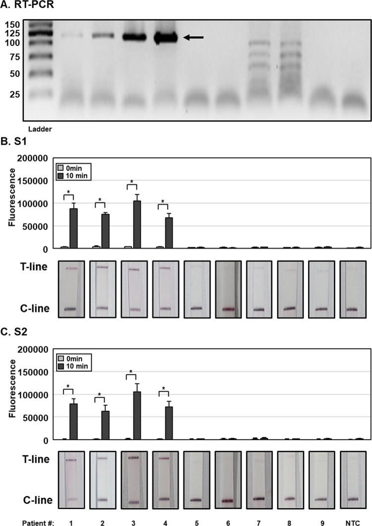
Severe fever with thrombocytopenia syndrome virus (SFTSV) infection is commonly reported in countries of Northeast Asia including China, Japan and South Korea. The majority of the SFTS patients are elderly and the average fatality rate is more than 10%. A rapid and sensitive diagnostic method to monitor and prevent SFTSV transmission remains an urgent clinical challenge. In this study, we developed a molecular diagnostic technique for detection of SFTSV using the CRISPR-Cas12a system combined with reverse transcription recombinase polymerase amplification (RT-RPA). Using this method, we successfully diagnosed SFTSV infections with the reaction time of 50 min from blood plasma without cross-reactivity to other viruses, supporting its application for rapid and sensitive diagnosis of SFTS.


Conflict of interest statement

No authors have competing interests

Figures

Fig 1
Fig 1. SFTSV DETECTR.
(A) Graphical workflow of SFTSV DETECTR. (B) Schematic presentation of positive and negative results obtained using a lateral flow strip. In a positive result, a band may appear at the C-line possibly due to the partial cleavage of the FB-reporter by Cas12a. FQ-reporter, fluorescence-quencher reporter; FB-reporter, FAM-Biotin lateral flow strip reporter; C-line, control line; T-line, test line.
Fig 2
Fig 2. Detection of lentiviruses pseudotyped with SFTSV S gene using DETECTR combined with fluorescence assay.
(A) A diagram of gRNAs and primer sets for SFTSV DETECTR. (B and C) Lentiviruses pseudotyped with and without SFTSV S gene (SPV and CPV, respectively) (100 CFU), IAV and IBV (100 PFU) and HCV and HEV (100 genome copies) were lysed via HUDSON and viral nucleic acids amplified via RT-RPA with a primer set specific for the SFTSV S gene. RT-RPA amplicons were detected with SFTSV DETECTR combined with fluorescence assay using gRNAs (A) S1 and (B) S2 corresponding to the S genes of all SFTSV genotypes. Fluorescence saturation occurred within 10 min. Values are presented as means ± s.d. (error bars) (n = 3 replicates; * p < 0.05 between samples, two-sample t-test). nt, nucleotides; PFU, plaque forming unit; CFU, colony forming unit; SPV, SFTSV pseudovirus; CPV, control pseudovirus.
Fig 3
Fig 3. Sensitivity of SFTSV DETECTR.
Different concentration of lentiviruses pseudotyped with SFTSV S gene (1.0 × 10−1 to 1.0 × 102 CFU per reaction) were lysed through HUDSON and viral nucleic acids amplified via RT-RPA with a primer set specific for the SFTSV S gene. RT-RPA amplicons were detected using SFTSV DETECTR combined with fluorescence assay or lateral flow using gRNAs (A) S1 and (B) S2 corresponding to the S genes of all SFTSV genotypes. Fluorescence saturation occurred within 10 min. Lateral flow assay results were evaluated 2 min after the strip was reacted with the sample. Values are presented as means ± s.d. (error bars) (n = 3 replicates; * p < 0.05 between samples, two-sample t-test). C-line, control line; T-line, test line; NTC, no template control. CFU, colony forming unit.
Fig 4
Fig 4. Application of SFTSV DETECTR to clinical samples.
(A) RNA was extracted from patient plasma samples and subjected to RT-PCR for SFTSV detection. PCR products (124 bp) were visualized using gel electrophoresis. (B, C) HUDSON-treated patient plasma samples were used to amplify viral nucleic acids using RT-RPA with a primer set specific for the SFTSV S gene. RT-RPA amplicons were detected using SFTSV DETECTR combined with fluorescence assay or lateral flow using gRNAs (A) S1 and (B) S2 corresponding to the S genes of all SFTSV genotypes. Fluorescence saturation occurred within 10 min. Lateral flow assay results were evaluated 2 min after the strip was reacted with the sample. Values are presented as means ± s.d. (error bars) (n = 3 replicates; * p < 0.05 between samples, two-sample t-test). C-line, control line; T-line, test line; NTC, no template control.

References

    1. Yu X-J, Liang M-F, Zhang S-Y, Liu Y, Li J-D, Sun Y-L, et al.. Fever with thrombocytopenia associated with a novel bunyavirus in China. New England Journal of Medicine. 2011;364(16):1523–32. doi: 10.1056/NEJMoa1010095 - DOI - PMC - PubMed
    1. Takahashi T, Maeda K, Suzuki T, Ishido A, Shigeoka T, Tominaga T, et al.. The first identification and retrospective study of severe fever with thrombocytopenia syndrome in Japan. The Journal of infectious diseases. 2014;209(6):816–27. doi: 10.1093/infdis/jit603 - DOI - PMC - PubMed
    1. Kim K-H, Yi J, Kim G, Choi SJ, Jun KI, Kim N-H, et al.. Severe fever with thrombocytopenia syndrome, South Korea, 2012. Emerging infectious diseases. 2013;19(11):1892. doi: 10.3201/eid1911.130792 - DOI - PMC - PubMed
    1. Raghavan R, Barker S, Cobos ME, Barker D, Teo E, Foley D, et al.. Potential spatial distribution of the newly introduced long-horned tick, Haemaphysalis longicornis in North America. Scientific reports. 2019;9(1):1–8. doi: 10.1038/s41598-018-37186-2 - DOI - PMC - PubMed
    1. Yun S-M, Park S-J, Park S-W, Choi W, Jeong HW, Choi Y-K, et al.. Molecular genomic characterization of tick-and human-derived severe fever with thrombocytopenia syndrome virus isolates from South Korea. PLoS neglected tropical diseases. 2017;11(9):e0005893. doi: 10.1371/journal.pntd.0005893 - DOI - PMC - PubMed

Publication types