. 2022 Aug;15(8):e011017.
doi: 10.1161/CIRCEP.122.011017. Epub 2022 Aug 2.

Advanced Techniques for Ethanol Ablation of Left Ventricular Summit Region Arrhythmias

Affiliations

Advanced Techniques for Ethanol Ablation of Left Ventricular Summit Region Arrhythmias

Apoor Patel et al. Circ Arrhythm Electrophysiol. 2022 Aug.

Abstract

Background: Coronary venous ethanol ablation (VEA) can be used as a strategy to treat ventricular arrhythmias arising from the left ventricular summit, but collateral flow and technical challenges cannulating intramural veins in complex venous anatomies can limit its use. Advanced techniques for VEA can capitalize on collateral vessels between target and nontarget sites to improve success.

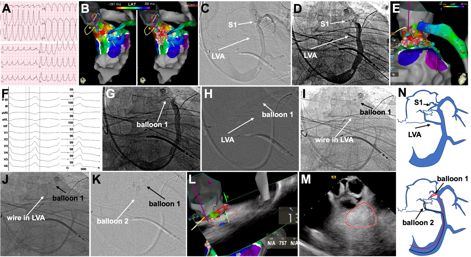
Methods: Of 55 patients with left ventricular summit ventricular arrhythmia, advanced techniques were used in 15 after initial left ventricular summit intramural vein mapping failed to show suitable targets for single vein, single-balloon VEA. All patients had previous radiofrequency ablation attempts. Techniques included: double-balloon for distal protection to block distal flow and target the proximal portion of a large intramural vein where best signal was proximal (n=6); balloons in 2 different left ventricular summit veins for a cross-fire multivein VEA (n=4); intramural collateral vein-to-vein cannulation to reach of targeted vein via collateral with antegrade ethanol and proximal balloon block (n=2); prolonged ethanol dwell time for vein sclerosis of large intramural vein and subsequent VEA (n=3); and intramural collateral VEA (n=1).

Results: Fifteen (8 females) patients (age 60.6±17.6 years) required advanced techniques. Procedure time was 210±49.9 minutes, fluoroscopy time was 25.3±14.1 minutes, and 113±17.9 cc of contrast was utilized. A median of 7 cc of ethanol was delivered (range, 4-15 cc). Intraprocedural radiofrequency ablation was delivered before ethanol in 9 out of 15 patients but failed. Ethanol achieved acute success in all 15 patients. Ethanol was used as the sole treatment in two patients. At a median follow-up of 194 days, one patient experienced recurrence.

Conclusions: Advanced techniques capitalizing on venous anatomy can enable successful VEA and selective targeting of arrhythmogenic sites, by blocking distal flow, utilization of collaterals between nontarget and target veins and multivein VEA. Understanding individual anatomy is critical for VEA success.

Keywords: catheter; coronary sinus; ethanol; sclerosis; veins.


Figures

Figure 1.
Figure 1.
Double balloon approach for distal protection: two examples. Case #1: A, activation map showing earliest activation in the AIV. B, ECG of PVC. C-D, AIV venograms in right (RAO) and left anterior oblique, caudal (LAOc). First septal branch (S1) is cannulated with octapolar branch with mid signal 44 ms pre-QRS (D inset). E-G, double balloon cannulation of S1. Wire signal in balloon 1 is 44 ms pre-QRS (E inset). VEA via balloon 1 terminated extrasystoles. Case #2. H-I, Activation maps with octapolar in S1 -signals shown in J (39 ms pre-QRS in electrodes 2–3). Wire signal in S1 was identical to octapolar (not shown). K-M, venograms in RAO, LAOc (octapolar in insets), showing large S1 beyond site of optimal signal. M, double balloon cannulation of S1 with wire via balloon 1 location in inset. N-O, schematic of double balloon approach for both cases. Abbreviations as in Figure 1.
Figure 2.
Figure 2.
Double-vein VEA. A, clinical VT. B, Activation maps and ineffective RF lesions in subaortic LV. C, Venogram showing S1 and LV annular (LVA) veins. D, octapolar in S1. E, location of octapolar relative to RF lesions. F, pace-mapping from wire in S1 showed a 99% match to the clinical VT. G, wire and balloon in S1. H, contrast injection in S1 with shunting towards LVA vein. I-J, second wire and balloon in LVA. K, simultaneous contrast through balloons in S1 and LVA vein stain targeted area. L–M, new echogenic area in subaortic LV. N, schematic of the approach.
Figure 3.
Figure 3.
Balloon blockade to assist septal cannulation and multi-vein VEA. A, activation map, with octapolar in AIV.B-C, venograms showing S1 and LVA vein in LAOc and RAO. D, best signal in AIV, close to S1. E, double balloon cannulation in GCV and AIV shows S1 and diagonal vein in-between balloons. F, AIV balloon facilitates wire and balloon cannulation of S1, where wire signal is 36 ms pre-QRS with optimal pace-map (G). H, wire location in 3D map. I-J, contrast through balloon in S1 opacifies LVA: ethanol lead to transient PVC elimination. K-N, wire and balloon cannulation of LVA show optimal signals (L inset), M-N, contrast and ethanol injection in LVA vein eliminated extrasystoles. O, schematic of the approach. Abbreviations as in Figure 1.
Figure 4.
Figure 4.
Double-vein and double-balloon approach. A, activation map of PVCs shown in B. C-D, two septal veins (S1, S2) shown in AIV venogram. D–E, octapolar in S1 (signal in inset) and its 3D location (E). F-G, octapolar in S2 (signal in inset) and its 3D location (G). H, wire (signal in inset) and balloon in S1, and octapolar in S2 (3D location in I). After VEA in S1, extrasystoles recurred and VEA was then delivered in S2 (J), leading to a morphology change (K). L, re-map of S1 after morphology change, best signal in electrodes 2–3. M, double-balloon for distal protection achieves success (N). O, echogenic area after VEA (3D location in P). O, schematic of the approach.
Figure 5.
Figure 5.
Intramural collateral VEA. A, AIV venogram showing 2 septal branches (S1, S2) communicated by collaterals. B-D, earliest site in the proximal AIV, in-between S1 and S2. E, telescopic approach to cannulate S1 and advance wire into intramural collateral, where signal is optimal (F). G contrast injection in intramural collateral leads to staining, and new echogenic area (H-I), and elimination of extrasystoles (M). K-L, schematic of the approach.
Figure 6.
Figure 6.
Collateral vein-to-vein cannulation and antegrade ethanol with proximal balloon block. A, AIV venogram (decapolar in AIV) with a large LVA and first septal (S1) vein. B, best site recorded in the very proximal portion of LVA vein (activation times identical to proximal AIV electrodes shown in E). C-D, 3D location of earliest sites, and location of ineffective RF applications. F, balloon and wire were advanced into S1, and via collaterals into the LVA vein, so that the balloon tip and wire face the AIV. G, optimal wire pace-map in ostial LVA vein. H-I, to prevent ethanol infusion into the AIV and GCV, a second large balloon is deployed and inflated in the AIV. Ethanol via LVA vein balloon 1 led to localized delivery and elimination of extrasystoles. J, schematic of the approach.
Figure 7.
Figure 7.
Prolonged ethanol dwelling time for distal vein sclerosis and localized VEA (3 cases). Case #1: A-B, venograms before and after RF ablation at AIV origin (shown in C), leading to AIV occlusion. D–E, wire and balloon in first septal vein (S1), show signal 27 ms pre-QRS and optimal pace-map. F, occlusion venogram of S1 shows extensive branching (arrowheads) beyond targeted tissue. G, after 15 min of ethanol dwell time, intramural branches are sclerosed with only localized staining. Repeat ethanol eliminated extrasystoles. Case #2: H, Initial AIV mapping of LVS PVCs (I). J, AIV venogram. RF was delivered in proximal AIV, without success. K, on a repeat procedure, there was total AIV occlusion with large first-to-second septal (S1,S2) collateral flow. L, signal in S1 was 28 ms pre-QRS. O, after 15 min of ethanol dwell time, only myocardial staining is seen, without collateral flow. Repeat ethanol eliminated extrasystoles. Case #3. P, AIV venogram with octapolar in large S1 (3D location in R) showing earliest signals in its mid-portion (Q). S-T, selective S1 venogram via long 2.5 × 50 mm balloon, showing a large portion of S1 beyond the targeted region. U-V, contrast injection in S1 after 15 min of ethanol dwell time, venous sclerosis and myocardial staining. Repeat ethanol eliminated extrasystoles.

Similar articles

Cited by

References

    1. Baher A, Shah DJ, Valderrabano M. Coronary venous ethanol infusion for the treatment of refractory ventricular tachycardia. Heart Rhythm. 2012;9(10):1637–1639. - PMC - PubMed
    1. Kreidieh B, Rodriguez-Manero M, P AS, Ibarra-Cortez SH, Dave AS, Valderrabano M. Retrograde Coronary Venous Ethanol Infusion for Ablation of Refractory Ventricular Tachycardia. Circ Arrhythm Electrophysiol. 2016;9(7):10.1161/CIRCEP.1116.004352 e004352. - DOI - PMC - PubMed
    1. Tavares L, Valderrábano M. Retrograde venous ethanol ablation for ventricular tachycardia. Heart Rhythm. 2019;16(3):478–483. - PubMed
    1. Da-Wariboko A, Lador A, Tavares L, et al. Double-balloon technique for retrograde venous ethanol ablation of ventricular arrhythmias in the absence of suitable intramural veins. Heart Rhythm. 2020;17(12):2126–2134. - PMC - PubMed
    1. Tavares L, Lador A, Fuentes S, et al. Intramural Venous Ethanol Infusion for Refractory Ventricular Arrhythmias. Outcomes of a Multicenter Experience. J Am Coll Cardiol EP. 2020:10.1016/j.jacep.2020.1007.1023. - DOI - PubMed

Publication types