. 2022 Aug 3;7(1):264.
doi: 10.1038/s41392-022-01057-0.

Targeting HECTD3-IKKα axis inhibits inflammation-related metastasis

Affiliations

Targeting HECTD3-IKKα axis inhibits inflammation-related metastasis

Fubing Li et al. Signal Transduct Target Ther. .

Abstract

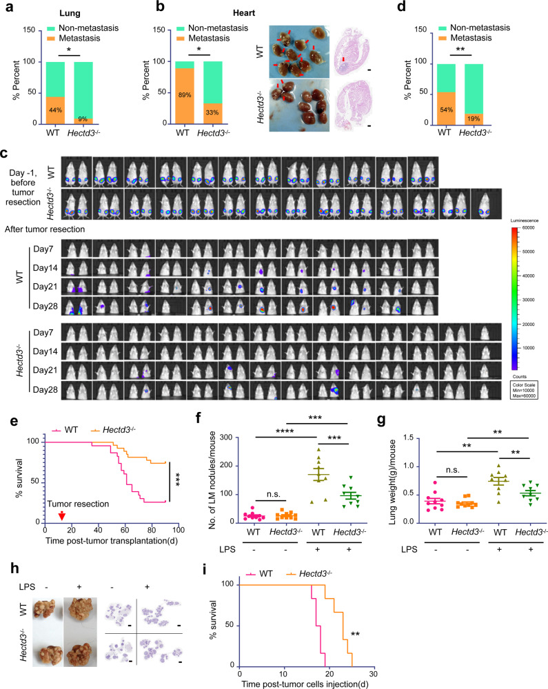
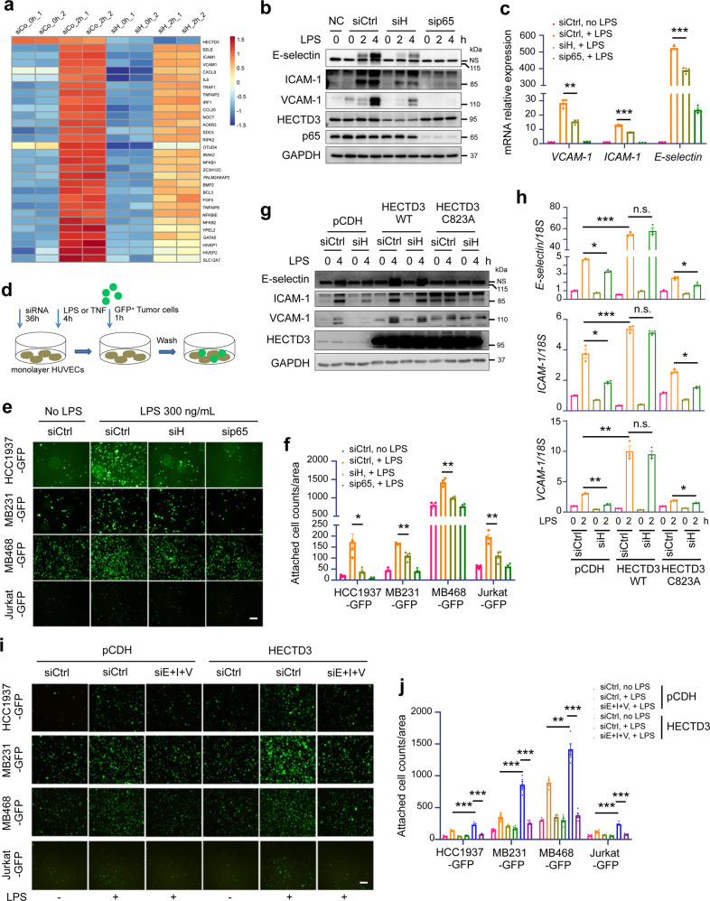
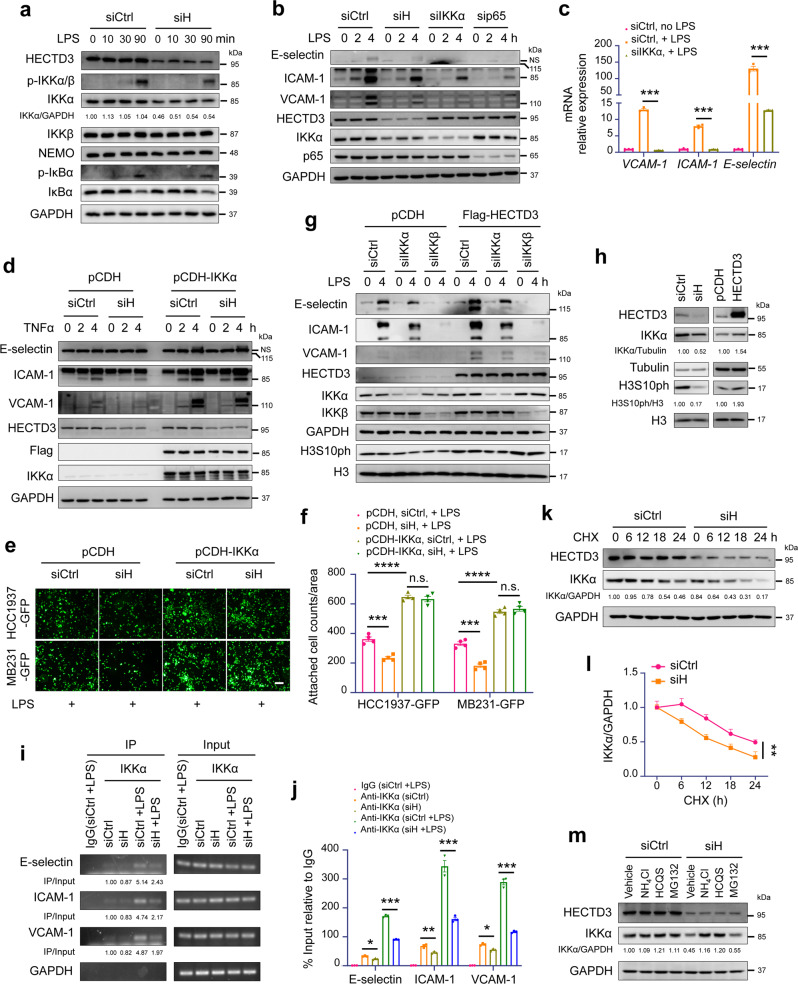
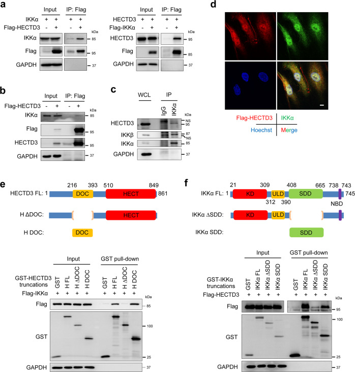
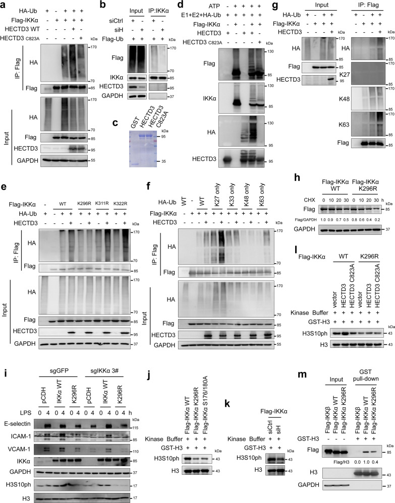
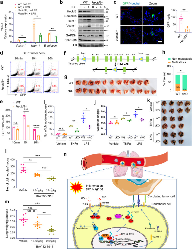
Metastasis is the leading cause of cancer-related death. The interactions between circulating tumor cells and endothelial adhesion molecules in distant organs is a key step during extravasation in hematogenous metastasis. Surgery is a common intervention for most primary solid tumors. However, surgical trauma-related systemic inflammation facilitates distant tumor metastasis by increasing the spread and adhesion of tumor cells to vascular endothelial cells (ECs). Currently, there are no effective interventions to prevent distant metastasis. Here, we show that HECTD3 deficiency in ECs significantly reduces tumor metastasis in multiple mouse models. HECTD3 depletion downregulates expression of adhesion molecules, such as VCAM-1, ICAM-1 and E-selectin, in mouse primary ECs and HUVECs stimulated by inflammatory factors and inhibits adhesion of tumor cells to ECs both in vitro and in vivo. We demonstrate that HECTD3 promotes stabilization, nuclear localization and kinase activity of IKKα by ubiquitinating IKKα with K27- and K63-linked polyubiquitin chains at K296, increasing phosphorylation of histone H3 to promote NF-κB target gene transcription. Knockout of HECTD3 in endothelium significantly inhibits tumor cells lung colonization, while conditional knockin promotes that. IKKα kinase inhibitors prevented LPS-induced pulmonary metastasis. These findings reveal the promotional role of the HECTD3-IKKα axis in tumor hematogenous metastasis and provide a potential strategy for tumor metastasis prevention.


Conflict of interest statement

The authors declare no competing interests.

Figures

Fig. 1
Fig. 1
Hectd3 knockout inhibits inflammation-induced tumor metastasis in mice.a A comparison of the incidence of lung metastases in WT (n = 9) versus Hectd3−/− (n = 11) mice with an FVB genetic background. PyMT-induced tumor cells were orthotopically injected into the fat pad of both groups of mice (5 × 105 cells per mouse). Primary tumors were removed 20 days later. Mice were sacrificed after 2 months, and the incidence of lung metastasis was recorded. b PyMT-induced breast tumor cells were inoculated as described above. The incidence of heart metastasis (left), representative heart metastasis nodule images and H&E staining (right) are shown. c 4T1-Luc2 cells were injected orthotopically into the fourth pair of fat pads of WT (n = 24) and Hectd3−/− (n = 27) BALB/c mice (bilateral, 1 × 105 cells per point). Eleven days after transplantation (Day -1), primary tumors were imaged using a bioluminescent IVIS system (upper). Twelve days after transplantation (Day 0), primary tumors were removed, then tumor metastasis was monitored weekly by imaging and representative bioluminescence images are shown (lower). d A comparison of the incidence of metastases in WT versus Hectd3−/− mice from panel c. e Kaplan–Meier survival curves of WT (n = 24) and Hectd3−/− (n = 27) mice which 4T1-Luc2 primary tumors were removed 12 days after transplantation. f WT and Hectd3−/− FVB mice were intravenously injected with or without LPS (1 mg/kg). 5 h later, PyMT-induced breast tumor cells were injected through the tail vein (2 × 105 cells per mouse). Each group contained 9–10 mice, and mice were sacrificed 20 days after injection of tumor cells. The graph shows the number of pulmonary metastasis nodules in each group of mice. g The weight of the whole lung with metastatic nodules in each group of mice from panel f. h Representative lung metastasis nodule images and corresponding H&E staining of the lungs in different groups of mice from panel f. i Kaplan–Meier survival curves of WT (n = 6) mice and Hectd3−/− (n = 6) mice pretreated with LPS and transplanted with PyMT-induced breast tumor cells through tail vein. Data represent three independent experiments for all of the above experiSments. Data are presented as the mean ± SEM, and statistics were calculated using the Chi-square test for a, b and d, two-way ANOVA for f, g, and log-rank test for e and i. *P < 0.05; **P < 0.01; ***P < 0.001; n.s., not significant. Scale bars are 500 μm for b and 2 mm for h
Fig. 2
Fig. 2
HECTD3 promotes adhesion of tumor cells to HUVECs by upregulating E-selectin, ICAM-1 and VCAM-1 expression in HUVECs. a RNA-sequencing analysis of gene expression in untreated and TNFα-treated control and HECTD3 knockdown HUVECs for 2 h. Heat map showing the expression of genes responsive to TNFα in untreated control HUVECs (siCo_0h), TNFα-treated control HUVECs (siCo_2h), untreated HECTD3 KD HUVECs (siH_0h) and TNFα-treated HECTD3 KD HUVECs (siH_2h). Each group has two experimental repeats. b Immunoblot analysis of adhesion molecules, like E-selectin, ICAM-1 and VCAM-1 in HUVECs knocking down HECTD3 or p65 using corresponding siRNA for 36 h, and stimulated with or without LPS (300 ng/mL) as indicated time. siControl (siCtrl) targeted nothing and siHECTD3 (siH) was a siRNA pool containing siHECTD3 1# and 2# here and in the following experiments. NC, negative control. NS, nonspecific band. c qRT-PCR analysis of adhesion molecules in HUVECs knocking down HECTD3 or p65 and stimulated with LPS (300 ng/mL) for 2 h. d Schematic representation of the in vitro adhesion assay. HECTD3 or p65 was knocked down in HUVECs, and cells were seeded into 6-well plates. HUVECs were treated with LPS (300 ng/ml) or TNFα for 4 h when the cells became fully confluent. Then, suspended GFP-labeled tumor cells were added and incubated for 1 h. Unattached cancer cells were washed away, and cancer cells adhered to HUVECs were quantified. e Representative images of the adhesion of GFP-labeled tumor cells to monolayer-cultured HUVECs transfected with the indicated siRNA and stimulated with or without LPS. f Bar graphs show the number of GFP-labeled tumor cells attached to monolayer-cultured HUVECs of panel e. g HUVECs stably overexpressing siRNA-resistant HECTD3, HECTD3 C823A mutant, and control (pCDH) were established. Immunoblots of these HUVECs knocked down endogenous HECTD3 or not and stimulated with LPS (300 ng/ml) for 4 h. h HUVECs stably overexpressing siRNA-resistant HECTD3, HECTD3 C823A mutant, and control (pCDH) were established. qRT-PCR analysis of these HUVECs knocked down endogenous HECTD3 or not and stimulated with LPS (300 ng/ml) for 2 h. i Representative images of the adhesion of GFP-labeled tumor cells to monolayer-cultured HUVECs in which HECTD3 was overexpressed or not. E-selectin, ICAM-1 and VCAM-1 were simultaneously knocked down by siE+I + V, a siRNA mixture of siE-selectin, siICAM-1 and siVCAM-1. j Quantitative data of panel i. Data represent 3 independent experiments for all of the above experiments. Data are presented as the mean ± SEM, and statistics were calculated using a two-tailed t-test for c, f, h, j. *P < 0.05; **P < 0.01; ***P < 0.001; n.s. not significant. Scale bars, 200 μm for e, i
Fig. 3
Fig. 3
HECTD3 increases the expression of adhesion molecules by stabilizing IKKα and recruiting nuclear IKKα to adhesion molecule gene promoters. a Immunoblot analysis of the expression of IKKα and activation degree of the NF-κB signal pathway. HUVECs knocking down HECTD3 were stimulated with LPS (300 ng/ml) for indicated time. b A comparison of the expression of the adhesion molecules in HUVEC knocking down HECTD3, IKKα and p65, and stimulated with or without LPS (300 ng/mL) as indicated time. c qRT-PCR analysis of adhesion molecules in HUVECs knocking down IKKα and stimulated with or without LPS (300 ng/mL) for 2 h. d IKKα overexpression largely rescued the HECTD3 KD caused reduction of E-selectin and ICAM-1, and partially rescue the reduction of VCAM-1 in HUVECs. HUVECs were treated with TNFα (10 ng/ml) for indicated time and different proteins were detected by WB. e IKKα overexpression largely rescued HECTD3 KD caused reduction of adhesion phenotype. Representative images of the adhesion of GFP-labeled tumor cells to monolayer-cultured HUVECs are shown. Scale bar, 200 μm. f Bar graphs show the number of GFP-labeled tumor cells attached to monolayer-cultured HUVECs from panel e. g IKKα and IKKβ was transiently knocked down in HECTD3-overexpressing HUVECs stimulated with LPS (300 ng/ml) and HECTD3 overexpression-induced increases of adhesion molecule expression were blocked when IKKα or IKKβ was depleted. h HECTD3 positively regulated IKKα and H3S10ph levels in HUVECs. Left: HECTD3 was knocked. Right: HECTD3 was stably overexpressed in HUVECs. i Chromatin immunoprecipitation (ChIP) assays were performed using an anti-IKKα antibody in HUVECs transfected with siControl or siHECTD3 and stimulated with LPS (300 ng/mL) for 1 h. j qPCR results of the samples in panel i. k HECTD3 knockdown by siRNA decreased IKKα protein stability in HUVECs. The cells were incubated with 50 μg/ml CHX for the indicated times and were collected for immunoblotting. Tubulin was used as the internal control. The band intensity of IKKα at each time point was quantified using ImageJ. The experiments were repeated three times, and a representative experimental result is presented. l Quantitative data of panel k. m HECTD3 knockdown induced IKKα protein degradation through lysosomes but not proteasomes. HUVECs were treated with lysosome inhibitors (NH4Cl, 10 mM and HCQS, 50 μM, overnight) or proteasome inhibitor (MG132, 20 μM for 6 h) after knocking down HECTD3. Data represent three independent experiments for all of the above experiments. Data are presented as the mean ± SEM, and statistics were calculated using two-tailed t-test for c, f, i, two-way ANOVA for l. *P < 0.05; **P < 0.01; ***P < 0.001
Fig. 4
Fig. 4
HECTD3 interacts with IKKα. a Exogenous HECTD3 interacts with IKKα in HEK293T cells. Flag-HECTD3 and IKKα (left) or Flag-IKKα and HECTD3 (right) plasmids were cotransfected into HEK293T cells. After Flag-tagged HECTD3 and IKKα proteins were immunoprecipitated with Flag-M2 beads, IKKα and HECTD3 were detected by immunoblotting. b FLAG-immunoprecipitation of FLAG-tagged HECTD3 from HUVECs. c Anti-IKKα antibody was used to immunoprecipitate IKKα from HUVECs. d Confocal microscopy images of Flag-HECTD3 and IKKα in HUVECs are shown. Scale bars, 10 μm. e Schematic diagram shows human HECTD3 and its truncation mutants (top). Flag-IKKα and GST-fused HECTD3 truncation mutants were coexpressed in HEK293T cells. By GST pull-down assay (bottom), GST-H DOC specifically pulled down Flag-IKKα. f Schematic diagram shows human IKKα and its truncation mutants (top). Flag-HECTD3 and GST-fused IKKα truncation mutants were coexpressed in HEK293T cells. By the GST pull-down assay (bottom), GST-IKKα SDD specifically pulled down Flag-HECTD3. Data represent three independent experiments for all of the above experiments
Fig. 5
Fig. 5
HECTD3 ubiquitinates IKKα with K27- and K63-linked polyubiquitin chains at K296 and increases IKKα protein stability and kinase activity. a Flag-IKKα, HA-Ub, and HECTD3 (WT) or HECTD3 C823A were coexpressed in HEK293T cells. Ubiquitinated Flag-IKKα proteins were immunoprecipitated with Flag-M2 beads and probed with anti-HA antibody. b Co-IP analysis of the ubiquitination of endogenous IKKα in HUVECs overexpressing stable Flag-ubiquitin (Flag-Ub). The cells were transfected with siRNA to knock down HECTD3. Anti-IKKα antibody was used for immunoprecipitation. The anti-Flag antibody was used to detect ubiquitinated IKKα. c Purified recombinant HECTD3 and HECTD3 C823A proteins from E. coli were detected by Coomassie blue staining. d HECTD3 ubiquitinates IKKα in vitro in an E3 ligase activity-dependent manner. ATP, HA-Ub, E1, UbcH5b, HECTD3 or HECTD3 C823A, and Flag-IKKα were mixed for ubiquitination assays. The Flag-IKKα protein was purified from HEK293 cells transfected with the plasmid encoding Flag-IKKα using Flag-M2 beads. e HECTD3 ubiquitinates IKKα at K296. HECTD3 failed to ubiquitinate Flag-IKKα K296R, similar to WT, K311R and K322R in HEK293T cells. f HECTD3 ubiquitinates IKKα with K27- and K63-linked polyubiquitin chains. WT, K27 only, and K63 only HA-Ub supported HECTD3-mediated Flag-IKKα ubiquitination. In contrast, K33 only and K48 only HA-Ub failed to do so. g Linkage-specific antibodies were used to validate the linkage of Flag-IKKα. h CHX chase assays were used to analyze the half-lives of Flag-IKKα WT and K296R mutant in HEK293T cells. i IKKα ubiquitination at K296 is essential for LPS to induce adhesion molecule expression in HUVECs. IKKα was stably knocked out in HUVECs using the CRISPR/Cas9 system. Immunoblotting of adhesion molecule expression in these cells restored the expression of IKKα by lentivirus encoding Flag-IKKα WT or Flag-IKKα K296R and stimulated with or without LPS (300 ng/mL) for indicated time (0–4 h). j The in vitro IKKα kinase assay contains purified Flag-IKKα WT, K296R, S175/180 A, GST-H3, and ATP. Flag-IKKα proteins were purified from HEK293T cells. k HECTD3 knockdown decreased IKKα activity toward histone H3. HECTD3 knockdown and Flag-IKKα overexpression were performed in HEK293T cells. Flag-IKKα proteins were purified for in vitro kinase assays toward GST-H3. l Overexpression of HECTD3, not HECTD3 C823A, increased IKKα activity toward histone H3. Flag-IKKα and Flag-IKKα K296R proteins were purified from HEK293T cells cotransfected with plasmids encoding Flag-IKKα or Flag-IKKα K296R with HECTD3 or HECTD3 C823A. In vitro kinase assays of Flag-IKKα WT and Flag-IKKα K296R toward GST-H3 were performed. m Flag-IKKα K296R decreased the interaction with H3 compared to Flag-IKKα WT. Cell lysates of HEK293T cells expressing Flag-IKKα WT or Flag-IKKα K296R were collected and incubated with purified GST-H3 protein for 30 min on ice. The GST pull-down assay was performed using glutathione sepharose beads. Data represent three independent experiments for all of the above experiments
Fig. 6
Fig. 6
Hectd3 promotes lung colonization of tumor cells under inflammatory conditions. a qRT-PCR analysis of adhesion molecules in WT and Hectd3 KO mECs stimulated with LPS (500 ng/ml) for 2 h. b A comparison of the expression of the adhesion molecules, IKKα and H3S10ph in mECs stimulated with or without LPS (500 ng/mL) as indicated time. c Representative frozen immunofluorescence images of GFP+ tumor cells in lungs of mice performed the tumor cell colonization assay in vivo and bar graph showing the number of GFP+ tumor cells in lungs of WT and Hectd3−/− mice (right). Counting rules: Eighty sections were randomly cut from each sample (the entire lung) and then scanned by a FluoView FV1000 confocal microscope after fluorescence staining. The total number of GFP-positive tumor cells in 80 sections was recorded. d The GFP+ tumor cells (5 × 106 cells per mouse) were injected through the tail vein into WT or Hectd3−/− mice pretreated with LPS (1 mg/kg) stimulation for 5 h. At 10 min, 10 h or 20 h after tumor cell injection, the mice were sacrificed and perfused with PBS and the whole lungs were digested to cell suspension to analyze GFP+ tumor cell colonization in the lung by FCM. e The rate of GFP+ tumor cell in the lung of panel d. f Schematic representation of the targeted allele and the conditional allele of Hectd3 KO. g B16-F10 cells were injected subcutaneously into Tie2-Cre+;Hectd3wt (n = 11) and Tie2-Cre+;Hectd3fl/fl (n = 12) C57BL/6 mice. Twelve days after transplantation, primary tumors were removed. The mice were sacrificed at day 45 and the incidence of lung metastasis was record. Representative lung metastasis nodule image. h The incidence of lung metastasis of panel g. i Tie2-Cre+;Hectd3wt and Tie2-Cre+;Hectd3fl/fl mice were intravenously injected with vehicle, TNFα (200 μg/kg) or LPS (1 mg/kg). 5 h later, B16-F10 cells were injected through the tail vein (2 × 105 cells per mouse). The mice were sacrificed 20 days after injection of tumor cells. The graph shows the number of pulmonary metastasis nodules in each group of mice. j The weight of the whole lung with metastatic nodules in each group of mice from panel i. k Representative lung metastasis nodule images of the lungs in different groups of mice from panel i. l 4T1-Luc2 cells were injected by tail vein into BALB/c mice (1 × 105 per mouse) that were pretreated with vehicle, BAY 32-5915 (12.5 mg/kg) or BAY 32-5915 (25mk/kg) for 24 h and LPS (1 mg/kg) for 5 h by intravenous injection. The mice were sacrificed 20 days after the injection of tumor cells. The number of mouse pulmonary metastasis nodules in the three groups is shown. m Analysis of the weight of whole lung with metastasis nodules in the three groups from l. n The hypothetical working model. Inflammatory factors, including LPS and TNFα, activate the NF-κB pathway and promote p65 nuclear translocation and transcription of adhesion molecules, including E-selectin, ICAM-1 and VCAM-1, in HUVECs. HECTD3 ubiquitinates IKKα with K27/K63-linked polyubiquitin chains at K269 to increase IKKα protein stability, kinase activity, and recruitment to NF-κB-responsive gene promoters, where IKKα phosphorylates histone H3 at Ser10 to increase the transcription of adhesion molecules. These adhesion molecules on the EC plasma membrane promote the adhesion of tumor cells to the endothelium, leading to extravasation, colonization and metastasis. IKKα and HECTD3-specific inhibitors may prevent cancer metastasis. The Figure was partly generated using Servier Medical Art, provided by Servier, licensed under a Creative Commons Attribution 3.0 unported license (http://creativecommons.org/licenses/by/3.0/). Data are presented as the mean ± SEM, and statistics were calculated using two-tailed t-test for a, c, e, Chi-square test for h, two-way ANOVA for i, j, l, m. *P < 0.05; **P < 0.01; ***P < 0.001; n.s., not significant. Scale bars, 100 μm for c

Similar articles

Cited by

References

    1. Mehlen P, Puisieux A. Metastasis: a question of life or death. Nat. Rev. Cancer. 2006;6:449–458. doi: 10.1038/nrc1886. - DOI - PubMed
    1. Braun S, et al. A pooled analysis of bone marrow micrometastasis in breast cancer. N. Engl. J. Med. 2005;353:793–802. doi: 10.1056/NEJMoa050434. - DOI - PubMed
    1. Koch M, et al. Detection of hematogenous tumor cell dissemination predicts tumor relapse in patients undergoing surgical resection of colorectal liver metastases. Ann. Surg. 2005;241:199–205. doi: 10.1097/01.sla.0000151795.15068.27. - DOI - PMC - PubMed
    1. Funaki S, et al. Novel approach for detection of isolated tumor cells in pulmonary vein using negative selection method: morphological classification and clinical implications. Eur. J. Cardiothorac. Surg. 2011;40:322–327. - PubMed
    1. McDonald B, et al. Systemic inflammation increases cancer cell adhesion to hepatic sinusoids by neutrophil mediated mechanisms. Int. J. Cancer. 2009;125:1298–1305. doi: 10.1002/ijc.24409. - DOI - PubMed

Publication types