. 2022 Aug 2;13(1):4493.
doi: 10.1038/s41467-022-32022-8.

Distinctive roles of translesion polymerases DinB1 and DnaE2 in diversification of the mycobacterial genome through substitution and frameshift mutagenesis

Affiliations

Distinctive roles of translesion polymerases DinB1 and DnaE2 in diversification of the mycobacterial genome through substitution and frameshift mutagenesis

Pierre Dupuy et al. Nat Commun. .

Abstract

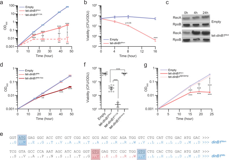
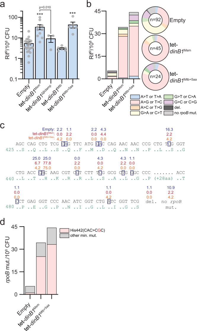
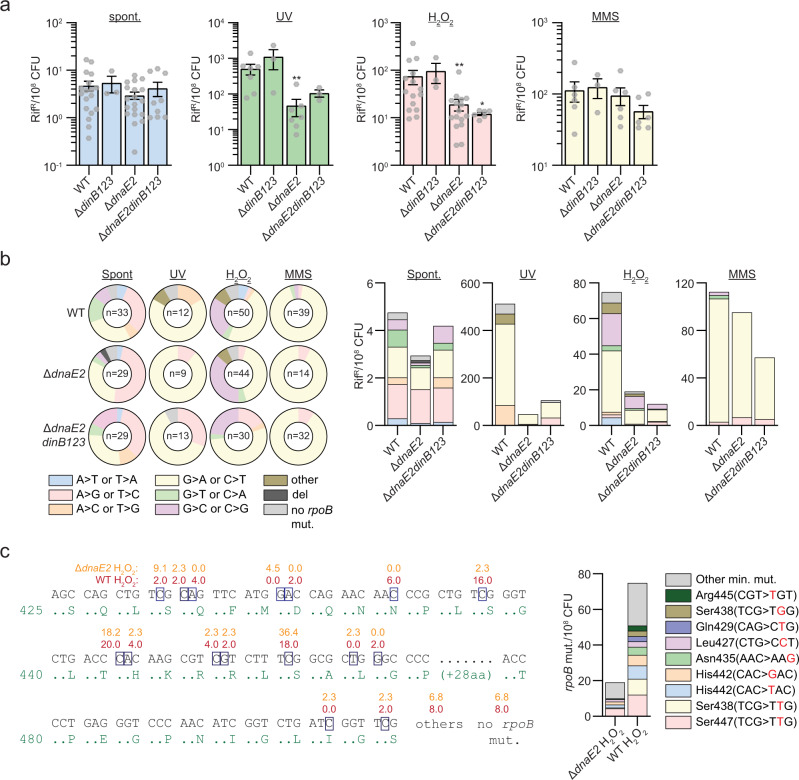
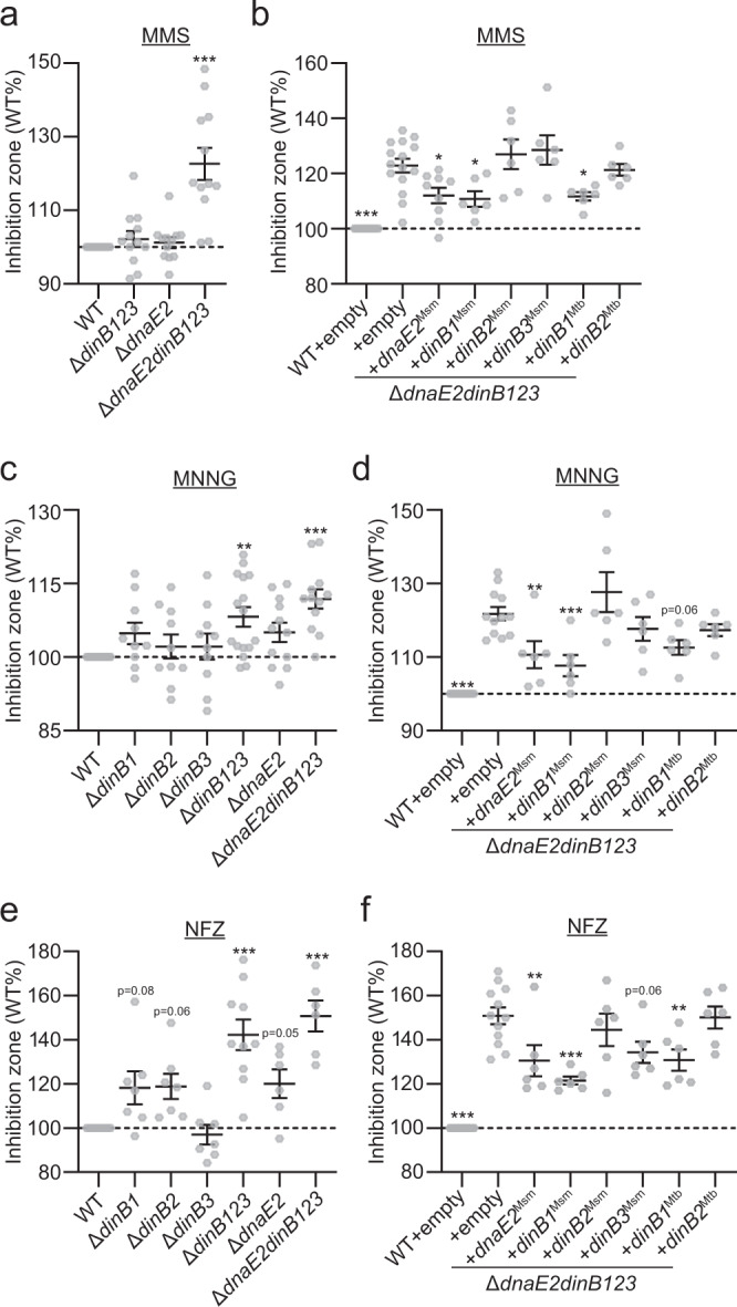
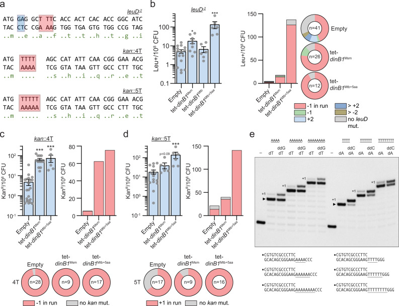
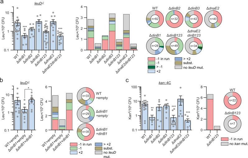
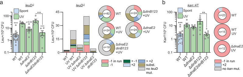
Antibiotic resistance of Mycobacterium tuberculosis is exclusively a consequence of chromosomal mutations. Translesion synthesis (TLS) is a widely conserved mechanism of DNA damage tolerance and mutagenesis, executed by translesion polymerases such as DinBs. In mycobacteria, DnaE2 is the only known agent of TLS and the role of DinB polymerases is unknown. Here we demonstrate that, when overexpressed, DinB1 promotes missense mutations conferring resistance to rifampicin, with a mutational signature distinct from that of DnaE2, and abets insertion and deletion frameshift mutagenesis in homo-oligonucleotide runs. DinB1 is the primary mediator of spontaneous -1 frameshift mutations in homo-oligonucleotide runs whereas DnaE2 and DinBs are redundant in DNA damage-induced -1 frameshift mutagenesis. These results highlight DinB1 and DnaE2 as drivers of mycobacterial genome diversification with relevance to antimicrobial resistance and host adaptation.


Conflict of interest statement

M.S.G. has received consulting fees from Vedanta Biosciences, PRL NYC, and Fimbrion Therapeutics and has equity in Vedanta biosciences. All other authors declare no competing interests.

Figures

Fig. 1
Fig. 1. DinB1Mtb activity requires five N-terminal amino acids omitted from the annotated ORF.
a, d, g Growth and b, f viability of strains carrying an inducible (tet = Anhydrotetracycline inducible promoter) DinB1 or its indicated derivatives (Msm = M. smegmatis, Mtb = annotated M. tuberculosis DinB1, Mtb+5aa = N terminal extended DinB1, dinB1 D113A = catalytically inactive M. smegmatis DinB1, Δβ clamp (ΔQESLF: 356–360 amino acids of M. smegmatis DinB1)) in presence of inducer. For a, the plotted OD value is the result of continuous dilution to maintain log phase growth (see methods). The viability in f was measured 24 h after inducer addition. c Anti-RecA/RpoB immunoblot from indicated strains with indicated times of inducer treatment. e Alignment of Msm and Mtb DinB1 N-termini with the potential start codons underlined. The blue boxed valine corresponds to the start codon of the published DinB1 noted as DinB1Mtb above, whereas the red boxed valine shows an alternative start codon of an extended DinB1 denoted as DinB1Mtb+5aa. Results shown are means (±SEM) of biological triplicates (a, b, d, g) or from biological replicates symbolized by gray dots (f). Stars above or under the means mark a statistical difference with the reference strain (empty vector) and lines connecting two strains show a statistical difference between them (*, P < 0.05; **, P < 0.01; ***, P < 0.001). p values were obtained on log-transformed data by one-way (f) or two-way (a, b, d, and g) ANOVA with a Bonferroni post-test.
Fig. 2
Fig. 2. DinB1 is an error-prone polymerase inducing antibiotic resistance through a characteristic mutagenic signature.
a Rifampicin resistance (rifR) frequency in indicated strains in presence of inducer. Results shown are means (±SEM) of data obtained from biological replicates symbolized by gray dots. Stars above bars mark a statistical difference with the reference strain (empty vector) and lines connecting two strains show a statistical difference between them (***, P < 0.001). p-values were obtained on log-transformed data by one-way ANOVA with a Bonferroni post-test. b Relative (pie chart) and absolute (bar chart) frequencies of nucleotide changes detected in rpoB of rifR clones from the indicated strains. The number of sequenced rifR is given in the center of each pie chart. c Location and relative frequency in % of mutated nucleotides in rpoB found in empty (blue), tet-dinB1Msm (red) or tet-dinB1Mtb+5aa (orange) rifR. d Absolute frequency of the main rpoB mutations found in indicated strains.
Fig. 3
Fig. 3. DnaE2 but not DinBs mediates stress-induced substitution mutagenesis.
a Rifampicin resistance (rifR) frequency in indicated strains and conditions. Results shown are means (±SEM) of data obtained from biological replicates symbolized by gray dots. Stars above bars mark a statistical difference with the reference strain (WT of each condition) (*, P < 0.05; **, P < 0.01). p-values were obtained on log-transformed data by one-way ANOVA with a Bonferroni post-test. b Relative (pie chart) and absolute (bar chart) frequencies of nucleotide changes detected in rpoB of rifR clones from the indicated strains. The number of sequenced rifR is given in the center of each pie chart. c Location and relative frequency of mutated nucleotides of rpoB found in rifR of ΔdnaE2 + H2O2 (orange) or WT + H2O2 (red). The bar chart shows the absolute frequency of the main rpoB mutations found in indicated strains.
Fig. 4
Fig. 4. Redundancy of DinB1 and DnaE2 in tolerance to alkylation damage and N-dG adducts.
Sensitivities of indicated strains to a and b MMS, c and d MNNG, or e and f NFZ were measured by disc diffusion assay. Translesion polymerases were expressed under their native promoter from a genome integrated vector in panels b, d, and f. Results shown are means (±SEM) of data obtained from biological replicates symbolized by gray dots. Stars above the means mark a statistical difference with the reference strain (WT or ΔdnaE2dinB123 + empty in complementation experiments) (*, P < 0.05; **, P < 0.01; ***, P < 0.001). p values were obtained on log-transformed data by one-way ANOVA with a Bonferroni post-test.
Fig. 5
Fig. 5. DinB1 promotes −1 and +1 frameshift mutations in homo-oligonucleotide runs.
a leuD and kan frameshift (FS) reporter assays. leuD and kan open reading frame N-termini in which 2 base pairs in the second leuD codon were removed (blue box) or 4T/5T runs (red box) were incorporated upstream the start codon of kan. Reversion can occur by FS mutations that restore the leuD or kan reading frames resulting in phenotypic leucine prototrophy (leu+) or kanamycin resistance (kanR). The red box in leuD shows the run of 3T in which the majority of detected FS in leu+ were found. b leu+ and c, d kanR frequencies in the indicated strains in presence of inducer. Results shown are means (±SEM) of data obtained from biological replicates symbolized by gray dots. Stars above the means mark a statistical difference with the reference strain (empty) (*, P < 0.05; ***, P < 0.001). p-values were obtained on log-transformed data by one-way ANOVA with a Bonferroni post-test. Relative (pie chart) and absolute (bar chart) frequencies of nucleotide changes detected in leuD of leu+ cells or in kan of kanR cells represented with colors: red = −1 FS in the b 3 T run, c 4 T run, or d 5 T run, other colors = FS outside of the run, and gray = no detected mutation. The number of sequenced leu+ or kanR colonies is given in the center of each pie chart. e DinB1 polymerase reaction mixtures containing 5’ 32P-labeled primer-template DNAs with A4, A6, A8, T4, T6, or T8 runs in the template strand (depicted below and included as indicated above the lanes) and 125 µM deoxynucleotides and dideoxynucleotides (as specified above the lanes) were incubated at 37 °C for 15 min. DinB1 was omitted from reactions in lanes –. The reaction products were analyzed by urea-PAGE and visualized by autoradiography. The +1 slippage products are indicated.
Fig. 6
Fig. 6. DinB1 is the primary mediator of spontaneous −1 frameshift mutations in homo-oligonucleotide runs.
a, b leu+ or c kanR frequency in the indicated strains and relative (pie chart) or absolute (bar chart) frequencies of nucleotide changes detected in leuD or kan coded by color. The number of sequenced leu+ or kanR colonies is given in the center of each pie chart. Results shown are means (± SEM) of data obtained from biological replicates symbolized by gray dots. Stars above bars mark a statistical difference with the reference strain (WT) and lines connecting two strains show a statistical difference between them (*, P < 0.05; **, P < 0.01; ***, P < 0.001). p-values were obtained on log-transformed data by one-way ANOVA with a Bonferroni post-test.
Fig. 7
Fig. 7. DnaE2 is the primary mediator of DNA damage-induced −1 frameshift mutations in homo-oligonucleotide runs.
a leu+ or b kanR frequency in indicated strains/conditions and relative (pie chart) or absolute (bar chart) frequencies of nucleotide changes detected in leuD or kan coded by color. The number of sequenced leu+ or kanR colonies is given in the center of each pie chart. Results shown are means (±SEM) of data obtained from biological replicates symbolized by gray dots. Stars above bars mark a statistical difference with the reference strain (WT + UV) and lines connecting two strains show a statistical difference between them (*, P < 0.05; **, P < 0.01; ***, P < 0.001). p values were obtained on log-transformed data by one-way ANOVA with a Bonferroni post-test.

Similar articles

Cited by

References

    1. Vaisman A, Woodgate R. Translesion DNA polymerases in eukaryotes: what makes them tick? Crit. Rev. Biochem. Mol. Biol. 2017;52:274–303. doi: 10.1080/10409238.2017.1291576. - DOI - PMC - PubMed
    1. Fuchs RP, Fujii S. Translesion DNA synthesis and mutagenesis in prokaryotes. Cold Spring Harb. Perspect. Biol. 2013;5:a012682. doi: 10.1101/cshperspect.a012682. - DOI - PMC - PubMed
    1. Fujii S, Fuchs RP. A comprehensive view of translesion synthesis in Escherichia coli. Microbiol. Mol. Biol. Rev. 2020;84:e00002–e00020. doi: 10.1128/MMBR.00002-20. - DOI - PMC - PubMed
    1. Reuven NB, Arad G, Maor-Shoshani A, Livneh Z. The mutagenesis protein UmuC Is a DNA polymerase activated by UmuD′, RecA, and SSB and is specialized for translesion replication *. J. Biol. Chem. 1999;274:31763–31766. doi: 10.1074/jbc.274.45.31763. - DOI - PubMed
    1. Tang M, et al. UmuD′2C is an error-prone DNA polymerase, Escherichia coli pol V. Proc. Natl Acad. Sci. 1999;96:8919–8924. doi: 10.1073/pnas.96.16.8919. - DOI - PMC - PubMed

Publication types