. 2023 Feb;44(2):454-464.
doi: 10.1038/s41401-022-00957-9. Epub 2022 Aug 2.

PPAR-γ alleviates the inflammatory response in TNF-α-induced fibroblast-like synoviocytes by binding to p53 in rheumatoid arthritis

Affiliations

PPAR-γ alleviates the inflammatory response in TNF-α-induced fibroblast-like synoviocytes by binding to p53 in rheumatoid arthritis

Xiao-Feng Li et al. Acta Pharmacol Sin. 2023 Feb.

Abstract

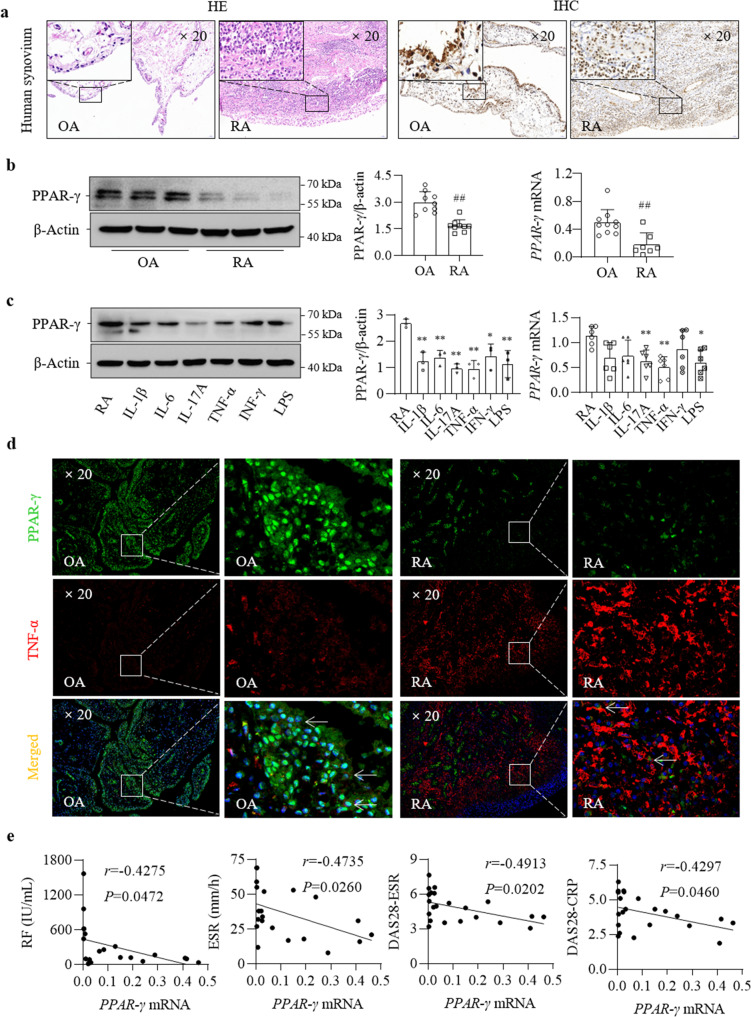
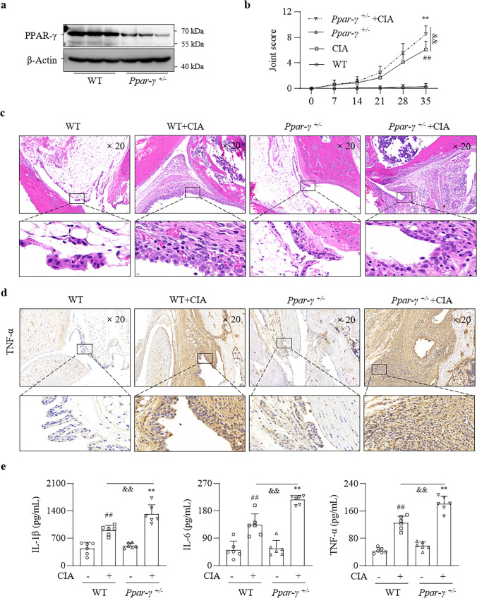
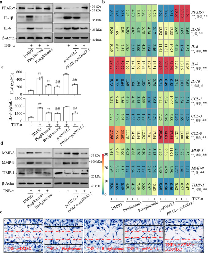
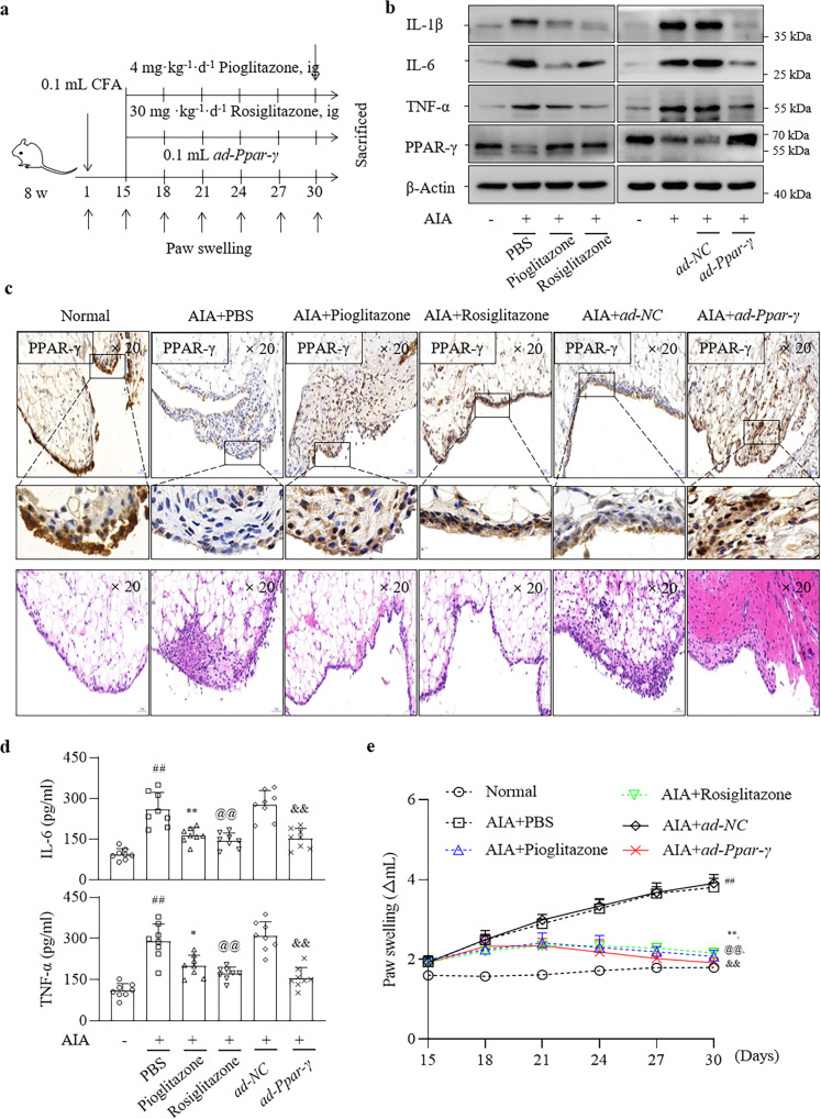
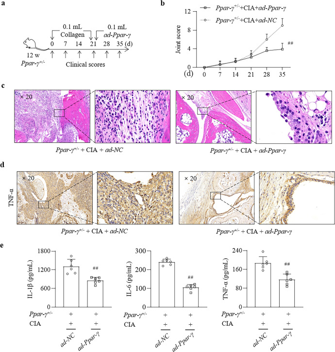
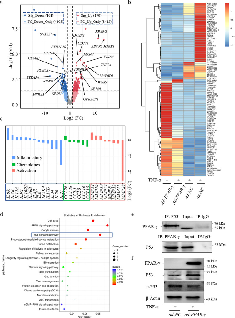
Rheumatoid arthritis (RA) is characterized by synovial inflammation, synoviocyte expansion and damage to cartilage and bone. We recently reported that peroxisome proliferator-activated receptor (PPAR)-γ inhibited the proliferation and activation of fibroblast-like synoviocytes (FLS), and was downregulated in RA synovial. In this study we investigated the role of PPAR-γ in RA and the underlying mechanisms. Adjuvant-induced arthritis (AIA) was induced in rats; from D15, AIA rats were orally administered pioglitazone (30 mg·kg-1·d-1) or rosiglitazone (4 mg·kg-1·d-1) for 14 days. Collagen-induced arthritis (CIA) was induced in wild-type and Ppar-γ+/- mice. We showed that the expression of PPAR-γ was significantly reduced, whereas that of TNF-α was markedly increased in human RA FLS. In CIA mice, knockdown of PPAR-γ expression (Ppar-γ+/-) aggravated the ankle inflammation. Similarly, T0070907 (a PPAR-γ antagonist) or si-PPAR-γ promoted the activation and inflammation of TNF-α-induced FLS in vitro. On the contrary, administration of PPAR-γ agonist pioglitazone or rosiglitazone, or injection of ad-Ppar-γ into the ankle of AIA rat in vivo induced overexpression of PPAR-γ, reduced the paw swelling and inflammation, and downregulated activation and inflammation of FLS in RA. Interesting, injection of ad-Ppar-γ into the ankle also reversed the ankle inflammation in Ppar-γ+/- CIA mice. We conducted RNA-sequencing and KEGG pathway analysis, and revealed that PPAR-γ overexpression was closely related to p53 signaling pathway in TNF-α-induced FLS. Co-IP study confirmed that p53 protein was bound to PPAR-γ in RA FLS. Taken together, PPAR-γ alleviates the inflammatory response of TNF-α-induced FLS by binding p53 in RA.

Keywords: PPAR-γ; adjuvant-induced arthritis; collagen-induced arthritis; fibroblast-like synoviocytes; inflammatory response; rheumatoid arthritis.


Conflict of interest statement

The authors declare no competing interests.

Figures

Fig. 1
Fig. 1. PPAR-γ and TNF-α expression showed opposite trends in RA FLS.
a Representative images showing PPAR-γ expression in the synovium by H&E staining and IHC (20×). PPAR-γ levels were examined by WB and RT–qPCR (b) after treatment with IL-1β (2 ng/mL), IL-6 (5 ng/mL), IL-17A (10 ng/mL), TNF-α (10 ng/mL), IFN-γ (10 ng/mL), and lipopolysaccharide (LPS; 1 μg/mL) (c). d PPAR-γ and TNF-α expression in the synovium was analyzed by IF analysis (20×). e Correlation analysis of PPAR-γ mRNA and RF, ESR, DAS28-ESR and DAS28-CRP levels in RA PBMCs. The values represent the mean ± SD of 3 different samples. ##P < 0.01 vs. OA group; *P < 0.05, **P < 0.01 vs. RA group.
Fig. 2
Fig. 2. Ppar-γ+/− CIA mice exhibited exacerbated joint inflammation.
a PPAR-γ expression in the mouse synovium. b Mouse joint scores. c Representative images showing H&E staining of the synovium (20×). d Representative images showing TNF-α expression by IHC (20×). e IL-1β, IL-6 and TNF-α protein levels in mouse blood were examined by ELISA. The values represent the mean ± SD of 10 different samples. ##P < 0.01 vs. WT group; **P < 0.01 vs. Ppar-γ+/− group; &&P < 0.01 vs. CIA group.
Fig. 3
Fig. 3. PPAR-γ overexpression inhibits inflammation and activation of TNF-α-induced FLS in RA.
a PPAR-γ, IL-1β and IL-6 protein expression in FLS was analyzed by WB. b PPAR-γ, IL-1β, IL-6, IL-8, IL-10, CCL-2, CCL-3, CCL-8, MMP-3, MMP-9 and TIMP-1 mRNA expression in FLS was analyzed by RT–qPCR. c IL-6 and IL-8 protein levels in RA FLS supernatant were examined by ELISA. d MMP-3, MMP-9 and TIMP-1 protein expression was analyzed by WB. e The migration of FLS was examined by wound-healing assays (20×). The values represent the mean ± SD of 3 different samples. ##P < 0.01 vs. RA group; *P < 0.05, **P < 0.01, @P < 0.05, @@P < 0.01 vs. TNF-α group; &&P < 0.01 vs. pcDNA3.1 group.
Fig. 4
Fig. 4. PPAR-γ overexpression suppresses inflammation in the joints of AIA rats.
a The schematic shows the delivery method of pioglitazone, rosiglitazone and ad-Ppar-γ in AIA rats. b IL-1β, IL-6, TNF-α and PPAR-γ protein expression in the synovium of rat knee joints was analyzed by WB. c Representative images of PPAR-γ in the synovium were examined by IHC and H&E staining (20×). d IL-6 and TNF-α protein levels in rat blood examined by ELISA. e Paw swelling. The values represent the mean ± SD of 10 different samples. ##P < 0.01 vs. normal group; *P < 0.05, **P < 0.01, @P < 0.05, @@P < 0.01 vs. AIA group; &&P < 0.01 vs. ad-NC group.
Fig. 5
Fig. 5. PPAR-γ overexpression suppresses inflammation in the joints of Ppar-γ+/− CIA mice.
a The schematic shows the delivery of ad-Ppar-γ in Ppar-γ+/− CIA mice. b Joint scores in CIA mice. c Representative images showing H&E staining in the synovium (20×). d Representative images showing TNF-α expression by IHC (20×). e IL-1β, IL-6 and TNF-α protein levels in mouse blood were examined by ELISA. The values represent the mean ± SD of 10 different samples. ##P < 0.01 vs. Ppar-γ+/− CIA group.
Fig. 6
Fig. 6. PPAR-γ binds to p53 in TNF-α-induced FLS.
a Volcano plot showing gene expression alterations in TNF-α-induced FLS treated with ad-PPAR-γ. b A heatmap showing representative significantly altered genes (P < 0.05) in TNF-α-induced FLS treated with ad-PPAR-γ. c A histogram showing inflammatory cytokine, chemokine and MMP genes in FLS. d KEGG pathway analysis showed that the p53 signaling pathway participates in PPAR-γ-mediated regulation of TNF-α-induced FLS. e The p53 protein bound to PPAR-γ, as shown by Co-IP. f PPAR-γ, p53 and p-p53 expression in TNF-α-induced FLS was analyzed by WB.

Similar articles

Cited by

References

    1. Smolen JS, Aletaha D, Barton A, Burmester GR, Emery P, Firestein GS, et al. Rheumatoid arthritis. Nat Rev Dis Prim. 2018;4:18001. doi: 10.1038/nrdp.2018.1. - DOI - PubMed
    1. de Oliveira PG, Farinon M, Sanchez-Lopez E, Miyamoto S, Guma M. Fibroblast-like synoviocytes glucose metabolism as a therapeutic target in rheumatoid arthritis. Front Immunol. 2019;10:1743. doi: 10.3389/fimmu.2019.01743. - DOI - PMC - PubMed
    1. Muntyanu A, Abji F, Liang K, Pollock RA, Chandran V, Gladman DD. Differential gene and protein expression of chemokines and cytokines in synovial fluid of patients with arthritis. Arthritis Res Ther. 2016;18:296. doi: 10.1186/s13075-016-1196-6. - DOI - PMC - PubMed
    1. Croft AP, Campos J, Jansen K, Turner JD, Marshall J, Attar M, et al. Distinct fibroblast subsets drive inflammation and damage in arthritis. Nature. 2019;570:246–51. doi: 10.1038/s41586-019-1263-7. - DOI - PMC - PubMed
    1. Loh C, Park SH, Lee A, Yuan R, Ivashkiv LB, Kalliolias GD. TNF-induced inflammatory genes escape repression in fibroblast-like synoviocytes: transcriptomic and epigenomic analysis. Ann Rheum Dis. 2019;78:1205–14. doi: 10.1136/annrheumdis-2018-214783. - DOI - PMC - PubMed

MeSH terms