. 2022 Aug;54(8):1098-1108.
doi: 10.1038/s12276-022-00813-0. Epub 2022 Aug 2.

Epigenetic regulator Cfp1 safeguards male meiotic progression by regulating meiotic gene expression

Affiliations

Epigenetic regulator Cfp1 safeguards male meiotic progression by regulating meiotic gene expression

Byeong Seong Ki et al. Exp Mol Med. 2022 Aug.

Abstract

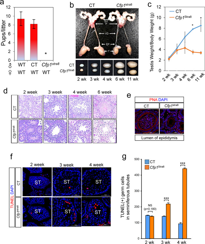
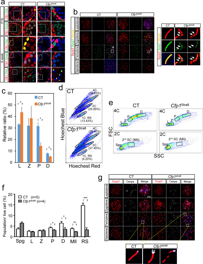
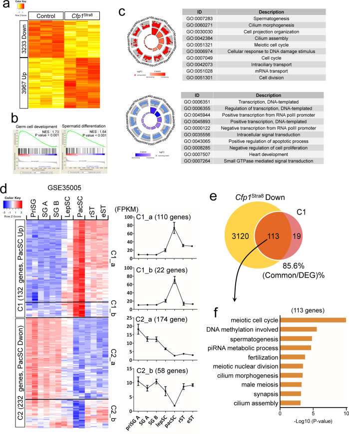
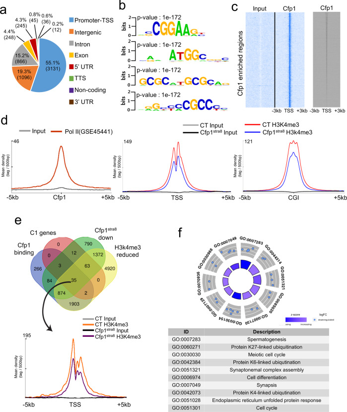
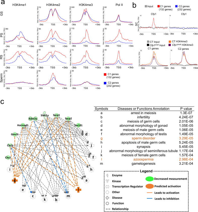
Meiosis occurs specifically in germ cells to produce sperm and oocytes that are competent for sexual reproduction. Multiple factors are required for successful meiotic entry, progression, and termination. Among them, trimethylation of histone H3 on lysine 4 (H3K4me3), a mark of active transcription, has been implicated in spermatogenesis by forming double-strand breaks (DSBs). However, the role of H3K4me in transcriptional regulation during meiosis remains poorly understood. Here, we reveal that mouse CXXC finger protein 1 (Cfp1), a component of the H3K4 methyltransferase Setd1a/b, is dynamically expressed in differentiating male germ cells and safeguards meiosis by controlling gene expression. Genetic ablation of mouse CFP1 in male germ cells caused complete infertility with failure in prophase I of the 1st meiosis. Mechanistically, CFP1 binds to genes essential for spermatogenesis, and its loss leads to a reduction in H3K4me3 levels and gene expression. Importantly, CFP1 is highly enriched within the promoter/TSS of target genes to elevate H3K4me3 levels and gene expression at the pachytene stage of meiotic prophase I. The most enriched genes were associated with meiosis and homologous recombination during the differentiation of spermatocytes to round spermatids. Therefore, our study establishes a mechanistic link between CFP1-mediated transcriptional control and meiotic progression and might provide an unprecedented genetic basis for understanding human sterility.


Conflict of interest statement

The authors declare no competing interests.

Figures

Fig. 1
Fig. 1. Male Cfp1Stra8 mice are infertile.
a Fertility test in control (CT) and Cfp1Stra8 male mice by mating with wild-type (WT) females for 6 months. The numbers on the Y-axis represent the average pup size/litter. *p value < 0.001. b Top: Anatomical analysis of male reproductive organs of CT and Cfp1Stra8 mice at 11 weeks of age. SV, seminal vesicle; P, prostate; VD, vas deferens; EP, epididymis; T, testis. Bottom: Morphological comparison. c Measurement of testes weight/body weight in CT (blue line) and Cfp1Stra8 (orange line) testes at various postnatal ages, from 2 weeks (2 weeks) to 11 weeks (11 weeks). n ≥ 4, *p value < 0.05. d H&E staining of cross sections of testes from 2-, 3-, 4-, and 6-week-old CT and Cfp1Stra8 male mice. Scale bars; 50 µm. e Immunostaining with PNA (red color) in the epididymis of CT and Cfp1Stra8 testes in 6-week-old mice. DNA was stained with DAPI (blue color). L, lumen. f TUNEL assay in CT and Cfp1Stra8 testes at 2, 3, and 4 weeks old. TUNEL-positive cells are shown in red, and DNA was stained with DAPI (blue color). ST, seminiferous tubule. g Quantification of TUNEL-positive cells in CT (blue bar) and Cfp1Stra8 testes (orange bar) (n ≥ 8). NS, no significant change; ***p value < 0.001.
Fig. 2
Fig. 2. Cfp1 loss leads to meiotic arrest in Cfp1Stra8 spermatocytes.
a Immunofluorescence with anti-Cfp1 (red), anti-Plzf (green, a spermatogonia marker), anti-Sycp3 (green, a spermatocyte marker), and PNA (green, a spermatid marker) in seminiferous tubules of control (CT) and Cfp1Stra8 testes at P18 and 6 weeks old. DNA was stained with DAPI (blue). b Arrows indicate dot-like stained PNA(+) Cfp1Stra8 spermatid chromosome spreading assay on spermatocytes of CT and Cfp1Stra8 mice at meiotic prophase I. Chromosomes were stained with anti-Sycp3 (red) and anti-Sycp1 (green) antibodies. White boxes define the field of enlarged images. Arrows indicate aberrant synapses in Cfp1Stra8 pachytene. c Quantification of prophase I substages in chromosome spread of CT (blue) and Cfp1Stra8 testes (orange) (n = 4/group). L, leptotene; Z, zygotene; P, pachytene; D, diplotene. *p value < 0.05. d Flow cytometry analysis of the spermatogenic population in 3 wks CT and Cfp1Stra8 testes using Hoechst 33342 dye (both blue and red laser). e Plots showing 4C, 2C, and 1C populations in CT and Cfp1Stra8. Measurement of 4C and 2C subpopulations by back-gating the germ cell population in D). L: leptotene, Z: zygotene, P: pachytene, D: diplotene, Spg: spermatogonia, 2nd SC (MII): secondary spermatocyte. f Quantification of germ cells in the flow cytometry analysis (Fig. 2d, e). *p value < 0.05; **p value < 0.01; ***p value < 0.001. g Representative images of chromosome spreads stained with anti-Sycp3 (red) and anti-Cenpa (white) antibodies in CT and Cfp1Stra8 spermatocytes. White boxes represent enlarged images.
Fig. 3
Fig. 3. Aberrant gene expression in Cfp1Stra8 testes.
a Heatmap showing differentially expressed genes (DEGs) between CT and Cfp1Stra8 P14 testes. Red and yellow indicate upregulated and downregulated genes, respectively. b Gene set enrichment analysis (GSEA) using the DEGs. NES; normalized enrichment score. c GOplot images showing GO terms with the DEGs. Each dot represents one gene in each GO term. Red and blue indicate downregulated and upregulated genes, respectively. d Heatmap showing DEGs during spermatogenesis. A publicly available RNA-Seq dataset (GSE35005) was used in the analysis. Note that 132 genes (C1) were upregulated, whereas 232 genes (C2) were downregulated in pachytene spermatocytes. Detailed subclassification of genes in C1 and C2. C1_a (110 genes), C1_b (22 genes), C2_a (174 genes), and C2_b (59 genes)] based on dynamic changes in their expression. priSG, primitive spermatogonia-A; SG-A, type A spermatogonia; SG-B, type B spermatogonia; LepSC, leptotene spermatocyte; PacSC, pachytene spermatocyte; rST, round spermatid; eST, elongated spermatid. e Venn diagram showing common genes between repressed genes in Cfp1Stra8 and C1 genes. Note that approximately 85% (113 out of 132) of genes upregulated at the pachytene stage (C1 genes) were repressed in Cfp1Stra8 testes. f GO term analysis with the 113 genes. Meiosis- and spermatogenesis-related GO terms were obtained with significant p values.
Fig. 4
Fig. 4. CFP1 is mainly enriched at the promoter/TSS in spermatocytes.
a Pie chart showing CFP1 enrichment in spermatocytes genome-wide. Note that more than half of the Cfp1 enrichment was detected at the promoter/TSS. The rest included intergenic regions (19.3%), introns (15.2%), exons (4.4%), UTRs (4.5%), and other genomic regions. UTR, untranslated region. TTS, transcription termination site. Analysis of de novo Cfp1-binding motifs using ChIP-Seq data. b Binding motifs containing cytosine (C) and guanine (G) dinucleotides were identified. c Heatmaps showing Cfp1 enrichment at transcription start sites (TSSs) and CpG islands. d Enrichment of RNA polymerase II (PolII) occupancy (GSE45441) at Cfp1-binding sites and H3K4me3 levels at TSSs and CGIs in Cfp1Stra8 spermatocytes. e Venn diagram showing the numbers of overlapping genes among Cfp1-binding sites, C1 genes, genes downregulated in Cfp1Stra8 testes and genes with reduced H3K4me3 levels in Cfp1Stra8 spermatocytes. f Mean tag density of H3K4me3 ChIP-Seq in 35 common genes is presented. GO term analysis with 35 common genes is illustrated.
Fig. 5
Fig. 5. Cfp1 function is critical for transcriptional activation of genes upregulated at the pachytene stage.
a Changes in H3K4me1/2/3 levels (GSE49624, GSE69946, GSE55471, GSE79227, and PRJNA281061) and PolII occupancy (GSE45441) in the C1 and C2 genes of germline stem cells (GS), spermatocytes at the pachytene stage (PS), round spermatids (RS) and sperm. Note that enrichment of H3K4me2/3 and PolII in C1 genes increased in PS and RS, whereas those in C2 genes decreased in the cell types. b Enrichment of Cfp1 binding (upper) and change of H3K4me3 levels in Cfp1Stra8 spermatocytes (lower) at TSS of genes in C1 and C2. SC, spermatocyte. c Ingenuity pathway analysis (IPA) of the 35 genes. Shapes and lines are color-coded based on predicted associations and functions as indicated in the legend box. The alphabet symbols represent associated disorders in reproduction with p values.

References

    1. Fayomi AP, Orwig KE. Spermatogonial stem cells and spermatogenesis in mice, monkeys and men. Stem Cell Res. 2018;29:207–214. doi: 10.1016/j.scr.2018.04.009. - DOI - PMC - PubMed
    1. Baudat F, Imai Y, de Massy B. Meiotic recombination in mammals: localization and regulation. Nat. Rev. Genet. 2013;14:794–806. doi: 10.1038/nrg3573. - DOI - PubMed
    1. Feng CW, Bowles J, Koopman P. Control of mammalian germ cell entry into meiosis. Mol. Cell Endocrinol. 2014;382:488–497. doi: 10.1016/j.mce.2013.09.026. - DOI - PubMed
    1. Godmann M, et al. Dynamic regulation of histone H3 methylation at lysine 4 in mammalian spermatogenesis. Biol. Reprod. 2007;77:754–764. doi: 10.1095/biolreprod.107.062265. - DOI - PubMed
    1. Borde V, et al. Histone H3 lysine 4 trimethylation marks meiotic recombination initiation sites. EMBO J. 2009;28:99–111. doi: 10.1038/emboj.2008.257. - DOI - PMC - PubMed