

doi: 10.1007/s40477-022-00708-w.
Epub 2022 Aug 2.
Tips for carotid ultrasound in the intensive care unit
Affiliations
- PMID: 35918601
- PMCID: PMC10063759
- DOI: 10.1007/s40477-022-00708-w
Item in Clipboard
Tips for carotid ultrasound in the intensive care unit
J Ultrasound.
2023 Mar.
Abstract
The ultrasonography of carotid arteries plays a key role in evaluating cerebrovascular disease. There are some useful considerations to perform it correctly in the intensive care unit, such as using different kind of transducer, Doppler mode optimization, and the correct interpretation of the findings.
Keywords: Carotid duplex; Carotid ultrasonography; POCUS; Point-of-care ultrasound.
© 2022. Società Italiana di Ultrasonologia in Medicina e Biologia (SIUMB).
Conflict of interest statement
The authors declare that they have no conflict of interest.
Figures

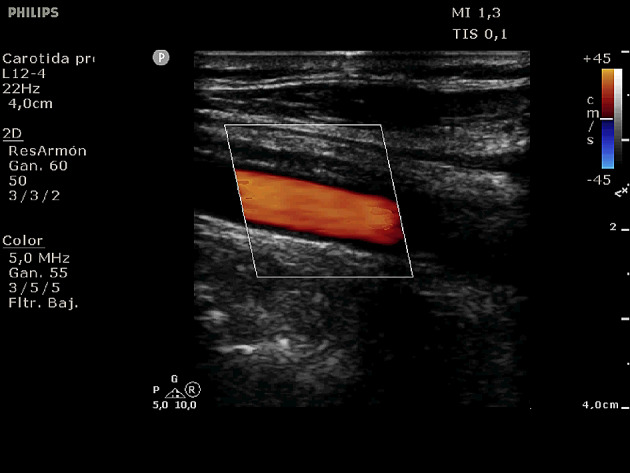
Use of a linear transducer in carotid Doppler ultrasound

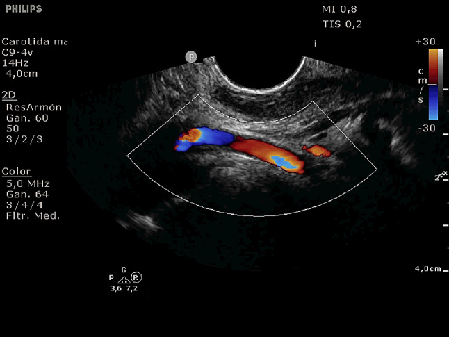
Visualization of carotid bifurcation in color Doppler mode with a linear transducer

Use of a phased array transducer in carotid Doppler ultrasound

Visualization of internal carotid artery in color Doppler mode with a phased array transducer

Use of a transoral endocavitary transducer in carotid Doppler ultrasound
Visualization of internal carotid artery in color Doppler mode with a transoral endocavitary transducer

Color Doppler mode at the level of the common carotid artery with box sides forming a 90° angulation in relation to the vessel with poor-signal
Optimization of the angle among box sides and the common carotid artery “manually tilting the vessel” and modifying angulation, thus obtaining good color Doppler signal

Pulsed wave Doppler mode at the level of the common carotid artery with sample volume at 90° of the vessel under study, with incorrectly low peak systolic and tele-diastolic velocities

Optimization of the angle between the sample volume and the direction of the common carotid artery “manually tilting the vessel”, modifying signal incidence (as tilted as possible) and rectifying the angle by aligning the dotted line with an arrow with the actual direction of the artery, always ensuring a 60° angle or less, thus obtaining actual peak systolic and tele-diastolic velocity values

B-mode showing carotid bifurcation. The external carotid artery is more anterior and smaller in caliber with respect to the ascending portion of the internal carotid artery

Low-resistance flow pattern typical of the internal carotid artery in pulsed wave Doppler mode

High-resistance flow pattern typical of the external carotid artery in pulsed wave Doppler mode

Filling defect at the level of the ascending portion of the right internal carotid artery associated with the presence of sonolucent plaque, producing a mosaic color pattern (aliasing) in color Doppler mode
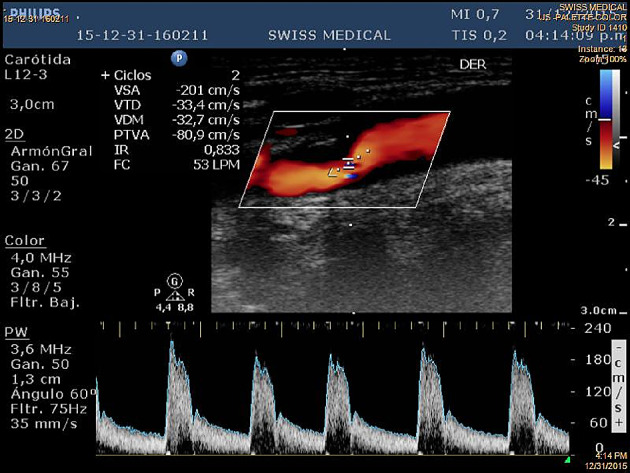
Pulsed wave Doppler mode at the level of aliasing area, showing increased peak systolic velocities consistent with stenosis > 50%

Lack of color Doppler signal at the level of the ascending portion of the internal carotid artery (arrow) suggesting occlusion at that level

Power Doppler mode shows the presence of a weak signal consistent with critical stenosis (arrow) at the level of the ascending portion of the internal carotid artery

Color Doppler mode showing kinking in the ascending portion of the internal carotid artery

Pulsed wave Doppler mode at the level of the right internal carotid artery with no flow evidence due to total occlusion, and left internal carotid artery with increased velocities due to compensatory hyperflow associated with collateral circulation at the level of the Circle of Willis
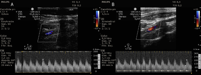
Pulsed wave Doppler mode at the level of both internal carotid arteries, showing increased generalized velocities associated with hyperdynamic circulation
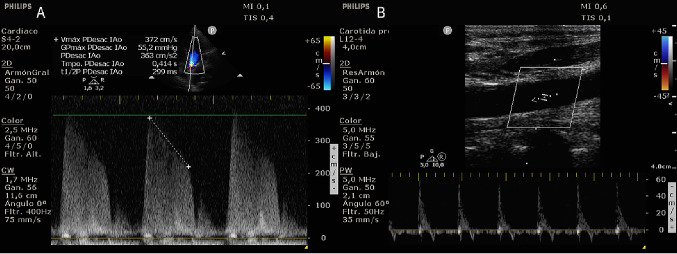
A Continuous Doppler mode through the aortic valve in modified apical view of 5 chambers showing significant aortic regurgitation. B Pulsed wave Doppler mode at the level of the common carotid artery showing reversal of diastolic flow associated with aortic insufficiency
References
-
- Cheong I, Otero Castro V, Tamagnone FM. Utility of transoral and transcranial ultrasonography in the diagnosis of internal carotid dissection: a case report. J Neurocrit Care. 2022 doi: 10.18700/jnc.210033. - DOI
MeSH terms
LinkOut - more resources
Full Text Sources
