

Long-term clinical prognosis of 335 infant single-gene positive FEVR cases
- PMID: 35918671
- PMCID: PMC9347171
- DOI: 10.1186/s12886-022-02522-8
Long-term clinical prognosis of 335 infant single-gene positive FEVR cases
Abstract
Purpose: To describe and analyze the clinical prognosis of infants diagnosed of familial exudative vitreoretinopathy (FEVR) with single gene mutation in long-term follow-up.
Methods: A retrospective case study was conducted on 355 FEVR infants with single positive gene.
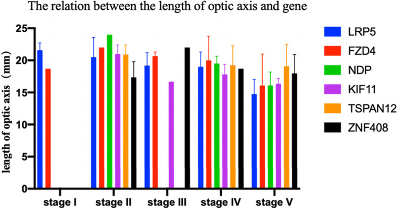
Result: Of the 335 single-gene positive infant FEVR cases (under 3 years old), 20% (n = 67) was diagnosed of strabismus at first visit. Staging of various genotypes was different (P < 0.001). Patients with NDP mutations presented the most severe clinical phenotypes and patients with ZNF408 mutations presented the mildest clinical phenotypes. Most infants underwent surgery under 1 year old (5th stage 75 of 108 [69.44%]). The axial length of different genotypes showed no significant difference (P = 0.2891). The 1st to 3rd stage cases were given intravitreal injection and/or retina photocoagulation with the last follow-up vision above 20/67. The 4th to 5th stage cases received the transcorneal vitrectomy with lensectomy or lens sparing vitrectomy (LSV), whose lens maintained transparent after LSV (11/14[78.58%]). After 2 to 10 years of follow-up, 37.96% (41/108) of post-surgery cases showed retinal funnel-like unfold and posterior pole unfold, 69.57% (16/ 23) of which received second surgery for closure of pupil with good prognosis. At the last follow-up, 20% (60/300) were with vision above 20/200.
Conclusion: LRP5 gene mutation was the most common mutation in FEVR patients. The severity of the clinical phenotype varied with different gene mutations. The main surgical methods for cases at Stage 4-5 were transcorneal vitrectomy with lensectomy or LSV. The earlier FEVR occurred, the worse prognosis would be. Active surgical intervention and lens sparing were necessary for cases at Stage 4-5.
Keywords: Clinical features; FEVR; Gene; Prognosis; Relation between phenotypes and genotypes.
© 2022. The Author(s).
Conflict of interest statement
The authors declare that the research was conducted in the absence of any commercial or financial relationships that could be construed as a potential conflict of interest.
The authors declare no conflicts of interest.
Figures


References
-
- Li F. Chinese Ophthalmology [M] (3rd edition) Beijing: People’s Medical Publishing House; 2014. pp. 24–25.
MeSH terms
Substances
Grants and funding
LinkOut - more resources
Full Text Sources
Medical
Miscellaneous
