

Post-mortem magnetic resonance imaging with computed tomography-guided biopsy for foetuses and infants: a prospective, multicentre, cross-sectional study
- PMID: 35918685
- PMCID: PMC9347089
- DOI: 10.1186/s12887-022-03519-4
Post-mortem magnetic resonance imaging with computed tomography-guided biopsy for foetuses and infants: a prospective, multicentre, cross-sectional study
Abstract
Background: Post-mortem imaging has been suggested as an alternative to conventional autopsy in the prenatal and postnatal periods. Noninvasive autopsies do not provide tissue for histological examination, which may limit their clinical value, especially when infection-related morbidity and mortality are suspected.
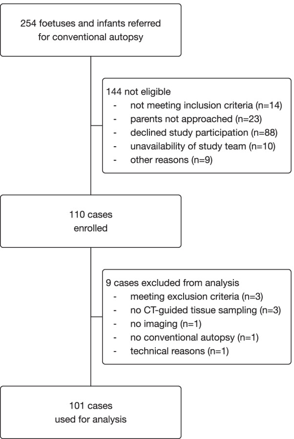
Methods: We performed a prospective, multicentre, cross-sectional study to compare the diagnostic performance of post-mortem magnetic resonance imaging with computed tomography-guided biopsy (Virtopsy®) with that of conventional autopsy in foetuses and infants. Cases referred for conventional autopsy were eligible for enrolment. After post-mortem imaging using a computed tomography scanner and a magnetic resonance imaging unit, computed tomography-guided tissue sampling was performed. Virtopsy results were compared with conventional autopsy in determining the likely final cause of death and major pathologies. The primary outcome was the proportion of cases for which the same cause of death was determined by both methods. Secondary outcomes included the proportion of false positive and false negative major pathological lesions detected by virtopsy and the proportion of computed tomography-guided biopsies that were adequate for histological examination.
Results: Overall, 101 cases (84 fetuses, 17 infants) were included. Virtopsy and autopsy identified the same cause of death in 91 cases (90.1%, 95% CI 82.7 to 94.5). The sensitivity and specificity of virtopsy for determining the cause of death were 96.6% (95% CI 90.6 to 98.8) and 41.7% (95% CI 19.3 to 68.0), respectively. In 32 cases (31.7%, 95% CI 23.4 to 41.3), major pathological findings remained undetected by virtopsy, and in 45 cases (44.6%, 95% CI 35.2 to 54.3), abnormalities were diagnosed by virtopsy but not confirmed by autopsy. Computed tomography-guided tissue sampling was adequate for pathological comments in 506 of 956 biopsies (52.7%) and added important diagnostic value in five of 30 cases (16.1%) with an unclear cause of death before autopsy compared with postmortem imaging alone. In 19 of 20 infective deaths (95%), biopsies revealed infection-related tissue changes. Infection was confirmed by placental examination in all fetal cases.
Conclusions: Virtopsy demonstrated a high concordance with conventional autopsy for the detection of cause of death but was less accurate for the evaluation of major pathologies. Computed tomography-guided biopsy had limited additional diagnostic value.
Trial registration: ClinicalTrials.gov (NCT01888380).
Keywords: Autopsy; Biopsy; Foetus; Infant; Magnetic resonance imaging; Minimally invasive; Post-mortem; Radiology; Virtopsy; Virtual autopsy.
© 2022. The Author(s).
Conflict of interest statement
The authors declare that they have no competing interests.
Figures
Similar articles
-
Minimally invasive autopsy for fetuses and children based on a combination of post-mortem MRI and endoscopic examination: a feasibility study.Health Technol Assess. 2019 Aug;23(46):1-104. doi: 10.3310/hta23460. Health Technol Assess. 2019. PMID: 31461397 Free PMC article.
-
Minimally invasive, imaging guided virtual autopsy compared to conventional autopsy in foetal, newborn and infant cases: study protocol for the paediatric virtual autopsy trial.BMC Pediatr. 2014 Jan 20;14:15. doi: 10.1186/1471-2431-14-15. BMC Pediatr. 2014. PMID: 24438163 Free PMC article. Clinical Trial.
-
Fetal postmortem imaging: an overview of current techniques and future perspectives.Am J Obstet Gynecol. 2020 Oct;223(4):493-515. doi: 10.1016/j.ajog.2020.04.034. Epub 2020 May 4. Am J Obstet Gynecol. 2020. PMID: 32376319 Review.
-
Post-mortem MRI versus conventional autopsy in fetuses and children: a prospective validation study.Lancet. 2013 Jul 20;382(9888):223-33. doi: 10.1016/S0140-6736(13)60134-8. Epub 2013 May 16. Lancet. 2013. PMID: 23683720
-
Dutch guideline for clinical foetal-neonatal and paediatric post-mortem radiology, including a review of literature.Eur J Pediatr. 2018 Jun;177(6):791-803. doi: 10.1007/s00431-018-3135-9. Epub 2018 Apr 19. Eur J Pediatr. 2018. PMID: 29675642 Free PMC article. Review.
Cited by
-
Causes of death in adults living with HIV in South Africa: A single-centre postmortem study.South Afr J HIV Med. 2025 May 9;26(1):1673. doi: 10.4102/sajhivmed.v26i1.1673. eCollection 2025. South Afr J HIV Med. 2025. PMID: 40469094 Free PMC article.
-
Diagnostic value of T1- and T2-weighted 3-Tesla MRI for postmortem detection and age stage classification of myocardial infarction.Forensic Sci Med Pathol. 2024 Mar;20(1):14-22. doi: 10.1007/s12024-023-00592-8. Epub 2023 Mar 2. Forensic Sci Med Pathol. 2024. PMID: 36862287 Free PMC article.
-
Current State of Robotics in Interventional Radiology.Cardiovasc Intervent Radiol. 2023 May;46(5):549-561. doi: 10.1007/s00270-023-03421-1. Epub 2023 Mar 31. Cardiovasc Intervent Radiol. 2023. PMID: 37002481 Free PMC article. Review.
-
The Current Status of Virtual Autopsy Using Combined Imaging Modalities: A Scoping Review.J Clin Med. 2025 Jan 25;14(3):782. doi: 10.3390/jcm14030782. J Clin Med. 2025. PMID: 39941453 Free PMC article. Review.
-
VIRTual autOPSY-applying CT and MRI for modern forensic death investigations.Front Radiol. 2025 May 12;5:1557636. doi: 10.3389/fradi.2025.1557636. eCollection 2025. Front Radiol. 2025. PMID: 40421097 Free PMC article. Review.
