. 2022 Aug 2;20(1):359.
doi: 10.1186/s12951-022-01526-2.

Docetaxel-loaded M1 macrophage-derived exosomes for a safe and efficient chemoimmunotherapy of breast cancer

Affiliations

Docetaxel-loaded M1 macrophage-derived exosomes for a safe and efficient chemoimmunotherapy of breast cancer

Yongmei Zhao et al. J Nanobiotechnology. .

Abstract

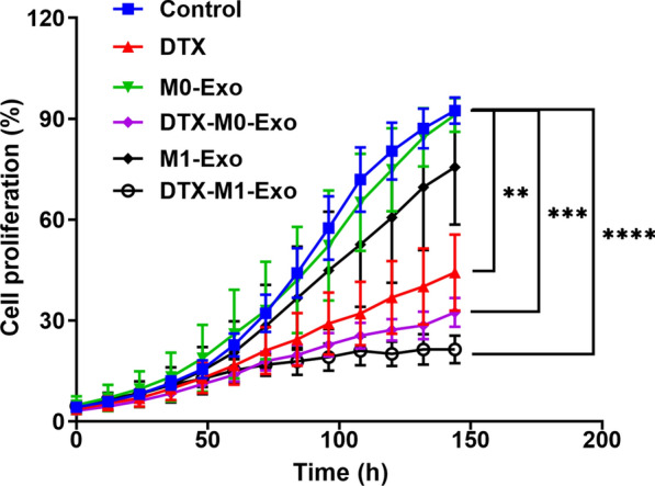
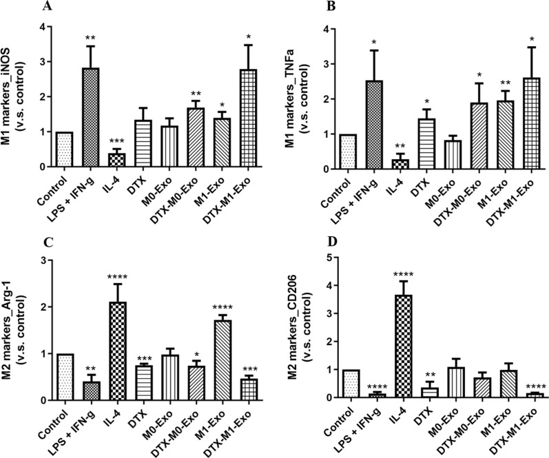
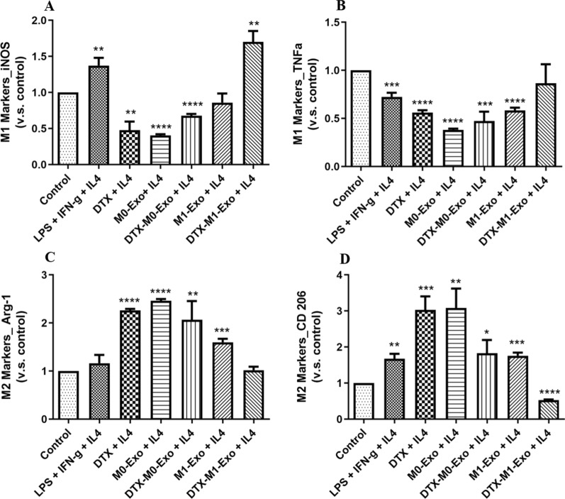
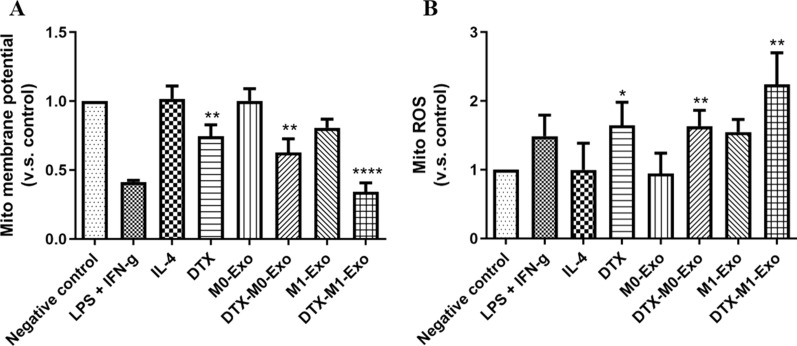
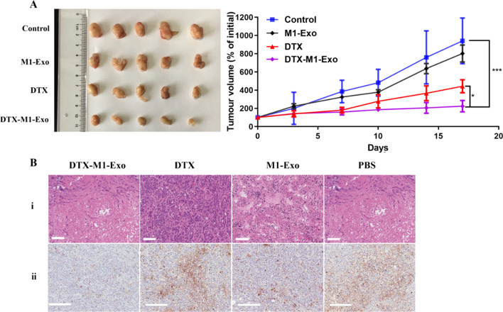
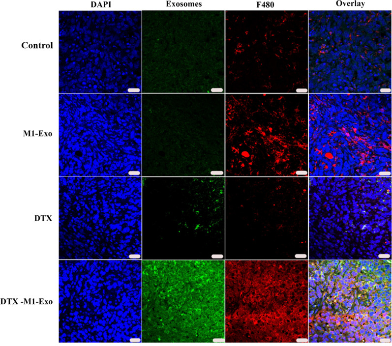
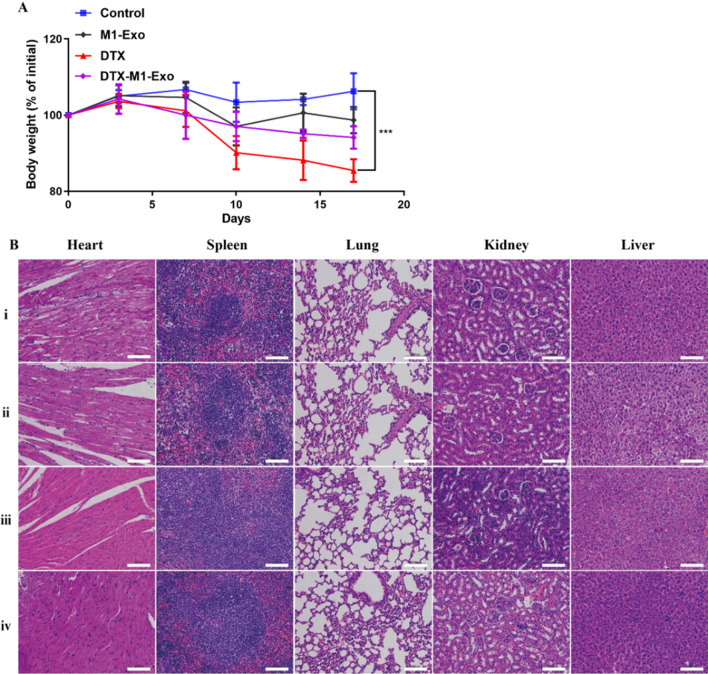
The conversion of tumor-promoting M2 macrophage phenotype to tumor-suppressing M1 macrophages is a promising therapeutic approach for cancer treatment. However, the tumor normally provides an abundance of M2 macrophage stimuli, which creates an M2 macrophage-dominant immunosuppressive microenvironment. In our study, docetaxel (DTX) as chemotherapeutic modularity was loaded into M1 macrophage-derived exosomes (M1-Exo) with M1 proinflammatory nature to establish DTX-M1-Exo drug delivery system. We found that DTX-M1-Exo induced naïve M0 macrophages to polarize to M1 phenotype, while failed to repolarize to M2 macrophages upon Interleukin 4 restimulation due to impaired mitochondrial function. This suggests that DTX-M1-Exo can achieve long-term robust M1 activation in immunosuppressive tumor microenvironment. The in vivo results further confirmed that DTX-M1-Exo has a beneficial effect on macrophage infiltration and activation in the tumor tissues. Thus, DTX-M1-Exo is a novel macrophage polarization strategy via combined chemotherapy and immunotherapy to achieve great antitumor therapeutic efficacy.

Keywords: Breast cancer; Exosomes; Macrophage polarization; Mitochondrial functions; Tumor immunity.


Conflict of interest statement

The authors declare that there is no conflict of interest.

Figures

Fig. 1
Fig. 1
Morphological characterization of exosomes with or without drug loading: TEM images of (A) M1-Exo and (B) DTX-M1-Exo
Fig. 2
Fig. 2
Cell cytotoxicity study of control, DTX, M0-Exo, DTX-M0-Exo, M1-Exo, and DTX-M1-Exo against 4T1 breast cancer cells. (** P < 0.01, *** P < 0.001, **** P < 0.0001 vs control)
Fig. 3
Fig. 3
The cellular uptake behavior of the exosomal vesicles (M1-Exo or M1-Exo) toward 4T1 cancer cells after 6 h incubation. In the CLSM images, cell nuclei were stained by DAPI (blue), while the exosomes were stained by fluorescent dye PKH67 (green). The scale bar is 20 µm
Fig. 4
Fig. 4
Macrophage phenotypes after treatment of IL-4, LPS + IFN-γ, DTX, M0-Exo, DTX-M0-Exo, M0-Exo, and DTX-M1-Exo, by measuring the expression of M1-macrophage markers (A) iNOS and (B) TNF-α, as well as M2-macrophage markers (C) Arg-1 and (D) CD206. (* P < 0.05, ** P < 0.01, *** P < 0.001, **** P < 0.0001 vs. the control group)
Fig. 5
Fig. 5
Macrophage repolarization capability of IL-4-induced M2 macrophages, followed by the treatment of LPS + IFN-γ, DTX, M0-Exo, DTX-M0-Exo, M0-Exo, and DTX-M1-Exo, by measuring the expression of M1-macrophage markers (A) iNOS and (B) TNF-α, as well as M2-macrophage markers (C) Arg-1 and (D) CD206. (* P < 0.05, ** P < 0.01, *** P < 0.001, **** P < 0.0001 vs. the control group)
Fig. 6
Fig. 6
Mitochondria function of macrophages with IL-4, LPS + IFN-γ, DTX, M0-Exo, DTX-M0-Exo, M0-Exo, and DTX-M1-Exo, determined by (A) mitochondrial membrane potential changes and (B) mitochondrial ROS induction. (* P < 0.05, ** P < 0.01, **** P < 0.0001)
Fig. 7
Fig. 7
Tumor growth inhibition of breast cancer 4T1in BALB/C mice. Mice were injected i.v. with PBS control, M1-Exo, DTX, and DTX-M1-Exo at concentration of 5 mg/kg DTX: (A) Tumor volume changes. (B) Representative histological images of slices of 4T1 tumor after final treatment using (i) H&E staining (scale bar = 100 μm) and (ii) Ki67 staining (scale bar = 200 μm)
Fig. 8
Fig. 8
The distribution of exosome formulations and macrophages in vivo. Representative fluorescent images showing nucleus (blue), PKH67-labelled exosomes (green), F480-labelled macrophages (red), and merged channels (from left to right). The scale bar denotes 20 μm
Fig. 9
Fig. 9
In vivo biosafety assessment. A Body weight changes. B Representative histological images of H&E-stained slices of each organ two days after treatment of (i) control, (ii) DTX, (iii) M1-Exo, and (iv) DTX-M1-Exo. The scale bar is 100 μm. (n = 3)

Similar articles

Cited by

References

    1. Yang L, Zhang Y. Tumor-associated macrophages: from basic research to clinical application. J Hematol Oncol. 2017;10(1):58. doi: 10.1186/s13045-017-0430-2. - DOI - PMC - PubMed
    1. Combes F, Meyer E, Sanders NN. Immune cells as tumor drug delivery vehicles. J Control Release. 2020;327:70–87. doi: 10.1016/j.jconrel.2020.07.043. - DOI - PubMed
    1. Verreck FAW, de Boer T, Langenberg DML, Hoeve MA, Kramer M, Vaisberg E, Kastelein R, Kolk A, de Waal-Malefyt R, Ottenhoff THM. Human IL-23-producing type 1 macrophages promote but IL-10-producing type 2 macrophages subvert immunity to (myco)bacteria. Proc Natl Acad Sci USA. 2004;101(13):4560. doi: 10.1073/pnas.0400983101. - DOI - PMC - PubMed
    1. Zheng P, Luo Q, Wang W, Li J, Wang T, Wang P, Chen L, Zhang P, Chen H, Liu Y, Dong P, Xie G, Ma Y, Jiang L, Yuan X, Shen L. Tumor-associated macrophages-derived exosomes promote the migration of gastric cancer cells by transfer of functional Apolipoprotein E. Cell Death Dis. 2018;9(4):434. doi: 10.1038/s41419-018-0465-5. - DOI - PMC - PubMed
    1. Obeid E, Nanda R, Fu YX, Olopade OI. The role of tumor-associated macrophages in breast cancer progression (review) Int J Oncol. 2013;43(1):5–12. doi: 10.3892/ijo.2013.1938. - DOI - PMC - PubMed