

Pathophysiology of mesangial expansion in diabetic nephropathy: mesangial structure, glomerular biomechanics, and biochemical signaling and regulation
- PMID: 35918708
- PMCID: PMC9347079
- DOI: 10.1186/s13036-022-00299-4
Pathophysiology of mesangial expansion in diabetic nephropathy: mesangial structure, glomerular biomechanics, and biochemical signaling and regulation
Abstract
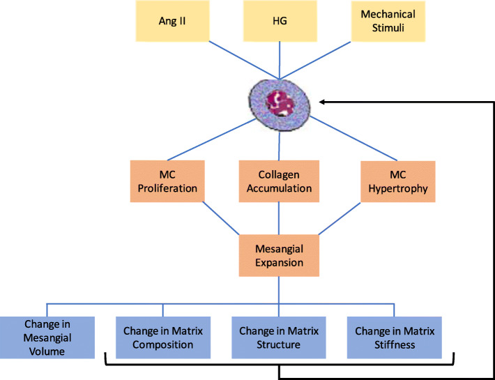
Diabetic nephropathy, a kidney complication arising from diabetes, is the leading cause of death in diabetic patients. Unabated, the growing epidemic of diabetes is increasing instances of diabetic nephropathy. Although the main causes of diabetic nephropathy have been determined, the mechanisms of their combined effects on cellular and tissue function are not fully established. One of many damages of diabetic nephropathy is the development of fibrosis within the kidneys, termed mesangial expansion. Mesangial expansion is an important structural lesion that is characterized by the aberrant proliferation of mesangial cells and excess production of matrix proteins. Mesangial expansion is involved in the progression of kidney failure in diabetic nephropathy, yet its causes and mechanism of impact on kidney function are not well defined. Here, we review the literature on the causes of mesangial expansion and its impacts on cell and tissue function. We highlight the gaps that still remain and the potential areas where bioengineering studies can bring insight to mesangial expansion in diabetic nephropathy.
Keywords: Chronic kidney disease; Collagen accumulation; Diabetic kidney disease; Extracellular matrix; Glomerulus; Mesangial cell; Mesangial matrix; Mesangium; Renal fibrosis.
© 2022. The Author(s).
Conflict of interest statement
The authors declare that they have no competing interests.
Figures


Similar articles
-
Expression of megsin mRNA, a novel mesangium-predominant gene, in the renal tissues of various glomerular diseases.J Am Soc Nephrol. 1999 Dec;10(12):2606-13. doi: 10.1681/ASN.V10122606. J Am Soc Nephrol. 1999. PMID: 10589701
-
The role of the BMP4/Smad1 signaling pathway in mesangial cell proliferation: A possible mechanism of diabetic nephropathy.Life Sci. 2019 Mar 1;220:106-116. doi: 10.1016/j.lfs.2019.01.049. Epub 2019 Jan 29. Life Sci. 2019. PMID: 30708099
-
Role of mesangial expansion in the pathogenesis of diabetic nephropathy.J Nephrol. 2001 Nov-Dec;14 Suppl 4:S51-7. J Nephrol. 2001. PMID: 11798146 Review.
-
Diabetic nephropathy. Mechanisms of mesangial matrix expansion.West J Med. 1995 Apr;162(4):318-21. West J Med. 1995. PMID: 7747496 Free PMC article.
-
Glomerular mesangial cell and podocyte injuries in diabetic nephropathy.Nephrology (Carlton). 2018 Oct;23 Suppl 4:32-37. doi: 10.1111/nep.13451. Nephrology (Carlton). 2018. PMID: 30298646 Review.
Cited by
-
Cellular phenotypic transitions in diabetic nephropathy: An update.Front Pharmacol. 2022 Nov 2;13:1038073. doi: 10.3389/fphar.2022.1038073. eCollection 2022. Front Pharmacol. 2022. PMID: 36408221 Free PMC article. Review.
-
Breviscapine alleviates podocyte injury by inhibiting NF-κB/NLRP3-mediated pyroptosis in diabetic nephropathy.PeerJ. 2023 Feb 13;11:e14826. doi: 10.7717/peerj.14826. eCollection 2023. PeerJ. 2023. PMID: 36815984 Free PMC article.
-
Studying the Roles of the Renin-Angiotensin System in Accelerating the Disease of High-Fat-Diet-Induced Diabetic Nephropathy in a db/db and ACE2 Double-Gene-Knockout Mouse Model.Int J Mol Sci. 2023 Dec 26;25(1):329. doi: 10.3390/ijms25010329. Int J Mol Sci. 2023. PMID: 38203500 Free PMC article.
-
The Nicotinamide/Streptozotocin Rodent Model of Type 2 Diabetes: Renal Pathophysiology and Redox Imbalance Features.Biomolecules. 2022 Sep 2;12(9):1225. doi: 10.3390/biom12091225. Biomolecules. 2022. PMID: 36139064 Free PMC article. Review.
-
Cytosolic Hmgb1 accumulation in mesangial cells aggravates diabetic kidney disease progression via NFκB signaling pathway.Cell Mol Life Sci. 2024 Sep 17;81(1):408. doi: 10.1007/s00018-024-05433-7. Cell Mol Life Sci. 2024. PMID: 39287634 Free PMC article.
References
Publication types
Grants and funding
LinkOut - more resources
Full Text Sources
