. 2022 Aug 2;13(1):391.
doi: 10.1186/s13287-022-03089-3.

PFKFB3-mediated glycometabolism reprogramming modulates endothelial differentiation and angiogenic capacity of placenta-derived mesenchymal stem cells

Affiliations

PFKFB3-mediated glycometabolism reprogramming modulates endothelial differentiation and angiogenic capacity of placenta-derived mesenchymal stem cells

Yang Zhang et al. Stem Cell Res Ther. .

Abstract

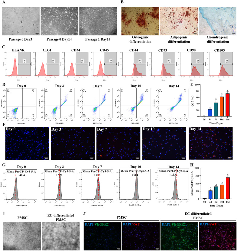
Background: Mesenchymal stem cells (MSCs) have a great potential ability for endothelial differentiation, contributing to an effective means of therapeutic angiogenesis. Placenta-derived mesenchymal stem cells (PMSCs) have gradually attracted attention, while the endothelial differentiation has not been fully evaluated in PMSCs. Metabolism homeostasis plays an important role in stem cell differentiation, but less is known about the glycometabolic reprogramming during the PMSCs endothelial differentiation. Hence, it is critical to investigate the potential role of glycometabolism reprogramming in mediating PMSCs endothelial differentiation.

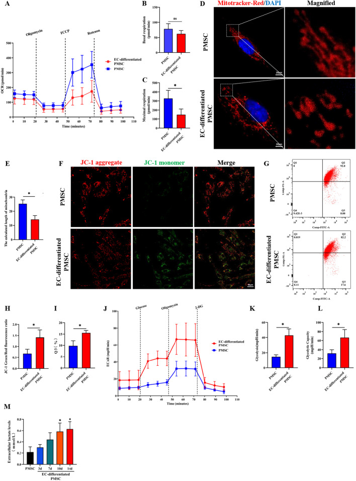
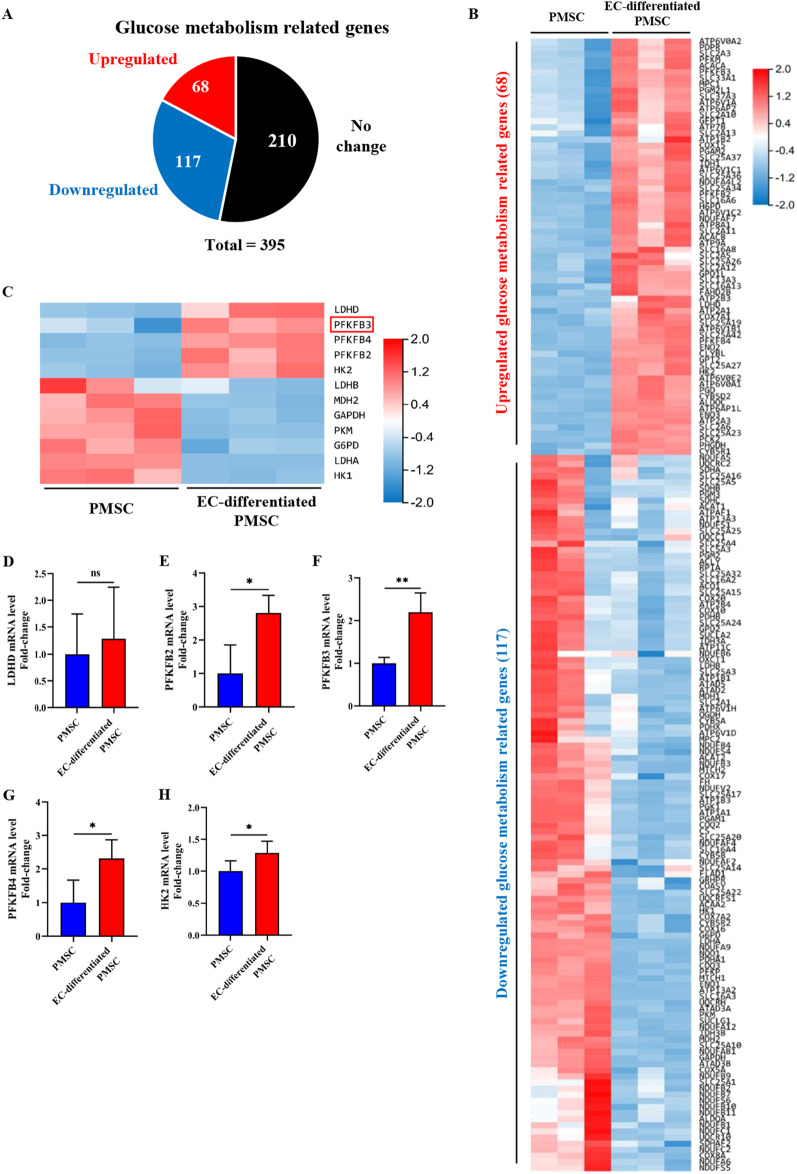
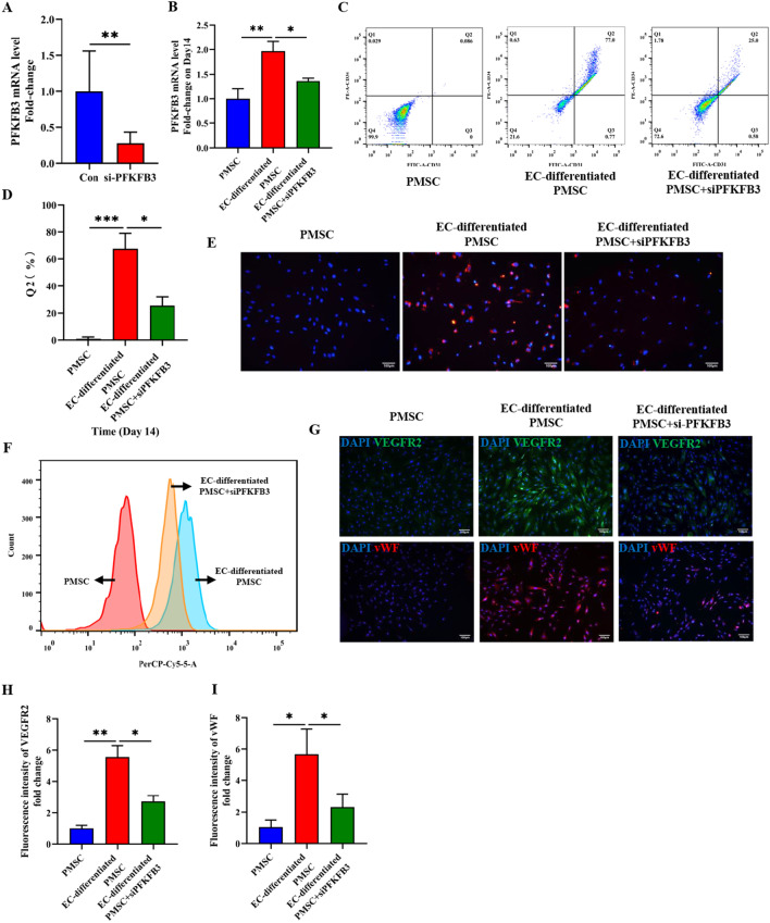
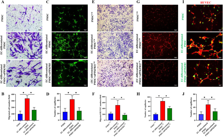
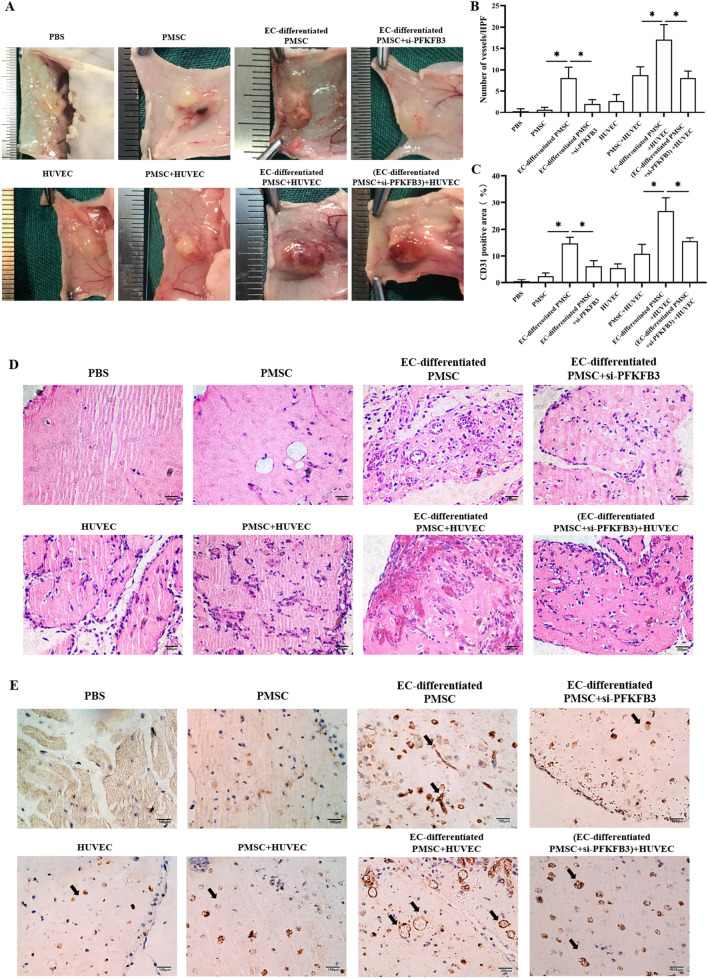
Methods: Dil-Ac-LDL uptake assay, flow cytometry, and immunofluorescence were all to verify the endothelial differentiation in PMSCs. Seahorse XF Extracellular Flux Analyzers, Mito-tracker red staining, Mitochondrial membrane potential (MMP), lactate secretion assay, and transcriptome approach were to assess the variation of mitochondrial respiration and glycolysis during the PMSCs endothelial differentiation. Glycolysis enzyme 6-phosphofructo-2-kinase/fructose-2,6-bisphosphatase 3 (PFKFB3) was considered a potential modulator for endothelial differentiation in PMSCs by small interfering RNA. Furthermore, transwell, in vitro Matrigel tube formation, and in vivo Matrigel plug assays were performed to evaluate the effect of PFKFB3-induced glycolysis on angiogenic capacities in this process.

Results: PMSCs possessed the superior potential of endothelial differentiation, in which the glycometabolic preference for glycolysis was confirmed. Moreover, PFKFB3-induced glycometabolism reprogramming could modulate the endothelial differentiation and angiogenic abilities of PMSCs.

Conclusions: Our results revealed that PFKFB3-mediated glycolysis is important for endothelial differentiation and angiogenesis in PMSCs. Our understanding of cellular glycometabolism and its regulatory effects on endothelial differentiation may propose and improve PMSCs as a putative strategy for clinical therapeutic angiogenesis.

Keywords: Angiogenesis; Endothelial differentiation; Glycolysis; Glycometabolism reprogramming; Mesenchymal stem cells; PFKFB3; Placenta.


Conflict of interest statement

The authors have declared that no competing interests exists.

Figures

Fig. 1
Fig. 1
Culture, identification, and endothelial differentiation of PMSCs. A PMSCs morphology was detected by an inverted microscope. Three and seven days after isolation, small fibroblast-like MSC colonies were visible. Cell culture was enriched in a population of cells characterized by a fibroblast-like spike appearance. Scale bar: 200 μm. B Successfully differentiated PMSCs were stained with Alizarin Red for osteogenic differentiation, Oil Red O for adipocytes, and Alcian blue, in which red calcium nodules, orange lipid droplets, and blue cartilage could be observed. Scale bar: 200 μm. C PMSCs surface markers. Flow cytometry demonstrated that the cells expressed CD44, CD73, CD90, and CD105 but not CD31, CD34, and CD45. D, E Identification of induced PMSCs by flow cytometry and quantitative analyses were also performed. The fractions of CD31+ CD34+ double-positive cells were about 10%, 50%, 60%, and 70%, at 3, 7, 10, and 14 days, respectively. F–H Dil-Ac-LDL uptake assay was to identify the induced PMSCs through immunofluorescence and flow cytometry, and quantitative analyses were also performed. Scale bar: 100 μm. I Morphological characteristics of cells after cultivated in the inducing and non-inducing groups. The cells developed into short spindle shapes in the inducing group. The cells in the non-inducing medium retained the typical spindle-shaped morphology. Scale bar: 200 μm. J Immunofluorescence staining of vWF and VEGFR-2 was performed in different groups. Scale bar: 100 μm. Data are shown as the mean ± SD from three independent experiments and the representative result is shown. *P < 0.05, **P < 0.01, ***P < 0.001 by Student’s t test. SD: standard deviation. EC-differentiated PMSC: the induced PMSCs group for endothelial differentiation
Fig. 2
Fig. 2
Glycometabolism reprogramming during the endothelial differentiation of PMSCs. A–C The OCR assay was used to observe the basal and maximal mitochondrial respiratory function. D, E Cells in different groups were stained with Mito-tracker red to evaluate the mitochondrial fragmentation, and the mitochondrial length was analyzed quantitatively. Scale bar: 20 μm. E–I MMP was evaluated by a confocal microscope and flow cytometry. Thereinto, the ratio of green/red puncta was calculated to assess the MMP changes, and quantitative analyses were also performed. Scale bar: 20 μm. J–L Glycolysis and glycolysis capacity was detected by ECAR assay. M Extracellular lactate release determination in PMSCs and endothelial differentiated PMSCs at Day 3, 7, 10, and 14. Data are shown as the mean ± SD from three independent experiments and the representative result is shown. *P < 0.05, **P < 0.01, ***P < 0.001 by Student’s t test. SD: standard deviation. EC-differentiated PMSC: the induced PMSCs group for endothelial differentiation
Fig. 3
Fig. 3
Bioinformatics analysis of whole transcriptome sequencing. A Volcano map of differentially expressed genes (DEGs) between PMSCs and induced PMSCs for endothelial differentiation. The x-axis is the log2 scale of the fold change of gene expression in MSCs and induced ECs (log2(fold change)). Negative values indicate downregulation; positive values indicate upregulation. The y-axis is the minus log10 scale of the adjusted p values (–log10 (p adj)), which indicates the significant level of expression difference. The red dots represent significantly upregulated genes, while the green dots represent significantly downregulated genes. B KEGG pathway enrichment analysis. C KEGG pathway classification analysis. D GO classification analysis. EC-differentiated PMSC: the induced PMSCs group for endothelial differentiation
Fig. 4
Fig. 4
Global identification of target genes modulating the glycometabolic conversion during the endothelial differentiation of PMSCs. A Pie graph from RNA sequencing (RNA-seq) showing the expression profile of glucose metabolism-related genes in induced PMSCs for endothelial differentiation, with upregulated glucose metabolism-related genes and downregulated glucose metabolism-related genes. B The upregulated genes and downregulated genes from RNA-seq analysis. Color scales represent fold changes relative to control cells. C Heat maps of glycometabolic genes in induced PMSCs for endothelial differentiation. Color scales in heat maps represent fold changes relative to the uninduced PMSCs. D–H Quantitation from quantitative RT-PCR (real-time polymerase chain reaction) assays of the glycolysis genes LDHD, HK2, PFKFB2, PFKFB3, and PFKFB4. Data are shown as the mean ± SD from three independent experiments and the representative result is shown. *P < 0.05, **P < 0.01, ***P < 0.001 by Student’s t test. SD: standard deviation. EC-differentiated PMSC: the induced PMSCs group for endothelial differentiation
Fig. 5
Fig. 5
Glycolytic enzyme PFKFB3 regulated the endothelial differentiation of PMSCs. A Confirmation of PFKFB3 knockdown by qRT-PCR. (B) Detection of PFKFB3 mRNA level on the 14th day of endothelial induction. C, D flow cytometry and quantitative analyses of the fractions of CD31+CD34+ double-positive cells in different groups on Day 14. E–F Dil-Ac-LDL uptake assay through immunofluorescence and flow cytometry, and quantitative analyses in different groups on Day 14. Scale bar: 100 μm. G Immunofluorescence staining of vWF and VEGFR-2 in different groups. H, I Quantitative analysis of VEGFR-2 and vWF fluorescence intensity. Scale bar: 100 μm. Data are shown as the mean ± SD from three independent experiments and the representative result is shown. *P < 0.05, **P < 0.01, ***P < 0.001 by Student’s t test. SD: standard deviation. EC-differentiated PMSC: the induced PMSCs group for endothelial differentiation. si-PFKFB3: PFKFB3 siRNA
Fig. 6
Fig. 6
PFKFB3 modulated the angiogenic and proangiogenic capacities of PMSCs during the endothelial differentiation in vitro. A, B Representative transwell photographs of the PMSCs migration and the migrated PMSCs number quantified in different groups (Scale bar: 200 μm). C, D Representative photographs of the PMSCs tube formation and the number of formative capillaries quantified in different groups (Scale bar: 100 μm). E, F Representative transwell photographs of the HUVEC migration and the migrated HUVEC number quantified in different groups (Scale bar: 200 μm). G, H Representative photographs of the HUVEC tube formation and the number of formative capillaries quantified in different groups (Scale bar: 200 μm). I, J Representative photographs of the tube formation in the PMSC-HUVEC coculture system and the number of formative capillaries quantified in different groups (Scale bar: 50 μm). Data are shown as the mean ± SD from three independent experiments and the representative result is shown. *P < 0.05, **P < 0.01, ***P < 0.001 by Student’s t test. SD: standard deviation. EC-differentiated PMSC: the induced PMSCs group for endothelial differentiation. si-PFKFB3: PFKFB3 siRNA
Fig. 7
Fig. 7
PFKFB3 modulated the angiogenic and proangiogenic capacities of PMSCs during the endothelial differentiation in Matrigel plugs in vivo. A Representative images of Matrigel plugs in different groups. B–C Quantitation of vessel number from the H&E-stained paraffin sections and quantitation of the human-specific CD31-positive percentage. D, E Representative images of macroscope view of explanted Matrigel plugs, H&E staining (Scale bar: 200 μm), and immunobiological human CD31 staining (Scale bar: 100 μm) of explanted Matrigel plugs. Data are shown as the mean ± SD from three independent experiments and the representative result is shown. *P < 0.05, **P < 0.01, ***P < 0.001 by Student’s t test. SD: standard deviation. EC-differentiated PMSC: the induced PMSCs group for endothelial differentiation. si-PFKFB3: PFKFB3 siRNA

Similar articles

Cited by

References

    1. Phng LK, Belting HG. Endothelial cell mechanics and blood flow forces in vascular morphogenesis. Semin Cell Dev Biol. 2021. - PubMed
    1. Mathew SA, Naik C, Cahill PA, Bhonde RR. Placental mesenchymal stromal cells as an alternative tool for therapeutic angiogenesis. Cell Mol Life Sci CMLS. 2020;77(2):253–265. doi: 10.1007/s00018-019-03268-1. - DOI - PMC - PubMed
    1. Laurenzana A, Fibbi G, Chillà A, Margheri G, Del Rosso T, Rovida E, et al. Lipid rafts: integrated platforms for vascular organization offering therapeutic opportunities. Cell Mol Life Sci CMLS. 2015;72(8):1537–1557. doi: 10.1007/s00018-014-1814-x. - DOI - PMC - PubMed
    1. Samura M, Hosoyama T, Takeuchi Y, Ueno K, Morikage N, Hamano K. Therapeutic strategies for cell-based neovascularization in critical limb ischemia. J Transl Med. 2017;15(1):49. doi: 10.1186/s12967-017-1153-4. - DOI - PMC - PubMed
    1. Tancharoen W, Aungsuchawan S, Pothacharoen P, Markmee R, Narakornsak S, Kieodee J, et al. Differentiation of mesenchymal stem cells from human amniotic fluid to vascular endothelial cells. Acta Histochem. 2017;119(2):113–121. doi: 10.1016/j.acthis.2016.11.009. - DOI - PubMed

Publication types

Substances

LinkOut - more resources