

Long-term peripheral retinal vascular behavior in retinopathy of prematurity patients treated with ranibizumab intravitreal injection as monotherapy using fluorescein angiography
- PMID: 35918740
- PMCID: PMC9344754
- DOI: 10.1186/s40942-022-00402-3
Long-term peripheral retinal vascular behavior in retinopathy of prematurity patients treated with ranibizumab intravitreal injection as monotherapy using fluorescein angiography
Abstract
Background: Few challenges are faced with the introduction of anti-VEGF agents as a modality of treatment for retinopathy of prematurity. The clinical behavior and time course of regression post injection differ compared to post laser ablation. This study aims to evaluate the long-term peripheral retinal vascularization outcome of Ranibizumab intravitreal injections monotherapy in the treatment of retinopathy of prematurity.
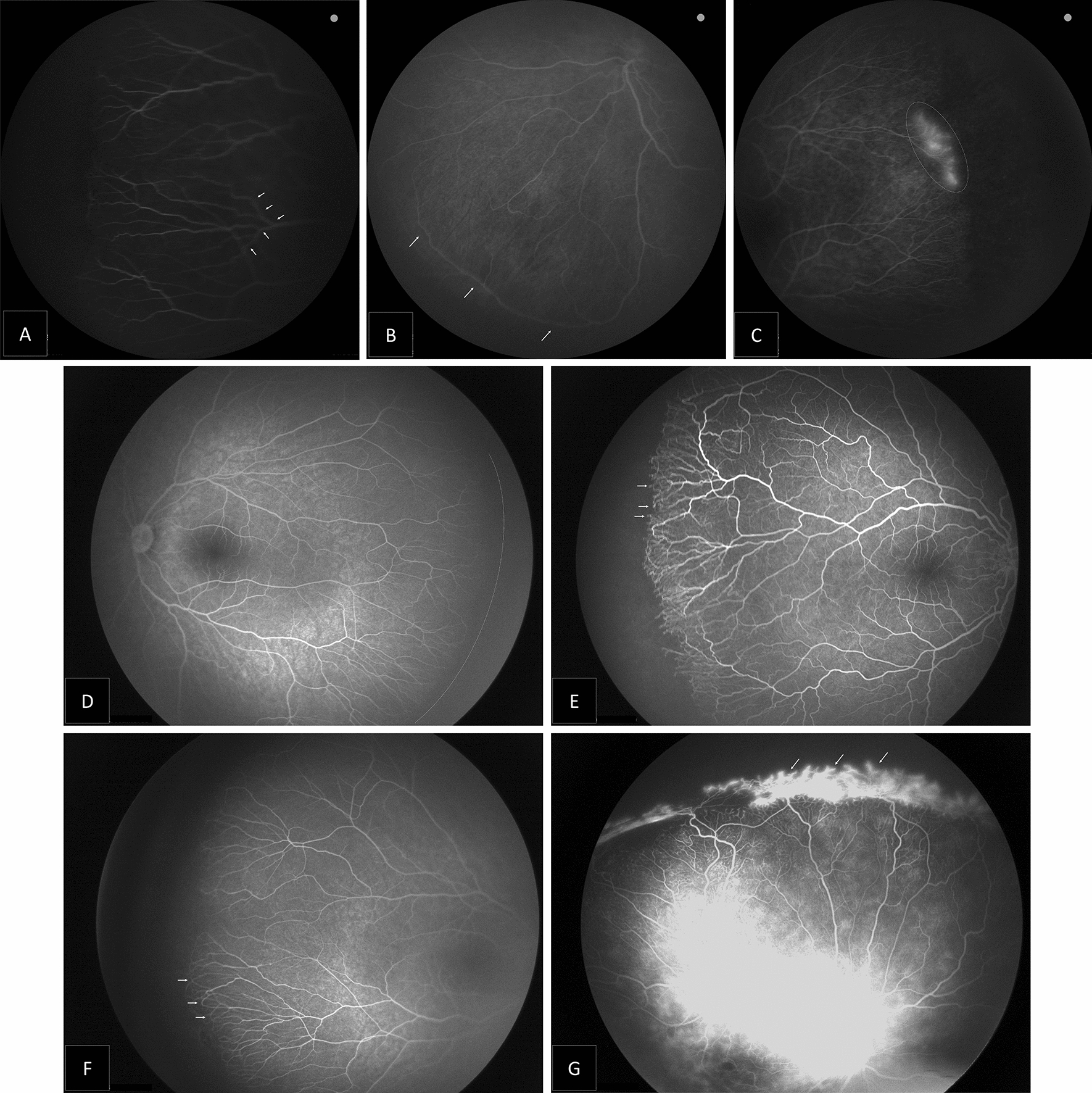
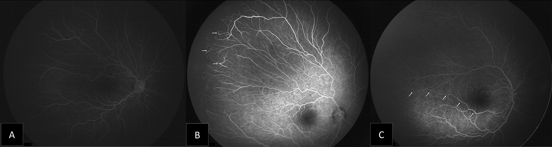
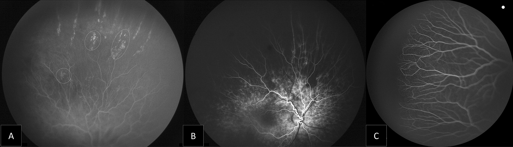
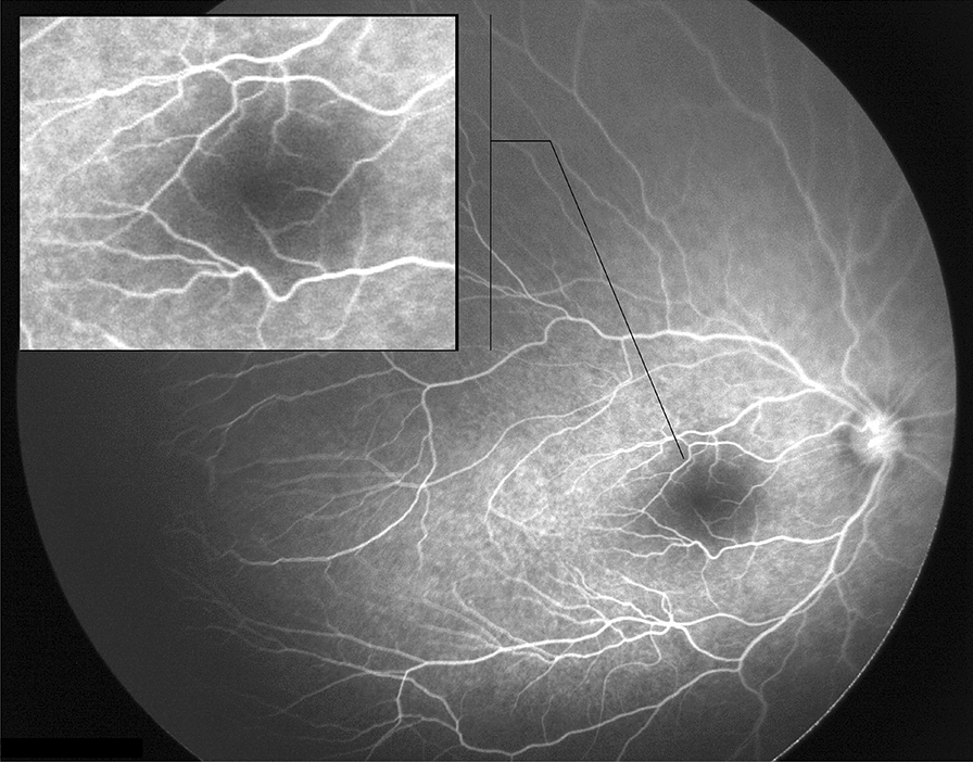
Method: Hospital-based quasi-experimental study. Include ROP patients who received intravitreal ranibizumab (IVR), as primary treatment for type 1 ROP. Patients were examined under general anaesthesia to ensure documentation of all junctions of vascular and avascular zones. Images were taken by RetCam III, Phoenix ICON and fluorescein angiography was performed to describe vascular behaviors.
Results: The mean gestational age was 24.67 weeks and the mean postmenstrual age at the time of intravitreal ranibizumab treatment was 36.3 weeks. Fluorescein angiography was performed at 155-288 weeks; most eyes showed two disk diameters of avascular peripheral retina. Only eyes with original aggressive ROP who required a second injection (six eyes) showed extensive peripheral avascular retina reaching zone I (13.64%). Neovascularization was evident in five eyes (11.36%), all with an original aggressive ROP and received multiple injections.
Conclusions: Ranibizumab treated babies with incomplete retinal vascularization require close and long-term follow-up visits to assess post injection vascular behavior. Peripheral retinal avascular zone of more than two-disc diameters was present in most of the patients evidenced by fluorescein angiography. Babies with initial diagnosis of aggressive ROP are more likely to have persistent peripheral neovascularization.
Keywords: Anti- VEGF; Fluorescein Angiography; Peripheral retinal vascularization; Ranibizumab; Reactivation of ROP; Regression of ROP; RetCam; Retinopathy of Prematurity.
© 2022. The Author(s).
Conflict of interest statement
The authors declare that they have no competing interests.
Figures
References
-
- McCannel C, Atebara N, Kim S, et al. Chapter 8: Retinopathy of Prematurity. Basic Clinical and Science Course Section 12: Retina and Vitreous. San Francisco, CA: AAO. 2018–2019; 175–187
-
- Early Treatment for Retinopathy of Prematurity Cooperative Group Revised indications for the treatment of retinopathy of prematurity: results of the early treatment for retinopathy of prematurity randomized trial. Arch Ophthalmol. 2003;121(12):1684–1694. doi: 10.1001/archopht.121.12.1684. - DOI - PubMed
LinkOut - more resources
Full Text Sources
