. 2022 Aug 2;21(1):158.
doi: 10.1186/s12943-022-01621-w.

BTApep-TAT peptide inhibits ADP-ribosylation of BORIS to induce DNA damage in cancer

Affiliations

BTApep-TAT peptide inhibits ADP-ribosylation of BORIS to induce DNA damage in cancer

Yanmei Zhang et al. Mol Cancer. .

Abstract

Background: Brother of regulator of imprinted sites (BORIS) is expressed in most cancers and often associated with short survival and poor prognosis in patients. BORIS inhibits apoptosis and promotes proliferation of cancer cells. However, its mechanism of action has not been elucidated, and there is no known inhibitor of BORIS.

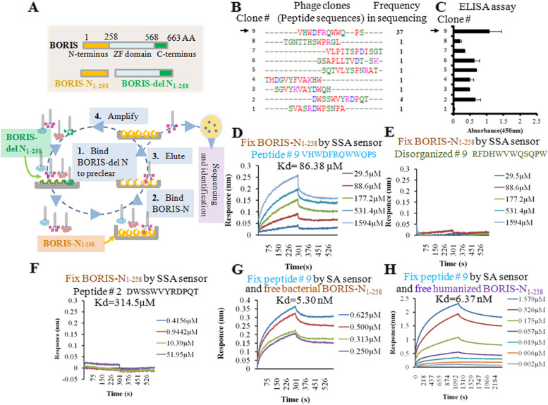
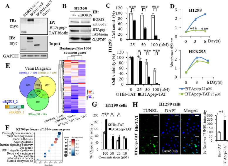
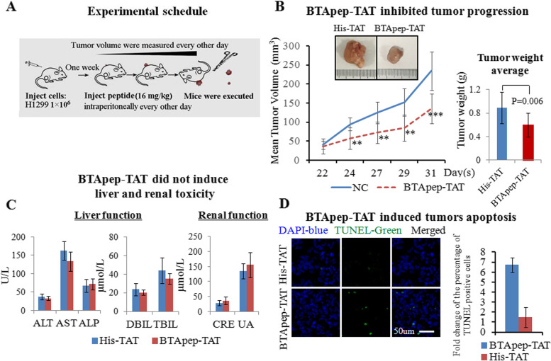
Methods: A phage display library was used to find the BORIS inhibitory peptides and BTApep-TAT was identified. The RNA sequencing profile of BTApep-TAT-treated H1299 cells was compared with that of BORIS-knockdown cells. Antitumor activity of BTApep-TAT was evaluated in a non-small cell lung cancer (NSCLC) xenograft mouse model. BTApep-TAT was also used to investigate the post-translational modification (PTM) of BORIS and the role of BORIS in DNA damage repair. Site-directed mutants of BORIS were constructed and used for investigating PTM and the function of BORIS.

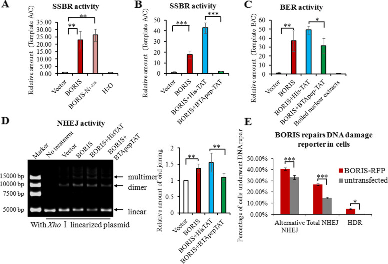
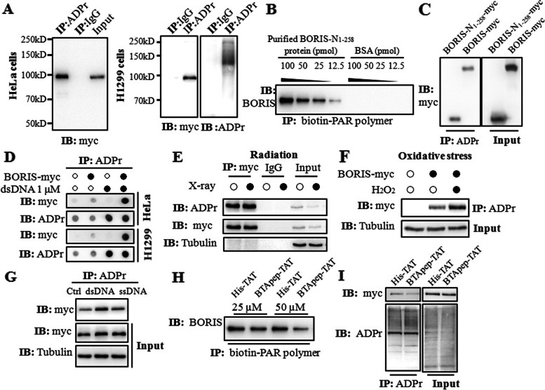
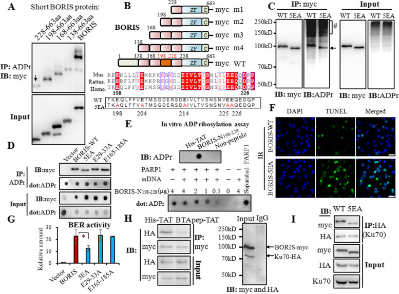
Results: BTApep-TAT induced DNA damage in cancer cells and suppressed NSCLC xenograft tumor progression. Investigation of the mechanism of action of BTApep-TAT demonstrated that BORIS underwent ADP ribosylation upon double- or single-strand DNA damage. Substitution of five conserved glutamic acid (E) residues with alanine residues (A) between amino acids (AAs) 198 and 228 of BORIS reduced its ADP ribosylation. Inhibition of ADP ribosylation of BORIS by a site-specific mutation or by BTApep-TAT treatment blocked its interaction with Ku70 and impaired the function of BORIS in DNA damage repair.

Conclusions: The present study identified an inhibitor of BORIS, highlighted the importance of ADP ribosylation of BORIS, and revealed a novel function of BORIS in DNA damage repair. The present work provides a practical method for the future screening or optimization of drugs targeting BORIS.

Keywords: ADP-ribosylation; BORIS; DNA damage; Non-small cell lung cancer; Targeted peptide.


Conflict of interest statement

We declare that we have no financial and personal relationships with other people or organizations that can inappropriately influence our work, and there is no professional or other personal interest of any nature or kind in any product, service and/or company that could be construed as influencing the position presented in, or the review of, the manuscript entitled.

Figures

Fig. 1
Fig. 1
Selection and characterization of the BORIS-binding peptide. (A) Procedure for the selection of BORIS-binding peptides. (B) The sequences and frequencies of the peptides enriched after elution. (C) ELISA testing the affinity of phages for the BORIS-N1-258 protein. (D) The peptide from phage clone 9 was used to determine the affinity of the interaction with BORIS-N1-258 protein by BLI. The panel shows the test of the BORIS-N1-258 protein immobilized on an SSA sensor and free peptide in solution. (E) Scrambled peptide 9 showing no affinity to BORIS-N1-258 in the BLI assay. (F) Peptide 2 showed a weak binding affinity (Kd) of 314.5 µM to BORIS-N1-258 in the BLI assay. (G) Peptide 9 immobilized on an SA sensor and free BORIS-N1-258 protein in solution. (H) The BORIS-N1-258 protein purified from HEK293 cells was used to examine the interaction with synthesized peptide 9. The test was performed by fixing peptide 9 on an SA sensor and releasing the humanized BORIS-N1-258 protein to solution in a BLI assay
Fig. 2
Fig. 2
BTApep-TAT induced DNA damage and cancer cell apoptosis. (A) Co-immunoprecipitation was performed to evaluate the interaction of BTApep-TAT-biotin with BORIS-N1-258, full length BORIS, and BORIS-del N1-258 in the cell lysates from transfected H1299 cells. (B) The level of BORIS in H1299 cells after siRNA-mediated knockdown was evaluated by BORIS antibody or the BTApep-TAT-biotin peptide. (C) Cells were incubated with graded concentrations of the peptides (25–100 µM) for three days. MTT assays and cell counting were performed to evaluate the effect of BTApep-TAT and the negative control peptide His-TAT on H1299 cells. (D) H1299 and HEK293 cells were treated with 25 µM BTApep-TAT or BTApep to examine the effect of BTApep-TAT on cancer cells and normal cells (E) Transcriptomes of H1299 cells with siBORIS knockdown or BTApep-TAT treatment were compared. The left panel shows an overlap between siBORIS knockdowns and BTApep-TAT treatment in a Venn diagram. Two siRNAs targeting BORIS were used to compare the common genes in the heatmap. (F) A bubble map showing the pathways associated with the genes common to BORIS knockdown and BTApep-TAT treatment, which are shown in Panel E. (G) Caspase 3/7 assay detected the peptide-induced apoptosis at peptide concentrations from 10 to 100 µM. (H) A TUNEL assay detected the DNA damage induced by 25 µM peptide
Fig. 3
Fig. 3
BTApep-TAT inhibited DNA damage repair governed by BORIS in cancer cells. (A) Artificial single-strand break DNA was incubated with crude nuclear extracts from HeLa cells transfected with full-length BORIS or the AA 1–258 N-terminus of BORIS (BORIS-N1-258). Crude nuclear extracts of empty vector-transfected cells or equal volumes of water were used as negative controls. The DNA repair activities were examined by qRT–PCR and are shown as the relative transcript amount of ligated target A templates. (B) SSBR activities were examined in BORIS-expressing crude nuclear extracts that were treated with 25 µM peptides. (C) BER activities were examined in BORIS-expressing crude nuclear extracts that were treated with 25 µM peptides. The boiled nuclear extracts from BORIS-expressing HeLa cells were used as another negative control. The DNA repair activities were examined by qRT-PCR and are shown as the relative transcript amount of ligated target B templates. (D) The Xho I-linearized plasmid was used as double-strand DNA break to evaluate NHEJ activity. The multimeric and dimeric forms of the plasmids were visualized on agarose gels, which are presented in the left panel. The right panel shows the statistical summary of the treatment results. (E) DNA damage repair reporters (GFP fluorescent-based DNA repair reporter system) were used to evaluate the effect of BORIS in cells. The percentage of cells that underwent DNA damage repair was compared between BORIS-RFP-transfected cells and cells without transfection. DNA damage repair, which includes alternative NHEJ, total NHEJ, and homology-directed repair (HDR), was analyzed by flow cytometry
Fig. 4
Fig. 4
BTApep-TAT inhibited the ADP ribosylation of BORIS in response to DNA damage. (A) ADP-ribosylation of BORIS was determined in BORIS-myc-transfected HeLa and H1299 cells using the ADPr antibody, which detected both poly-ADPr and mono-ADP-ribosylation. (B) The purified BORIS-N1-258 protein or BSA was diluted and incubated with 5 pmol of biotin-PAR polymers immobilized on streptavidin beads. Specific interaction was observed with the BORIS-N1-258 protein, but not with BSA. (C) ADP-ribosylation of BORIS-N1-258-myc was determined in transfected H1299 cells. The levels of ADP ribosylation were nearly identical for BORIS-N1-258 and full-length BORIS. (D) The plasmids of BORIS-myc or empty vector were transfected into H1299 and HeLa cells. The crude nuclear extracts were supplemented with 1 μM dsDNA. BORIS was ADP-ribosylated in both H1299 and HeLa cells, and ADP-ribosylation was enhanced upon dsDNA induction. (E) DNA damage was induced in H1299 cells by 30 Gy X-ray irradiation. (F) DNA damage was induced in H1299 cells by treatment with H2O2 at a concentration of 500 µM for 10 min. (G) The levels of ADP ribosylation of BORIS-myc in H1299 cells were compared between dsDNA and ssDNA treatments. (H) ADP ribosylation of BORIS-N1-258 was examined by an in vitro ADP-ribosylation assay. BTApep-TAT treatment significantly suppressed ADP ribosylation of BORIS-N1-258. (I) ADP ribosylation of BORIS-myc in H1299 cells after treatment with 25 µM BTApep-TAT or His-TAT was examined by immunoprecipitation of ADP-ribosylated protein and immunoblotting against the myc tag
Fig. 5
Fig. 5
BTApep-TAT inhibited the ADP ribosylation at residues 198–228 of BORIS and inhibited the interaction of BORIS with Ku70. (A) Truncations of variable lengths of amino acids between AA 2 and 227 in the N-terminus of BORIS were constructed and transfected into H1299 cells. ADP ribosylation of the short BORIS protein, which contains residues 228–663 of BORIS, was reduced compared with that of BORIS 198–663. (B) Schematic representation of the truncated BORIS proteins and the details of the homology of BORIS in Mus musculus (Mus.), Rattus norvegicus (Rattus.) and Homo sapiens (Homo.). Conserved amino acids are shown in red boxes. Five conserved glutamic acid residues were mutated to alanine residues in the BORIS-5EA mutant. (C) The 5EA quintuple mutant of BORIS tagged with myc (5EA) or BORIS-myc (WT) was transfected into H1299 cells. The cell lysates were used to evaluate the interactions of ADP-ribosylated and myc-tagged proteins. (D) ADP-ribosylation of the 5EA quintuple mutant and other mutants (which contained substitutions of glutamic acid residues with alanine residues in the 29–33 or 165–185 regions) were compared by precipitation with an anti-ADP-ribose antibody and visualization by dot blot or Western blot assays with a myc antibody. (E) The BORIS-N198-228 peptide was subjected to PARP1-mediated PARylation in vitro, which was activated by recombinant PARP1 and ssDNA. After the reaction, the peptides and PARP1 were separated by centrifugal filtration and analyzed by the dot blot assay. (F) DNA damage induced by 30 Gy X-ray irradiation was examined in BORIS-WT- and BORIS-5EA-transfected H1299 cells by TUNEL assay. (G) The comparison of BER activity of the mutants is presented as a histogram. (H) BORIS-myc and Ku70-HA were co-transfected into H1299 cells, and the cells were incubated with BTApep-TA or His-TAT to investigate the interaction between BORIS and Ku70. (I) BORIS-myc/BORIS-5EA-myc and Ku70-HA were co-transfected into H1299 cells, and the interaction between BORIS and Ku70 was examined by immunoprecipitation
Fig. 6
Fig. 6
BTApep-TAT inhibited the progression of xenograft tumors. (A) The experimental schedule of the xenograft tumor and treatment experiments. (B) The growth of the NSCLC xenograft in animals treated with BTApep-TAT was compared with those treated with His-TAT. The left panel shows the changes in the tumor volume during the observation and treatment period. The right panel presents the differencee in the tumor weight between the two treatment groups at the end of the experiment. (C) Hepatic and renal functions after peptide treatments were examined by serum-based tests. (D) The extent of DNA damage was evaluated by TUNEL assays. The left panel shows representative images of the TUNEL assays captured by microscopy, and the right panel shows a statistical summary of the comparison of TUNEL-positive cells between the treatment groups

Similar articles

Cited by

References

    1. Debruyne DN, Dries R, Sengupta S, Seruggia D, Gao Y, Sharma B, Huang H, Moreau L, McLane M, Day DS, et al. BORIS promotes chromatin regulatory interactions in treatment-resistant cancer cells. Nature. 2019;572:676–680. doi: 10.1038/s41586-019-1472-0. - DOI - PMC - PubMed
    1. Asano T, Hirohashi Y, Torigoe T, Mariya T, Horibe R, Kuroda T, Tabuchi Y, Saijo H, Yasuda K, Mizuuchi M, et al. Brother of the regulator of the imprinted site (BORIS) variant subfamily 6 is involved in cervical cancer stemness and can be a target of immunotherapy. Oncotarget. 2016;7:11223–11237. doi: 10.18632/oncotarget.7165. - DOI - PMC - PubMed
    1. Martin-Kleiner I. BORIS in human cancers – a review. Eur J Cancer. 2012;48:929–935. doi: 10.1016/j.ejca.2011.09.009. - DOI - PubMed
    1. Loukinov D. Targeting CTCFL/BORIS for the immunotherapy of cancer. Cancer Immunol Immunother. 2018;67:1955–1965. doi: 10.1007/s00262-018-2251-8. - DOI - PMC - PubMed
    1. Loukinov DI, Pugacheva E, Vatolin S, Pack SD, Moon H, Chernukhin I, Mannan P, Larsson E, Kanduri C, Vostrov AA, et al. BORIS, a novel male germ-line-specific protein associated with epigenetic reprogramming events, shares the same 11-zinc-finger domain with CTCF, the insulator protein involved in reading imprinting marks in the soma. Proc Natl Acad Sci U S A. 2002;99:6806–6811. doi: 10.1073/pnas.092123699. - DOI - PMC - PubMed