Retrospective Review of Radiographic Imaging of Tibial Bony Stress Injuries in Adolescent Athletes With Positive MRI Findings: A Comparative Study
- PMID: 35918903
- PMCID: PMC9950998
- DOI: 10.1177/19417381221109537
Retrospective Review of Radiographic Imaging of Tibial Bony Stress Injuries in Adolescent Athletes With Positive MRI Findings: A Comparative Study
Abstract
Background: It is difficult to diagnose and grade bony stress injury (BSI) in the athletic adolescent population without advanced imaging. Radiographs are recommended as a first imaging modality, but have limited sensitivity and, even when findings are present, advanced imaging is often recommended.
Hypothesis: It was hypothesized that the significance of radiographs is underestimated for BSI in the adolescent with positive clinical examination and history findings.
Study design: Case series.
Level of evidence: Level 4.
Methods: A total of 80 adolescent athletes with a history of shin pain underwent clinical examination by an orthopaedic surgeon. On the day of clinical examination, full-length bilateral tibial radiographs and magnetic resonance imaging (MRI) scans were obtained. MRI scans were reviewed using Fredericson grading for BSI. At the completion of the study, radiographic images were re-evaluated by 2 musculoskeletal (MSK) radiologists, blinded to MRI and clinical examination results, who reviewed the radiographs for evidence of BSI. Radiographic results were compared with clinical examination and MRI findings. Sensitivity, specificity, negative predictive value, and positive predictive value were calculated based on comparison with MRI.
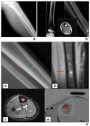
Results: All radiographs were originally read as normal. Of the tibia studied, 80% (127 of 160) showed evidence of BSI on MRI. None of the original radiographs demonstrated a fracture line on initial review by the orthopaedic surgeons. Retrospective review by 2 MSK radiologists identified 27% of radiographs (34 of 127) with evidence of abnormality, which correlated with clinical examination and significant findings on MRI. Review of radiographs found evidence of new bone on 0 of 28 Fredericson grade 0, 0 of 19 Fredericson grade I, 11 of 80 (13.7%) Fredericson grade II, 18 of 28 (64%) Fredericson grade III, and 5 of 5 (100%) Fredericson grade IV. Sensitivity of radiographs showed evidence of new bone on 27% (34 of 127) of initial radiographs, with presence more common with greater degree of BSI, as 23 of 33 (70%) were higher-grade injuries (III of IV) of BSI. Specificity and positive predictive value were 100%, while negative predictive value was 17%.
Conclusion: These findings highlight the importance of initial radiographs in identifying high-grade BSI. As radiographs are readily available in most office settings of sports medicine physicians, this information can influence the management of adolescent athletic BSI without the need to delay treatment to obtain an MRI.
Clinical relevance: Adolescent athletes with radiographic evidence of BSI should be treated in a timely and more conservative manner, given the likelihood of higher-grade BSI. In addition, clinicians knowledgeable of the radiographic findings of high-grade BSI should feel more confident that a negative initial radiograph is not likely to be a high-grade BSI and can modify their treatment plans accordingly.
Keywords: MRI; adolescent athlete; bony stress injury; new bone; radiograph; radiography.
Conflict of interest statement
The following author declared potential conflicts of interest: C.J.G.J. has received consulting fee or honorarium from Musculoskeletal Transplant Foundation and reports patents from Meniscofix.
Figures


Similar articles
-
MRI of tibial stress fractures: relationship between Fredericson classification and time to recovery in pediatric athletes.Pediatr Radiol. 2020 Nov;50(12):1735-1741. doi: 10.1007/s00247-020-04760-8. Epub 2020 Aug 18. Pediatr Radiol. 2020. PMID: 32809066
-
Validation of the Shin Pain Scoring System: A Novel Approach for Determining Tibial Bone Stress Injuries.Orthop J Sports Med. 2019 Oct 30;7(10):2325967119877803. doi: 10.1177/2325967119877803. eCollection 2019 Oct. Orthop J Sports Med. 2019. PMID: 31696132 Free PMC article.
-
Significance of radiographic abnormalities in patients with tibial stress injuries: correlation with magnetic resonance imaging.Skeletal Radiol. 2007 Jul;36(7):633-40. doi: 10.1007/s00256-006-0272-4. Epub 2007 Mar 27. Skeletal Radiol. 2007. PMID: 17387472
-
Does Magnetic Resonance Imaging Grading Correlate With Return to Sports After Bone Stress Injuries? A Systematic Review and Meta-analysis.Am J Sports Med. 2022 Mar;50(3):834-844. doi: 10.1177/0363546521993807. Epub 2021 Mar 15. Am J Sports Med. 2022. PMID: 33720786
-
Current Evidence Regarding Diagnostic Imaging Methods for Pediatric Lumbar Spondylolysis: A Report From the Scoliosis Research Society Evidence-Based Medicine Committee.Spine Deform. 2017 Mar;5(2):97-101. doi: 10.1016/j.jspd.2016.10.006. Spine Deform. 2017. PMID: 28259272 Review.
Cited by
-
Exploring the early diagnostic value of MRI for type I stress fractures: a retrospective analysis based on imaging manifestations.Front Surg. 2025 Mar 17;12:1333714. doi: 10.3389/fsurg.2025.1333714. eCollection 2025. Front Surg. 2025. PMID: 40166621 Free PMC article.
-
Stress fractures and overuse injuries in children and adolescents.J Pediatr Soc North Am. 2024 Apr 9;7:100029. doi: 10.1016/j.jposna.2024.100029. eCollection 2024 May. J Pediatr Soc North Am. 2024. PMID: 40433260 Free PMC article.
References
-
- Baleani M. Compressive behavior of cortical bone in young humans.J Biomechanics. 2008;16:41.
-
- Beck BR. Tibial stress injury: relationship of radiographic, nuclear medicine bone scanning, MR imaging, and CT severity grades to clinical severity and time to healing. Radiology. 2012;263:311-318. - PubMed
-
- Bennell KL, Brukner PD. Epidemiology and site specificity of stress fractures. Clin Sports Med. 1997;16:179-196. - PubMed
-
- Berger FH, de Jonge MC, Maas M. Stress fractures in the lower extremity. The importance of increasing awareness amongst radiologists. Eur J Radiol. 2007;62:16-26. - PubMed
MeSH terms
LinkOut - more resources
Full Text Sources
Medical
Research Materials

