

Clinical and imaging characteristics of outer retinal folds in eyes with retinitis
- PMID: 35918957
- PMCID: PMC9672790
- DOI: 10.4103/ijo.IJO_70_22
Clinical and imaging characteristics of outer retinal folds in eyes with retinitis
Abstract
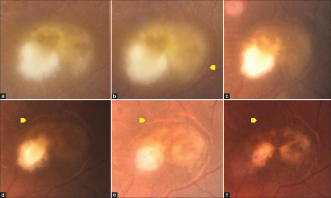
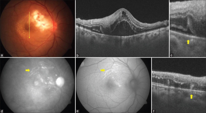
Purpose: To describe clinical and imaging characteristics of the outer retinal folds (ORF) in cases of retinitis, retinochoroiditis, and chorioretinitis.
Methods: Retrospective review of retinitis cases with presence of ORFs either at presentation or during follow up.
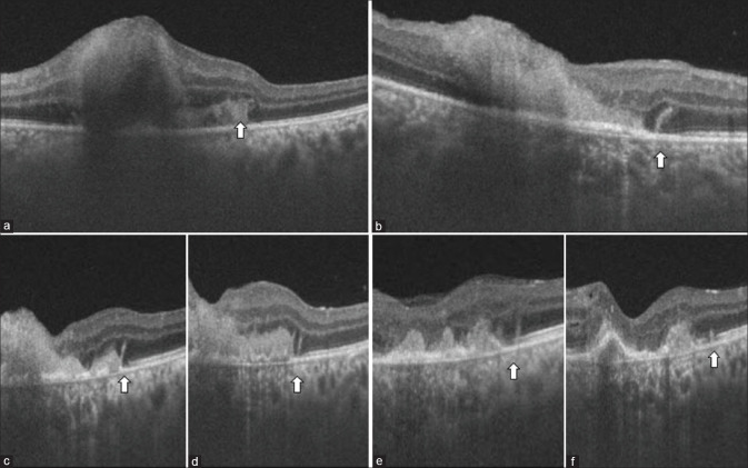
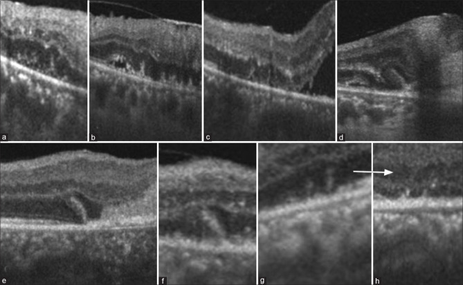
Results: ORFs were seen adjacent to retinitis lesions in 16 eyes of 14 cases (retinitis post-febrile illness n = 10, toxoplasma retinochoroiditis n = 2, fungal chorioretinitis n = 2) either at presentation (n = 2) or during follow up (n = 14). Optical coherence tomography (OCT) appearance was outer retinal vertical stout lesions involving ellipsoid, external limiting membrane, and outer nuclear layer. All the cases had a presence of past or concurrent subretinal fluid and/or subretinal hyperreflective material when ORF was seen. ORF resolved with variable outer retinal atrophy over a mean period of 2.86 months.
Conclusion: ORF is observed in cases of retinitis with subretinal fluid either at presentation or during resolution. It is not specific to any etiological disease. Differentiation of this sign from vertical outer retinal stripes in viral retinitis on OCT is important to avoid misinterpretation.
Keywords: Chorioretinitis; optical coherence tomography; outer retinal folds; retinitis; retinochoroiditis.
Conflict of interest statement
None
Figures
Similar articles
-
Spectral domain optical coherence tomography findings in patients with acute syphilitic posterior placoid chorioretinopathy.Retina. 2014 Feb;34(2):373-84. doi: 10.1097/IAE.0b013e3182993f11. Retina. 2014. PMID: 23860561
-
SWEPT-SOURCE OPTICAL COHERENCE TOMOGRAPHY ANGIOGRAPHY IN RICKETTSIAL RETINITIS.Retin Cases Brief Rep. 2019 Fall;13(4):348-351. doi: 10.1097/ICB.0000000000000603. Retin Cases Brief Rep. 2019. PMID: 28614137
-
Multimodal imaging in a case of bilateral outer retinitis associated with mumps infection.Int Ophthalmol. 2018 Feb;38(1):339-343. doi: 10.1007/s10792-016-0417-y. Epub 2016 Dec 27. Int Ophthalmol. 2018. PMID: 28028739
-
EN FACE OPTICAL COHERENCE TOMOGRAPHY AND OPTICAL COHERENCE TOMOGRAPHY ANGIOGRAPHY OF MULTIPLE EVANESCENT WHITE DOT SYNDROME: New Insights Into Pathogenesis.Retina. 2016 Dec;36 Suppl 1:S178-S188. doi: 10.1097/IAE.0000000000001255. Retina. 2016. PMID: 28005676
-
Punctate Inner Retinal Toxoplasmosis: Case Series and Review of Literature.Ocul Immunol Inflamm. 2022 Apr 3;30(3):546-555. doi: 10.1080/09273948.2021.1980815. Epub 2021 Oct 8. Ocul Immunol Inflamm. 2022. PMID: 34623927 Review.
Cited by
-
Post-fever Retinitis With a Positive Weil-Felix Test: A Study From a Tertiary Center in South India.Cureus. 2024 Jan 29;16(1):e53162. doi: 10.7759/cureus.53162. eCollection 2024 Jan. Cureus. 2024. PMID: 38420096 Free PMC article.
References
-
- Invernizzi A, Cozzi M, Staurenghi G. Optical coherence tomography and optical coherence tomography angiography in uveitis:A review. Clin Exp Ophthalmol. 2019;47:357–71. - PubMed
-
- Pichi F, Sarraf D, Arepalli S, Lowder CY, Cunningham E T, Jr, Neri P, et al. The application of optical coherence tomography angiography in uveitis and inflammatory eye diseases. Prog Retin Eye Res. 2017;59:178–201. - PubMed
-
- Pichi F, Invernizzi A, Tucker WR, Munk MR. Optical coherence tomography diagnostic signs in posterior uveitis. Prog Retin Eye Res. 2020;75:100797. - PubMed
-
- Fuchs E. Classification of retinitis. Arch Ophthalmol. 1930;3:393–402.
MeSH terms
LinkOut - more resources
Full Text Sources
Miscellaneous
