. 2022 Aug;70(8):3116-3121.
doi: 10.4103/ijo.IJO_120_22.

Holographic elysium of a 4D ophthalmic anatomical and pathological metaverse with extended reality/mixed reality

Affiliations

Holographic elysium of a 4D ophthalmic anatomical and pathological metaverse with extended reality/mixed reality

Prasanna V Ramesh et al. Indian J Ophthalmol. 2022 Aug.

Abstract

Extended reality is one of the leading cutting-edge technologies, which has not yet fully set foot into the field of ophthalmology. The use of extended reality technology especially in ophthalmic education and counseling will revolutionize the face of teaching and counseling on a whole new level. We have used this novel technology and have created a holographic museum of various anatomical structures such as the eyeball, cerebral venous system, cerebral arterial system, cranial nerves, and various parts of the brain in fine detail. These four-dimensional (4D) ophthalmic holograms created by us (patent pending) are cost-effectively constructed with TrueColor confocal images to serve as a new-age immersive 4D pedagogical and counseling tool for gameful learning and counseling, respectively. According to our knowledge, this concept has not been reported in the literature before.

Keywords: 4D Ophthalmology; Cerebral Circulation; Counseling; Extended Reality; Mixed Reality; Pedagogy.


Conflict of interest statement

None

Figures

Figure 1
Figure 1
Image showing the user wearing the head-mounted HoloLens 2 device to experience the mixed reality (MR) 4D holograms of the brain, cerebral circulation, and the eyes with extraocular muscles
Figure 2
Figure 2
The HoloLens 2 user is seen experiencing the visual auras of various anatomical and pathological 4D models of the eyeball in space. The pathological retina hologram models are provided in color, autofluorescence, and infrared modes, thus giving the freedom of choice to the user for multimodal learning by touch and eye-tracking. The user is also seen holding the desired holograms after selection
Figure 3
Figure 3
Image shows the 4D holograms of the normal anatomy of the eyes in (a) cross-sectional view, (b) gonioscopic view, (c) electron microscopic view (for visualizing the trabecular meshwork) and (d) anteroposterior view
Figure 4
Figure 4
(a) Image shows the real-time demonstration of the flecked retina 4D holograms created by us in the HoloLens 2, from a third person’s point of view. (b) Image shows the flecked retina 4D holograms in color, autofluorescence, and infrared modes, as viewed through the HoloLens 2
Figure 5
Figure 5
(a) Image shows the real-time demonstration of the retinitis pigmentosa 4D holograms created by us in the HoloLens 2, from a third person’s point of view. (b) Image shows the flecked retina 4D holograms in color, autofluorescence, and infrared modes, as viewed through the HoloLens 2
Figure 6
Figure 6
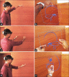
(a, c, and e) Image shows the real-time demonstration of the cerebral venous system 4D hologram created by us in the HoloLens 2, from a third person’s point of view. (b, d, and f) The image shows the cerebral venous system 4D hologram as viewed through the HoloLens 2 from different angles
Figure 7
Figure 7
(a–c) Image shows the hologram of the cerebral arterial circulation as viewed through the HoloLens 2 from different angles
Figure 8
Figure 8
(a–d) The image shows the holograms of the cerebrum, cerebellum, brainstem, cranial nerves with their nucleus and visual pathway anatomy from different views
Figure 9
Figure 9
The image shows the holograms of the extraocular muscles of both eyes simultaneously as viewed through the HoloLens 2
Figure 10
Figure 10
“Microsoft Mesh App” on Microsoft Store seen as a hologram, when viewed through HoloLens 2
Figure 11
Figure 11
“Microsoft Mesh App” icon seen as a hologram, when viewed through HoloLens 2
Figure 12
Figure 12
Image shows the holographic view of the (a) content options (red arrow) and (b) “OneDrive Cloud” icon (red arrow) after downloading the “Microsoft Mesh App” for viewing the different holograms in HoloLens 2

Comment in

Similar articles

Cited by

References

    1. Ong CW, Tan MCJ, Lam M, Koh VTC. Applications of extended reality in ophthalmology:Systematic review. J Med Internet Res. 2021;23:e24152. - PMC - PubMed
    1. Iskander M, Ogunsola T, Ramachandran R, McGowan R, Al-Aswad LA. Virtual reality and augmented reality in ophthalmology:A contemporary prospective. Asia-Pac J Ophthalmol. 2021;10:244–52. - PMC - PubMed
    1. HoloLens 2. [Last accessed on 2022 Jan 14]. Available from: https://medtrixhealthcare.com/holoLens-2blog-post .
    1. Ramesh PV, Aji K, Joshua T, Ramesh SV, Ray P, Raj PM, et al. Immersive photoreal new-age innovative gameful pedagogy for e-ophthalmology with 3D augmented reality. Indian J Ophthalmol. 2022;70:275–80. - PMC - PubMed
    1. Sostel. Eye tracking-Mixed Reality. [Last accessed on 2022 Jan 14]. Available from: https://docs.microsoft.com/en-us/windows/mixed-reality/design/eye-tracking .