

A software program for automated compressive vertebral fracture detection on elderly women's lateral chest radiograph: Ofeye 1.0
- PMID: 35919046
- PMCID: PMC9338385
- DOI: 10.21037/qims-22-433
A software program for automated compressive vertebral fracture detection on elderly women's lateral chest radiograph: Ofeye 1.0
Abstract
Background: Because osteoporotic vertebral fracture (OVF) on chest radiographs is commonly missed in radiological reports, we aimed to develop a software program which offers automated detection of compressive vertebral fracture (CVF) on lateral chest radiographs, and which emphasizes CVF detection specificity with a low false positivity rate.
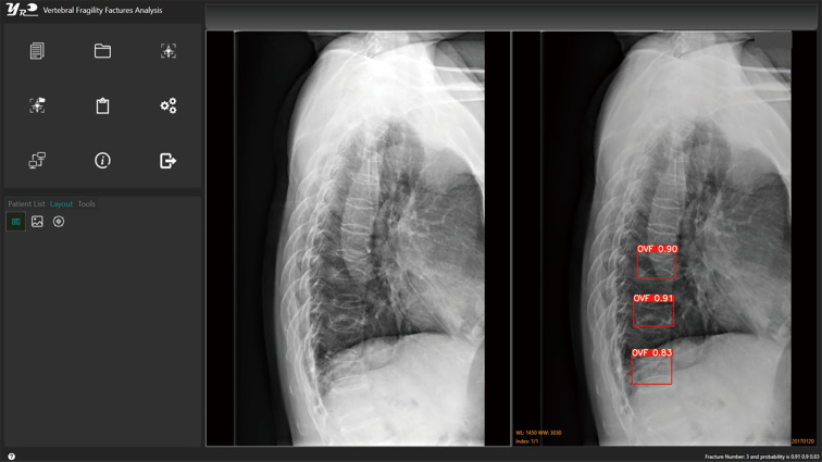
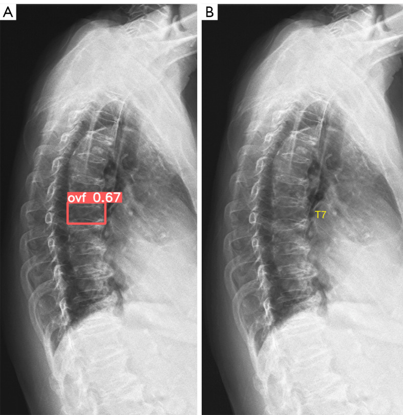
Methods: For model training, we retrieved 3,991 spine radiograph cases and 1,979 chest radiograph cases from 16 sources, with among them in total 1,404 cases had OVF. For model testing, we retrieved 542 chest radiograph cases and 162 spine radiograph cases from four independent clinics, with among them 215 cases had OVF. All cases were female subjects, and except for 31 training data cases which were spine trauma cases, all the remaining cases were post-menopausal women. Image data included DICOM (Digital Imaging and Communications in Medicine) format, hard film scanned PNG (Portable Network Graphics) format, DICOM exported PNG format, and PACS (Picture Archiving and Communication System) downloaded resolution reduced DICOM format. OVF classification included: minimal and mild grades with <20% or ≥20-25% vertebral height loss respectively, moderate grade with ≥25-40% vertebral height loss, severe grade with ≥40%-2/3 vertebral height loss, and collapsed grade with ≥2/3 vertebral height loss. The CVF detection base model was mainly composed of convolution layers that include convolution kernels of different sizes, pooling layers, up-sampling layers, feature merging layers, and residual modules. When the model loss function could not be further decreased with additional training, the model was considered to be optimal and termed 'base-model 1.0'. A user-friendly interface was also developed, with the synthesized software termed 'Ofeye 1.0'.
Results: Counting cases and with minimal and mild OVFs included, base-model 1.0 demonstrated a specificity of 97.1%, a sensitivity of 86%, and an accuracy of 93.9% for the 704 testing cases. In total, 33 OVFs in 30 cases had a false negative reading, which constituted a false negative rate of 14.0% (30/215) by counting all OVF cases. Eighteen OVFs in 15 cases had OVFs of ≥ moderate grades missed, which constituted a false negative rate of 7.0% (15/215, i.e., sensitivity 93%) if only counting cases with ≥ moderate grade OVFs missed. False positive reading was recorded in 13 vertebrae in 13 cases (one vertebra in each case), which constituted a false positivity rate of 2.7% (13/489). These vertebrae with false positivity labeling could be readily differentiated from a true OVF by a human reader. The software Ofeye 1.0 allows 'batch processing', for example, 100 radiographs can be processed in a single operation. This software can be integrated into hospital PACS, or installed in a standalone personal computer.
Conclusions: A user-friendly software program was developed for CVF detection on elderly women's lateral chest radiographs. It has an overall low false positivity rate, and for moderate and severe CVFs an acceptably low false negativity rate. The integration of this software into radiological practice is expected to improve osteoporosis management for elderly women.
Keywords: Osteoporosis; artificial intelligence; chest; deep learning; radiograph; vertebral fracture.
2022 Quantitative Imaging in Medicine and Surgery. All rights reserved.
Conflict of interest statement
Conflicts of Interest: All authors have completed the ICMJE uniform disclosure form (available at https://qims.amegroups.com/article/view/10.21037/qims-22-433/coif). YXJW serves as the Editor-in-Chief of Quantitative Imaging in Medicine and Surgery. YXJW is the founder of Yingran Medicals Ltd, which develops medical image-based diagnostics software, including Ofeye. BHX and MSYZ contributed to the development of Ofeye 1.0. The other authors have no conflicts of interest to declare.
Figures
Similar articles
-
Improving osteoporotic vertebral deformity detection on chest frontal view radiograph by adjusted X-ray beam positioning.J Orthop Translat. 2021 May 5;28:169-178. doi: 10.1016/j.jot.2021.04.001. eCollection 2021 May. J Orthop Translat. 2021. PMID: 34036040 Free PMC article.
-
Can a Deep-learning Model for the Automated Detection of Vertebral Fractures Approach the Performance Level of Human Subspecialists?Clin Orthop Relat Res. 2021 Jul 1;479(7):1598-1612. doi: 10.1097/CORR.0000000000001685. Clin Orthop Relat Res. 2021. PMID: 33651768 Free PMC article.
-
CT detects more osteoporotic endplate depressions than radiograph: a descriptive comparison of 76 vertebrae.Osteoporos Int. 2022 Jul;33(7):1569-1577. doi: 10.1007/s00198-022-06391-1. Epub 2022 Apr 4. Osteoporos Int. 2022. PMID: 35368223
-
Interpretation of osteoporotic vertebral deformity on frontal view radiographs of the chest and abdomen: a pictorial review.Quant Imaging Med Surg. 2021 Jan;11(1):423-442. doi: 10.21037/qims-2020-28. Quant Imaging Med Surg. 2021. PMID: 33392042 Free PMC article. Review.
-
Radiological diagnosis of prevalent osteoporotic vertebral fracture on radiographs: an interim consensus from a group of experts of the ESSR osteoporosis and metabolism subcommittee.Skeletal Radiol. 2024 Dec;53(12):2563-2574. doi: 10.1007/s00256-024-04678-4. Epub 2024 Apr 25. Skeletal Radiol. 2024. PMID: 38662094 Free PMC article. Review.
Cited by
-
Deep learning in the radiologic diagnosis of osteoporosis: a literature review.J Int Med Res. 2024 Apr;52(4):3000605241244754. doi: 10.1177/03000605241244754. J Int Med Res. 2024. PMID: 38656208 Free PMC article. Review.
-
Artificial Intelligence in Spine Surgery: Imaging-Based Applications for Diagnosis and Surgical Techniques.Curr Rev Musculoskelet Med. 2025 Oct;18(10):398-405. doi: 10.1007/s12178-025-09972-9. Epub 2025 Apr 30. Curr Rev Musculoskelet Med. 2025. PMID: 40304942 Free PMC article. Review.
-
Opportunistic Screening Techniques for Analysis of CT Scans.Curr Osteoporos Rep. 2023 Feb;21(1):65-76. doi: 10.1007/s11914-022-00764-5. Epub 2022 Nov 26. Curr Osteoporos Rep. 2023. PMID: 36435912 Free PMC article. Review.
-
Generalizability of Deep Learning Classification of Spinal Osteoporotic Compression Fractures on Radiographs Using an Adaptation of the Modified-2 Algorithm-Based Qualitative Criteria.Acad Radiol. 2023 Dec;30(12):2973-2987. doi: 10.1016/j.acra.2023.04.023. Epub 2023 Jul 10. Acad Radiol. 2023. PMID: 37438161 Free PMC article.
-
New Horizons: Artificial Intelligence Tools for Managing Osteoporosis.J Clin Endocrinol Metab. 2023 Mar 10;108(4):775-783. doi: 10.1210/clinem/dgac702. J Clin Endocrinol Metab. 2023. PMID: 36477337 Free PMC article.
References
-
- Johansson L, Sundh D, Magnusson P, Rukmangatharajan K, Mellström D, Nilsson AG, Lorentzon M. Grade 1 Vertebral Fractures Identified by Densitometric Lateral Spine Imaging Predict Incident Major Osteoporotic Fracture Independently of Clinical Risk Factors and Bone Mineral Density in Older Women. J Bone Miner Res 2020;35:1942-51. 10.1002/jbmr.4108 - DOI - PubMed
-
- Lentle BC, Berger C, Probyn L, Brown JP, Langsetmo L, Fine B, Lian K, Shergill AK, Trollip J, Jackson S, Leslie WD, Prior JC, Kaiser SM, Hanley DA, Adachi JD, Towheed T, Davison KS, Cheung AM, Goltzman D; CaMos Research Group. Comparative Analysis of the Radiology of Osteoporotic Vertebral Fractures in Women and Men: Cross-Sectional and Longitudinal Observations from the Canadian Multicentre Osteoporosis Study (CaMos). J Bone Miner Res 2018;33:569-79. 10.1002/jbmr.3222 - DOI - PubMed
-
- Kadowaki E, Tamaki J, Iki M, Sato Y, Chiba Y, Kajita E, Kagamimori S, Kagawa Y, Yoneshima H. Prevalent vertebral deformity independently increases incident vertebral fracture risk in middle-aged and elderly Japanese women: the Japanese Population-based Osteoporosis (JPOS) Cohort Study. Osteoporos Int 2010;21:1513-22. 10.1007/s00198-009-1113-9 - DOI - PubMed
LinkOut - more resources
Full Text Sources