. 2022 Aug;12(8):4176-4189.
doi: 10.21037/qims-21-1133.

Three-dimensional ultrashort echo time magnetic resonance imaging in assessment of idiopathic pulmonary fibrosis, in comparison with high-resolution computed tomography

Affiliations

Three-dimensional ultrashort echo time magnetic resonance imaging in assessment of idiopathic pulmonary fibrosis, in comparison with high-resolution computed tomography

Xiaoyan Yang et al. Quant Imaging Med Surg. 2022 Aug.

Abstract

Background: We aimed to evaluate the image quality, feasibility, and diagnostic performance of three-dimensional ultrashort echo time magnetic resonance imaging (3D UTE-MRI) to assess idiopathic pulmonary fibrosis (IPF) compared with high-resolution computed tomography (HRCT) and half-Fourier single-shot turbo spin-echo (HASTE) MRI.

Methods: A total of 36 patients with IPF (34 men; mean age: 62±8 years, age range: 43 to 78 years) were prospectively included and underwent HRCT and chest MRI on the same day. Chest MRI was performed with a free-breathing 3D spiral UTE pulse sequence and HASTE sequence on a 1.5 T MRI. Two radiologists independently evaluated the image quality of the HRCT, HASTE, and 3D UTE-MRI. They assessed the representative imaging features of IPF, including honeycombing, reticulation, traction bronchiectasis, and ground-glass opacities. Image quality of the 3D UTE-MRI, HASTE, and HRCT were assessed using a 5-point visual scoring method. Kappa and weighted kappa analysis were used to measure intra- and inter-observer and inter-method agreements. Sensitivity (SE), specificity (SP), and accuracy (AC) were used to assess the performance of 3D UTE-MRI for detecting image features of IPF and monitoring the extent of pulmonary fibrosis. Linear regressions and Bland-Altman plots were generated to assess the correlation and agreement between the assessment of the extent of pulmonary fibrosis made by the 2 observers.

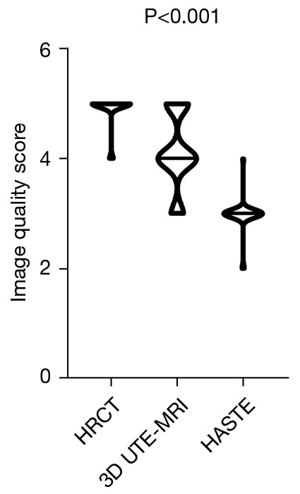
Results: The image quality of HRCT was higher than that of HASTE and UTE-MRI (HRCT vs. UTE-MRI vs. HASTE: 4.9±0.3 vs. 4.1±0.7 vs. 3.0±0.3; P<0.001). Interobserver agreement of HRCT, HASTE, and 3D UTE-MRI when assessing pulmonary fibrosis was substantial and excellent (HRCT: 0.727≤ κ ≤1, P<0.001; HASTE: 0.654≤ κ ≤1, P<0.001; 3D UTE-MRI: 0.719≤ κ ≤0.824, P<0.001). In addition, reticulation (SE: 97.1%; SP: 100%; AC: 97.2%; κ =0.654), honeycombing (SE: 83.3%; SP: 100%; AC: 86.1%; κ =0.625) patterns, and traction bronchiectasis (SE: 94.1%; SP: 100%; AC: 94.4%, κ =0.640) were also well-visualized on 3D UTE-MRI, which was significantly superior to HASTE. Compared with HRCT, the sensitivity of 3D UTE-MRI to detect signs of pulmonary fibrosis (n=35) was 97.2%. The interobserver agreement in elevation of the extent of pulmonary fibrosis with HRCT and 3D UTE-MRI was R2=0.84 (P<0.001) and R2=0.84 (P<0.001), respectively. The extent of pulmonary fibrosis assessed with 3D UTE-MRI [median =9, interquartile range (IQR): 6.25 to 10.00] was lower than that from HRCT (median =12, IQR: 9.25 to 13.00; U=320.00, P<0.001); however, they had a positive correlation (R=0.72, P<0.001).

Conclusions: As a radiation-free non-contrast enhanced imaging method, although the image quality of 3D UTE-MRI is inferior to that of HRCT, it has high reproducibility to identify the imaging features of IPF and evaluate the extent of pulmonary fibrosis.

Keywords: Lungs; high-resolution computed tomography (HRCT); idiopathic pulmonary fibrosis (IPF); three-dimensional ultrashort echo time magnetic resonance imaging (3D UTE-MRI).


Conflict of interest statement

Conflicts of Interest: All authors have completed the ICMJE uniform disclosure form (available at https://qims.amegroups.com/article/view/10.21037/qims-21-1133/coif). TB is employed by Siemens Healthcare GmbH and JA is employed by Siemens Shenzhen Magnetic Resonance Ltd. The other authors have no conflicts of interest to declare.

Figures

Figure 1
Figure 1
The flowchart of inclusion and exclusion criteria of patients in this study. IPF, idiopathic pulmonary fibrosis; MRI, magnetic resonance imaging.
Figure 2
Figure 2
A violin plot graph shows the image quality scores of HRCT, 3D UTE-MRI, and HASTE. Median: the line that crosses the box; data density: smoothed histograms alone the data point. 3D UTE-MRI, 3D ultrashort echo time magnetic resonance imaging; HASTE, half-Fourier single-shot turbo spin-echo; HRCT, high-resolution computed tomography.
Figure 3
Figure 3
A 77-year-old male patient with interstitial pulmonary fibrosis. Images were obtained with HRCT (A), 3D UTE-MRI (B), and HASTE (C). Honeycombing (green arrow), reticular patterns (red arrow), and traction bronchiectasis (blue arrow) are seen in the left and/or right lower lobes. 3D UTE-MRI and HRCT image qualities are equal (IQS: 5), which were higher than the HASTE image quality (IQS: 4). The extent of pulmonary fibrosis on images obtained with 3D UTE-MRI (IQS: 16) was equal to those obtained with HRCT (IQS: 16). The transverse 3D UTE-MRI images are MPRs. HRCT, high-resolution computed tomography; 3D UTE-MRI, 3D ultrashort echo time magnetic resonance imaging; HASTE, half-Fourier single-shot turbo spin-echo; MPRs, multiplanar reconstructions; IQS, image quality score.
Figure 4
Figure 4
A 68-year-old male patient with interstitial pulmonary fibrosis and emphysema. Images obtained with HRCT (A), and 3D UTE-MRI (B) show honeycombing (green arrow), reticular patterns (red arrow), and traction bronchiectasis (blue arrow) in the left and/or right lower lobes. The image quality of the 3D UTE-MRI (IQS: 4) was lower than that of the HRCT (IQS: 5). The extent of pulmonary fibrosis on images obtained with 3D UTE-MRI (IQS: 10) was lower than those obtained with HRCT (IQS: 14). The images obtained with HRCT also showed that the pulmonary emphysema (yellow arrow) was heterogeneously distributed in the low attenuated areas of both lungs (C). However, pulmonary emphysema was not seen in the lung segments of the 3D UTE-MRI images (D). The transverse 3D UTE-MRI images are MPRs. HRCT, high-resolution computed tomography; 3D UTE-MRI, 3D ultrashort echo time magnetic resonance imaging; IQS, image quality score; MPRs, multiplanar reconstructions.
Figure 5
Figure 5
The extent of pulmonary fibrosis obtained with reader one plotted as a function of reader two for IPF patients with both HRCT (R2=0.84, P<0.001) and 3D UTE-MRI (R2=0.84, P<0.001) (A,B). Bland-Altman plot of the agreement analysis of the extent of pulmonary fibrosis between reader one and reader two from HRCT and 3D UTE-MRI. Mean difference for HRCT (Reader 1, Reader 2) =0.86, 95% limits of agreement =−3.48 to 1.75 (C). Mean difference for 3D UTE-MRI (Reader 1, Reader 2) =−1.28, 95% limits of agreement =−4.00 to 1.44 (D). IPF, idiopathic pulmonary fibrosis; HRCT, high-resolution computed tomography; 3D UTE-MRI, 3D ultrashort echo time magnetic resonance imaging; LOA, limits of agreement.
Figure 6
Figure 6
Correlation between the extent of pulmonary fibrosis with HRCT and 3D UTE-MRI (R=0.72, P<0.001). HRCT, high-resolution computed tomography; 3D UTE-MRI, 3D ultrashort echo time magnetic resonance imaging.
Figure 7
Figure 7
A bar chart showing the discrepancies in the extent of pulmonary fibrosis obtained with an average of two readers between HRCT (blue) and 3D UTE-MRI (red) for 36 patients with IPF. HRCT, high-resolution computed tomography; 3D UTE-MRI, 3D ultrashort echo time magnetic resonance imaging; IPF, idiopathic pulmonary fibrosis.

References

    1. Raghu G, Remy-Jardin M, Myers JL, Richeldi L, Ryerson CJ, Lederer DJ, et al. Diagnosis of Idiopathic Pulmonary Fibrosis. An Official ATS/ERS/JRS/ALAT Clinical Practice Guideline. Am J Respir Crit Care Med 2018;198:e44-68. 10.1164/rccm.201807-1255ST - DOI - PubMed
    1. Ciet P, Tiddens HA, Wielopolski PA, Wild JM, Lee EY, Morana G, Lequin MH. Magnetic resonance imaging in children: common problems and possible solutions for lung and airways imaging. Pediatr Radiol 2015;45:1901-15. 10.1007/s00247-015-3420-y - DOI - PMC - PubMed
    1. Hatabu H, Gaa J, Tadamura E, Edinburgh KJ, Stock KW, Garpestad E, Edelman RR. MR imaging of pulmonary parenchyma with a half-Fourier single-shot turbo spin-echo (HASTE) sequence. Eur J Radiol 1999;29:152-9. 10.1016/S0720-048X(98)00167-3 - DOI - PubMed
    1. Puderbach M, Eichinger M, Gahr J, Ley S, Tuengerthal S, Schmähl A, Fink C, Plathow C, Wiebel M, Müller FM, Kauczor HU. Proton MRI appearance of cystic fibrosis: comparison to CT. Eur Radiol 2007;17:716-24. 10.1007/s00330-006-0373-4 - DOI - PubMed
    1. Puderbach M, Eichinger M, Haeselbarth J, Ley S, Kopp-Schneider A, Tuengerthal S, Schmaehl A, Fink C, Plathow C, Wiebel M, Demirakca S, Müller FM, Kauczor HU. Assessment of morphological MRI for pulmonary changes in cystic fibrosis (CF) patients: comparison to thin-section CT and chest x-ray. Invest Radiol 2007;42:715-25. 10.1097/RLI.0b013e318074fd81 - DOI - PubMed