

UTE-T2* versus conventional T2* mapping to assess posterior cruciate ligament ultrastructure and integrity-an in-situ study
- PMID: 35919061
- PMCID: PMC9338370
- DOI: 10.21037/qims-22-251
UTE-T2* versus conventional T2* mapping to assess posterior cruciate ligament ultrastructure and integrity-an in-situ study
Abstract
Background: Clinical-standard morphologic magnetic resonance imaging (MRI) is limited in the refined diagnosis of posterior cruciate ligament (PCL) injuries. Quantitative MRI sequences such as ultrashort echo-time (UTE)-T2* mapping or conventional T2* mapping have been theorized to quantify ligament (ultra-) structure and integrity beyond morphology. This study evaluates their diagnostic potential in identifying and differentiating partial and complete PCL injuries in a standardized graded injury model.
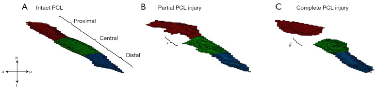
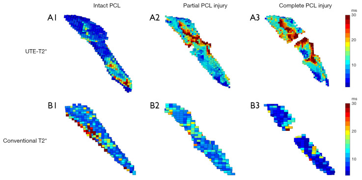
Methods: Ten human cadaveric knee joint specimens were imaged on a clinical 3.0 T MRI scanner using morphologic, conventional T2* mapping, and UTE-T2* mapping sequences before and after standardized arthroscopic partial and complete PCL transection. Following manual segmentation, quantitative T2* and underlying texture features (i.e., energy, homogeneity, and variance) were analyzed for each specimen and PCL condition, both for the entire PCL and its subregions. For statistical analysis, Friedman's test followed by Dunn's multiple comparison test was used against the level of significance of P≤0.01.
Results: For the entire PCL, T2* was significantly increased as a function of injury when acquired with the UTE-T2* sequence [entire PCL: 11.1±3.1 ms (intact); 10.9±4.6 ms (partial); 14.3±4.9 ms (complete); P<0.001], but not when acquired with the conventional T2* sequence [entire PCL: 10.0±3.2 ms (intact); 11.4±6.2 ms (partial); 15.5±7.8 ms (complete); P=0.046]. The PCL subregions and texture variables showed variable changes indicative of injury-associated disorganization.
Conclusions: In contrast to the conventional T2* mapping, UTE-T2* mapping is more receptive in the detection of structural damage of the PCL and allows quantitative assessment of ligament (ultra-)structure and integrity that may help to improve diagnostic differentiation of distinct injury states. Once further substantiated beyond the in-situ setting, UTE-T2* mapping may refine diagnostic evaluation of PCL injuries and -possibly- monitor ligament healing, ageing, degeneration, and inflammation.
Keywords: Magnetic resonance imaging (MRI); knee joint instability; posterior cruciate ligament (PCL); quantitative imaging; ultrashort echo-time (UTE)-T2*.
2022 Quantitative Imaging in Medicine and Surgery. All rights reserved.
Conflict of interest statement
Conflicts of Interest: All authors have completed the ICMJE uniform disclosure form (available at https://qims.amegroups.com/article/view/10.21037/qims-22-251/coif). The authors have no conflicts of interest to declare.
Figures
Similar articles
-
Quantitative differentiation of tendon and ligament using magnetic resonance imaging ultrashort echo time T2* mapping of normal knee joint.Acta Radiol. 2022 Nov;63(11):1489-1496. doi: 10.1177/02841851211043834. Epub 2021 Sep 24. Acta Radiol. 2022. PMID: 34558315
-
Quantitative Magnetic Resonance Imaging UTE-T2* Mapping of Cartilage and Meniscus Healing After Anatomic Anterior Cruciate Ligament Reconstruction.Am J Sports Med. 2014 Aug;42(8):1847-56. doi: 10.1177/0363546514532227. Epub 2014 May 8. Am J Sports Med. 2014. PMID: 24812196 Free PMC article.
-
Quantifiable Imaging Biomarkers for Evaluation of the Posterior Cruciate Ligament Using 3-T Magnetic Resonance Imaging: A Feasibility Study.Orthop J Sports Med. 2016 Apr 8;4(4):2325967116639044. doi: 10.1177/2325967116639044. eCollection 2016 Apr. Orthop J Sports Med. 2016. PMID: 27104206 Free PMC article.
-
Quantitative mapping of acute and chronic PCL pathology with 3 T MRI: a prospectively enrolled patient cohort.J Exp Orthop. 2019 May 28;6(1):22. doi: 10.1186/s40634-019-0188-2. J Exp Orthop. 2019. PMID: 31139976 Free PMC article.
-
Injuries to the posterior cruciate ligament and posterolateral instabilities of the knee.Chang Gung Med J. 2002 May;25(5):288-97. Chang Gung Med J. 2002. PMID: 12141701 Review.
Cited by
-
Ultrashort echo time pulse sequences for visualization of deep peripheral fasciae and epimysium in porcine models with histologic correlations.Quant Imaging Med Surg. 2023 Dec 1;13(12):8447-8461. doi: 10.21037/qims-23-687. Epub 2023 Oct 11. Quant Imaging Med Surg. 2023. PMID: 38106251 Free PMC article.
-
Magnetic resonance imaging of cruciate ligament disorders: current updates.EFORT Open Rev. 2025 Jul 1;10(7):475-486. doi: 10.1530/EOR-2024-0093. EFORT Open Rev. 2025. PMID: 40591678 Free PMC article. Review.
-
Ultrashort-T2* mapping at 7 tesla using an optimized pointwise encoding time reduction with radial acquisition (PETRA) sequence at standard and extended echo times.PLoS One. 2025 Apr 17;20(4):e0310590. doi: 10.1371/journal.pone.0310590. eCollection 2025. PLoS One. 2025. PMID: 40245029 Free PMC article.
-
Assessment of ultrashort echo time (UTE) T2* mapping at 3T for the whole knee: repeatability, the effects of fat suppression, and knee position.Quant Imaging Med Surg. 2023 Dec 1;13(12):7893-7909. doi: 10.21037/qims-23-459. Epub 2023 Nov 22. Quant Imaging Med Surg. 2023. PMID: 38106304 Free PMC article.
-
Multilesion Segmentations in Patients with Intracerebral Hemorrhage: Reliability of ICH, IVH and PHE Masks.Tomography. 2023 Jan 11;9(1):89-97. doi: 10.3390/tomography9010008. Tomography. 2023. PMID: 36648995 Free PMC article.
References
-
- Arøen A, Sivertsen EA, Owesen C, Engebretsen L, Granan LP. An isolated rupture of the posterior cruciate ligament results in reduced preoperative knee function in comparison with an anterior cruciate ligament injury. Knee Surg Sports Traumatol Arthrosc 2013;21:1017-22. 10.1007/s00167-012-2132-1 - DOI - PubMed
LinkOut - more resources
Full Text Sources