

Coronary-specific quantification of myocardial deformation by strain echocardiography may disclose the culprit vessel in patients with non-ST-segment elevation acute coronary syndrome
- PMID: 35919124
- PMCID: PMC9242069
- DOI: 10.1093/ehjopen/oeac010
Coronary-specific quantification of myocardial deformation by strain echocardiography may disclose the culprit vessel in patients with non-ST-segment elevation acute coronary syndrome
Abstract
Aims: To compare the diagnostic accuracy of speckle tracking echocardiography technique using territorial longitudinal strain (TLS) for the detection of culprit vessel vs. vessel-specific wall motion score index (WMSI) in non-ST-segment elevation-acute coronary syndrome (NSTE-ACS) patients scheduled for invasive coronary angiography (ICA).
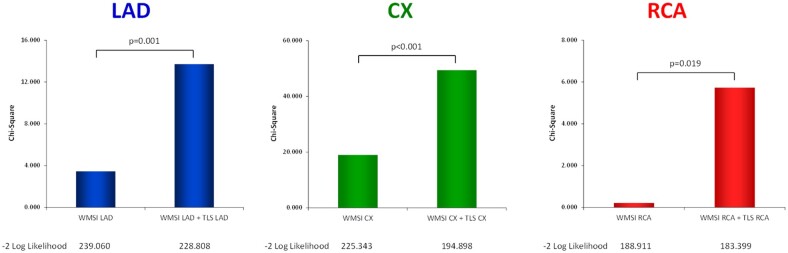
Methods and results: One hundred and eighty-three patients (mean age: 66 ± 12 years, male: 71%) diagnosed with NSTE-ACS underwent echocardiography evaluation at hospital admission and ICA within 24 h. Culprit vessels were left anterior descending (LAD), left circumflex (CX), and right coronary arteries (RCAs) in 38.5%, 39.6%, and 21.4%, respectively. An increase of affected vessels [1-, 2-, and 3-vessel coronary artery disease (CAD)] was associated with increased WMSI and TLS values. There was a statistically significant difference of both WMSI-LAD, WMSI-CX, WMSI-RCA and TLS-LAD, TLS-CX, TLS-RCA of myocardial segments with underlying severe CAD compared to no CAD (P = 0.001 and P < 0.001, respectively). Moreover, a significant difference of TLS-LAD, TLS-CX, TLS-RCA, and WMSI-CX of myocardial segments with an underlying culprit vessel compared to non-culprit vessels (P < 0.001, P < 0.001, P = 0.022, and P < 0.001, respectively) was identified. WMSI-LAD and WMSI-RCA did not show statistical significant differences. A regression model revealed that the combination of WMSI + TLS was more accurate compared to WMSI alone in detecting the culprit vessel (LAD, P = 0.001; CX, P < 0.001; and RCA, P = 0.019).
Conclusion: Territorial longitudinal strain allows an accurate identification of the culprit vessel in NSTE-ACS patients. In addition to WMSI, TLS may be considered as part of routine echocardiography for better clinical assessment in this subset of patients.
Keywords: 2D speckle tracking echocardiography; Culprit lesion; Non-ST-elevation myocardial infarction; Territorial longitudinal strain.
© The Author(s) 2022. Published by Oxford University Press on behalf of the European Society of Cardiology.
Figures


Comment on
-
Territorial longitudinal strain discloses the culprit vessel in a patient with non-ST-segment elevation acute coronary syndrome.Eur Heart J Case Rep. 2022 Mar 3;6(3):ytac097. doi: 10.1093/ehjcr/ytac097. eCollection 2022 Mar. Eur Heart J Case Rep. 2022. PMID: 35350722 Free PMC article. No abstract available.
Similar articles
-
Correlation between left ventricular global and regional longitudinal systolic strain and impaired microcirculation in patients with acute myocardial infarction.Echocardiography. 2012 Nov;29(10):1181-90. doi: 10.1111/j.1540-8175.2012.01784.x. Epub 2012 Aug 3. Echocardiography. 2012. PMID: 22862151
-
Predictive value of global and territorial longitudinal strain imaging in detecting significant coronary artery disease in patients with myocardial infarction without persistent ST-segment elevation.Echocardiography. 2019 Mar;36(3):512-520. doi: 10.1111/echo.14275. Epub 2019 Feb 25. Echocardiography. 2019. PMID: 30803009
-
Usefulness of layer-specific strain for identifying complex CAD and predicting the severity of coronary lesions in patients with non-ST-segment elevation acute coronary syndrome: Compared with Syntax score.Int J Cardiol. 2016 Nov 15;223:1045-1052. doi: 10.1016/j.ijcard.2016.08.277. Epub 2016 Aug 18. Int J Cardiol. 2016. PMID: 27592047
-
Strain Rate Changes during Stress Echocardiography Are the Most Accurate Predictors of Significant Coronary Artery Disease in Patients with Previously Treated Acute Coronary Syndrome.Diagnostics (Basel). 2023 May 19;13(10):1796. doi: 10.3390/diagnostics13101796. Diagnostics (Basel). 2023. PMID: 37238281 Free PMC article.
-
Longitudinal 2D strain can help diagnose coronary artery disease in patients with suspected non-ST-elevation acute coronary syndrome but apparent normal global and segmental systolic function.Int J Cardiol. 2017 Jun 1;236:91-94. doi: 10.1016/j.ijcard.2017.02.068. Epub 2017 Feb 22. Int J Cardiol. 2017. PMID: 28258851
Cited by
-
Assessment and management of heart failure in patients with chronic kidney disease.Heart Fail Rev. 2024 Mar;29(2):379-394. doi: 10.1007/s10741-023-10346-x. Epub 2023 Sep 20. Heart Fail Rev. 2024. PMID: 37728751 Free PMC article. Review.
-
Evaluation of changes in the global longitudinal strain for left ventricular function before and after eight weeks of taking high dose of atorvastatin in patients with coronary slow flow phenomenon.BMC Cardiovasc Disord. 2024 Sep 27;24(1):522. doi: 10.1186/s12872-024-04198-y. BMC Cardiovasc Disord. 2024. PMID: 39333856 Free PMC article.
-
Improvement of Left Ventricular Global Longitudinal Strain after 6-Month Therapy with GLP-1RAs Semaglutide and Dulaglutide in Type 2 Diabetes Mellitus: A Pilot Study.J Clin Med. 2023 Feb 16;12(4):1586. doi: 10.3390/jcm12041586. J Clin Med. 2023. PMID: 36836121 Free PMC article.
-
Deep learning improves test-retest reproducibility of regional strain in echocardiography.Eur Heart J Imaging Methods Pract. 2024 Oct 23;2(4):qyae092. doi: 10.1093/ehjimp/qyae092. eCollection 2024 Oct. Eur Heart J Imaging Methods Pract. 2024. PMID: 39449961 Free PMC article.
-
Multimodality Imaging of Sudden Cardiac Death and Acute Complications in Acute Coronary Syndrome.J Clin Med. 2022 Sep 26;11(19):5663. doi: 10.3390/jcm11195663. J Clin Med. 2022. PMID: 36233531 Free PMC article. Review.
References
-
- Benjamin EJ, Virani SS, Callaway CW, Chamberlain AM, Chang AR, Cheng S, Chiuve SE, Cushman M, Delling FN, Deo R, de Ferranti SD, Ferguson JF, Fornage M, Gillespie C, Isasi CR, Jiménez MC, Jordan LC, Judd SE, Lackland D, Lichtman JH, Lisabeth L, Liu S, Longenecker CT, Lutsey PL, Mackey JS, Matchar DB, Matsushita K, Mussolino ME, Nasir K, O'Flaherty M, Palaniappan LP, Pandey A, Pandey DK, Reeves MJ, Ritchey MD, Rodriguez CJ, Roth GA, Rosamond WD, Sampson UKA, Satou GM, Shah SH, Spartano NL, Tirschwell DL, Tsao CW, Voeks JH, Willey JZ, Wilkins JT, Wu JH, Alger HM, Wong SS, Muntner P; American Heart Association Council on Epidemiology and Prevention Statistics Committee and Stroke Statistics Subcommittee. Heart disease and stroke statistics—2018 update: a report from the American Heart Association. Circulation 2018;137:e67–e492. - PubMed
-
- Boeddinghaus J, Nestelberger T, Twerenbold R, Neumann JT, Lindahl B, Giannitsis E, Sörensen NA, Badertscher P, Jann JE, Wussler D, Puelacher C, Rubini Giménez M, Wildi K, Strebel I, Du Fay de Lavallaz J, Selman F, Sabti Z, Kozhuharov N, Potlukova E, Rentsch K, Miró Ò, Martin-Sanchez FJ, Morawiec B, Parenica J, Lohrmann J, Kloos W, Buser A, Geigy N, Keller DI, Osswald S, Reichlin T, Westermann D, Blankenberg S, Mueller C; APACE, BACC, and TRAPID-AMI Investigators. Impact of age on the performance of the ESC 0/1h algorithms for early diagnosis of myocardial infarction. Eur Heart J 2018;39:3780–3794. - PubMed
-
- Collet JP, Thiele H, Barbato E, Barthélémy O, Bauersachs J, Bhatt DL, Dendale P, Dorobantu M, Edvardsen T, Folliguet T, Gale CP, Gilard M, Jobs A, Jüni P, Lambrinou E, Lewis BS, Mehilli J, Meliga E, Merkely B, Mueller C, Roffi M, Rutten FH, Sibbing D, Siontis GCM.. ESC Scientific Document Group. 2020 ESC Guidelines for the management of acute coronary syndromes in patients presenting without persistent ST-segment elevation. Eur Heart J 2021 Apr 7;42(14):1289–1367.
-
- Guaricci AI, Pontone G, Fusini L, De Luca M, Cafarelli FP, Guglielmo M, Baggiano A, Beltrama V, Muscogiuri G, Mushtaq S, Conte E, Guglielmi G, Andreini D, Brunetti ND, Di Biase M, Bartorelli AL, Pepi M.. Additional value of inflammatory biomarkers and carotid artery disease in prediction of significant coronary artery disease as assessed by coronary computed tomography angiography. Eur Heart J Cardiovasc Imaging 2017;18:1049–1056. - PubMed
